

A single cell death is disruptive to spontaneous Ca2+ activity in astrocytes
- PMID: 35966204
- PMCID: PMC9364045
- DOI: 10.3389/fncel.2022.945737
A single cell death is disruptive to spontaneous Ca2+ activity in astrocytes
Abstract
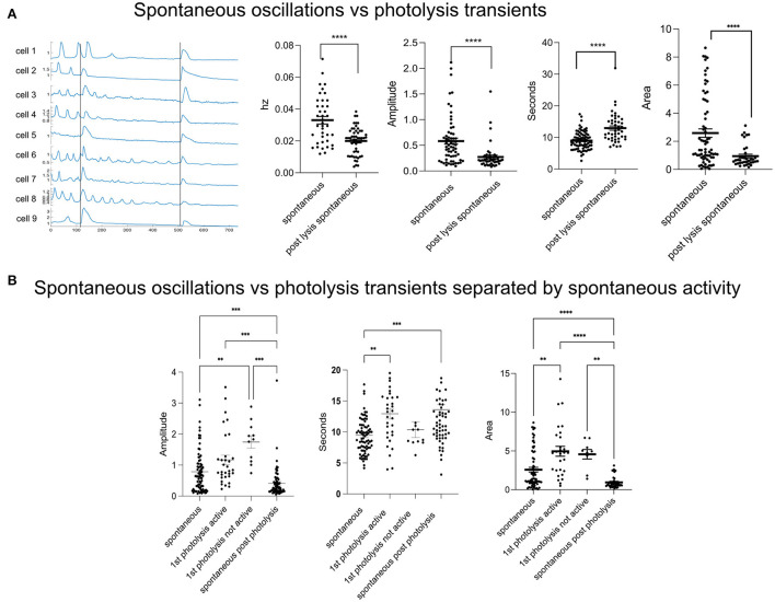
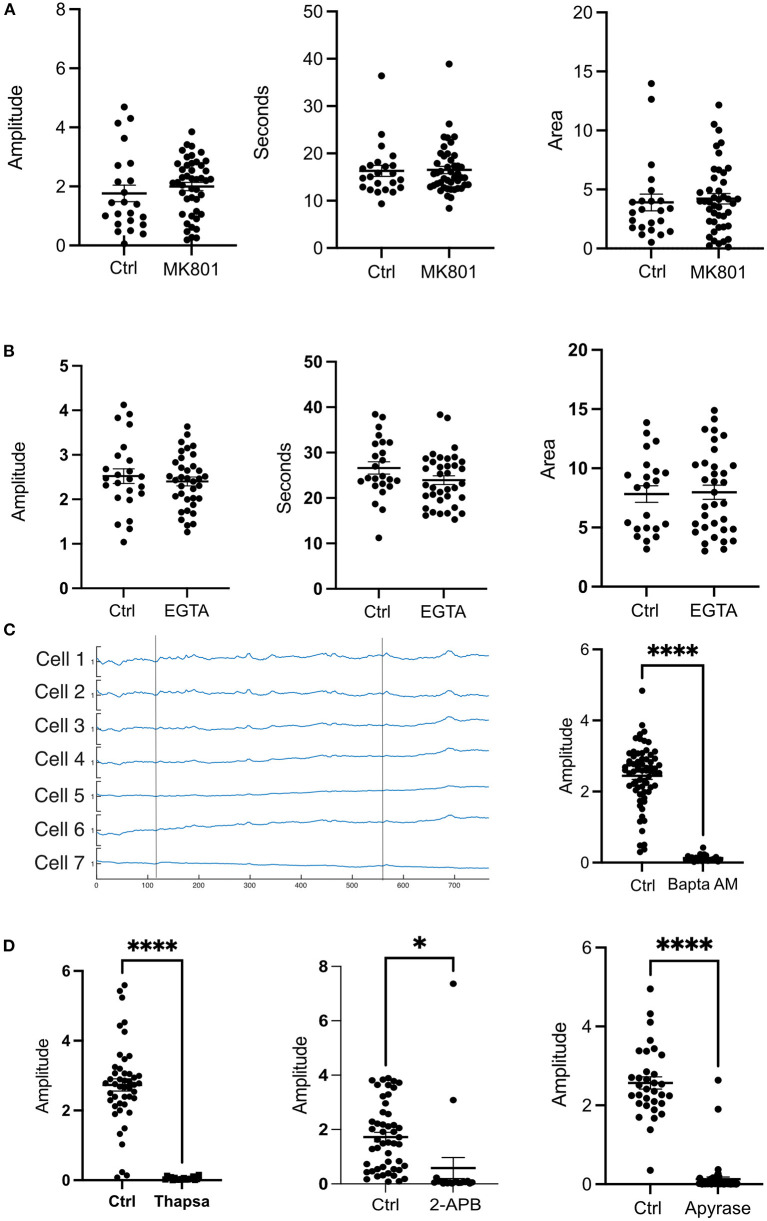
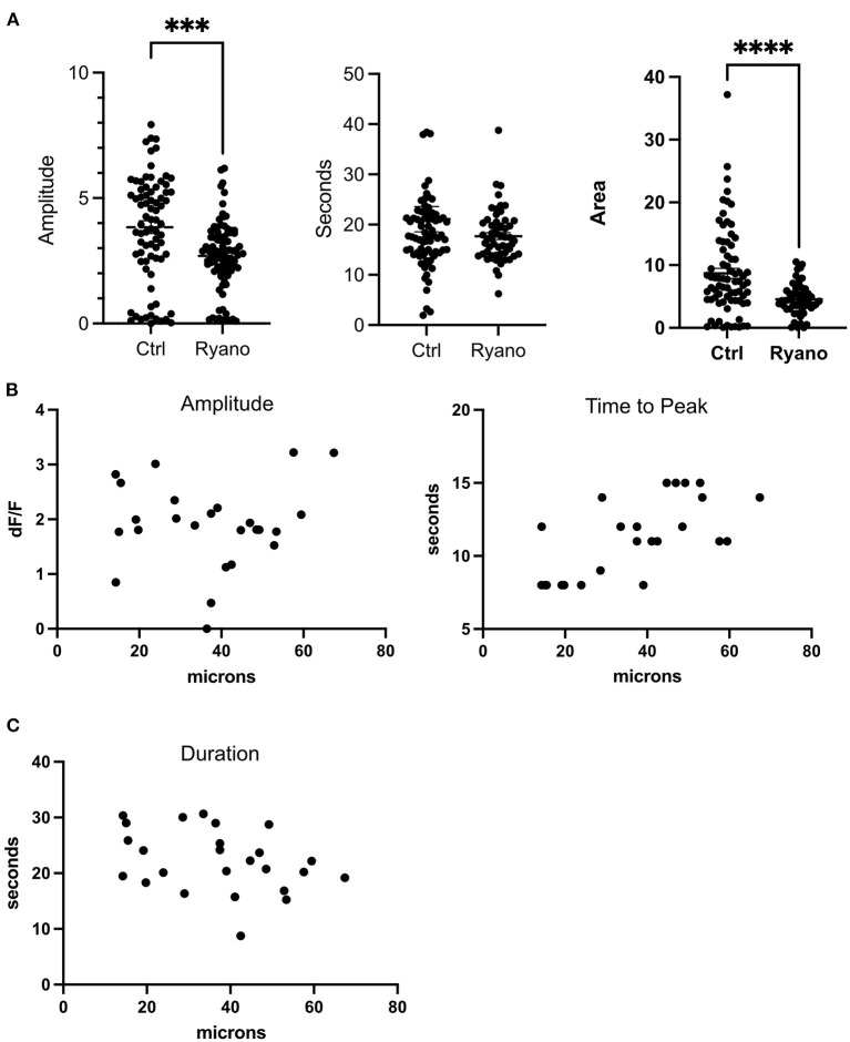
Astrocytes in the brain are rapidly recruited to sites of injury where they phagocytose damaged material and take up neurotransmitters and ions to avoid the spreading of damaging molecules. In this study we investigate the calcium (Ca2+) response in astrocytes to nearby cell death. To induce cell death in a nearby cell we utilized a laser nanosurgery system to photolyze a selected cell from an established astrocyte cell line (Ast1). Our results show that the lysis of a nearby cell is disruptive to surrounding cells' Ca2+ activity. Additionally, astrocytes exhibit a Ca2+ transient in response to cell death which differs from the spontaneous oscillations occurring in astrocytes prior to cell lysis. We show that the primary source of the Ca2+ transient is the endoplasmic reticulum.
Keywords: Ca2+; astrocyte; calcium; cell death; laser; photolysis; spontaneous activity.
Copyright © 2022 Gomez-Godinez, Li, Kuang, Liu, Shi and Berns.
Conflict of interest statement
The authors declare that the research was conducted in the absence of any commercial or financial relationships that could be construed as a potential conflict of interest.
Figures
Similar articles
-
Calcium Dynamics in Astrocytes During Cell Injury.Front Bioeng Biotechnol. 2020 Aug 27;8:912. doi: 10.3389/fbioe.2020.00912. eCollection 2020. Front Bioeng Biotechnol. 2020. PMID: 32984268 Free PMC article.
-
The role of Ca2+ in the generation of spontaneous astrocytic Ca2+ oscillations.Neuroscience. 2003;120(4):979-92. doi: 10.1016/s0306-4522(03)00379-8. Neuroscience. 2003. PMID: 12927204
-
Astrocyte Ca2+ Waves and Subsequent Non-Synchronized Ca2+ Oscillations Coincide with Arteriole Diameter Changes in Response to Spreading Depolarization.Int J Mol Sci. 2021 Mar 26;22(7):3442. doi: 10.3390/ijms22073442. Int J Mol Sci. 2021. PMID: 33810538 Free PMC article.
-
Local energy on demand: Are 'spontaneous' astrocytic Ca2+-microdomains the regulatory unit for astrocyte-neuron metabolic cooperation?Brain Res Bull. 2018 Jan;136:54-64. doi: 10.1016/j.brainresbull.2017.04.011. Epub 2017 Apr 24. Brain Res Bull. 2018. PMID: 28450076 Review.
-
Calcium oscillations encoding neuron-to-astrocyte communication.J Physiol Paris. 2002 Apr-Jun;96(3-4):193-8. doi: 10.1016/s0928-4257(02)00006-2. J Physiol Paris. 2002. PMID: 12445896 Review.
Cited by
-
Calcium Signaling in Astrocytes and Its Role in the Central Nervous System Injury.Mol Neurobiol. 2025 May 26. doi: 10.1007/s12035-025-05055-5. Online ahead of print. Mol Neurobiol. 2025. PMID: 40419752 Review.
References
LinkOut - more resources
Full Text Sources
Miscellaneous