

Outcomes of a Modified Arthroscopic-assisted Reconstruction Technique for Lateral Ankle Instability
- PMID: 35966426
- PMCID: PMC9365499
- DOI: 10.1055/s-0041-1741446
Outcomes of a Modified Arthroscopic-assisted Reconstruction Technique for Lateral Ankle Instability
Abstract
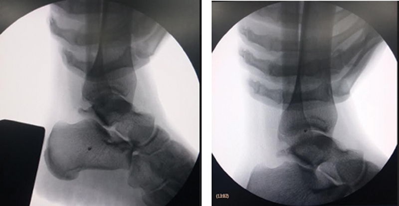
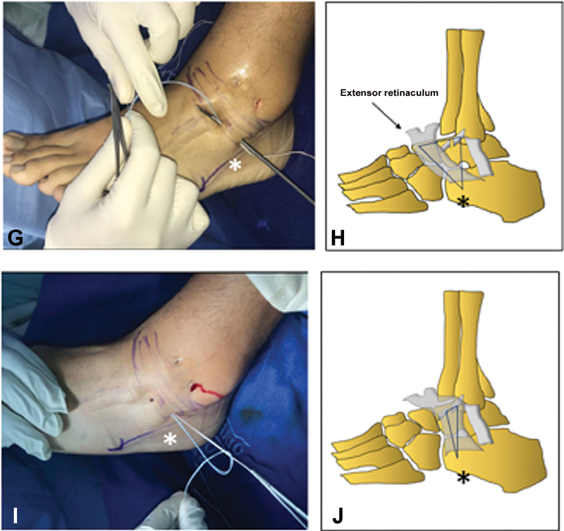
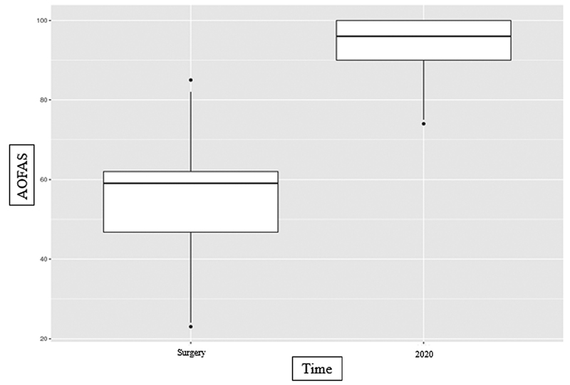
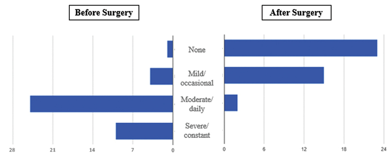
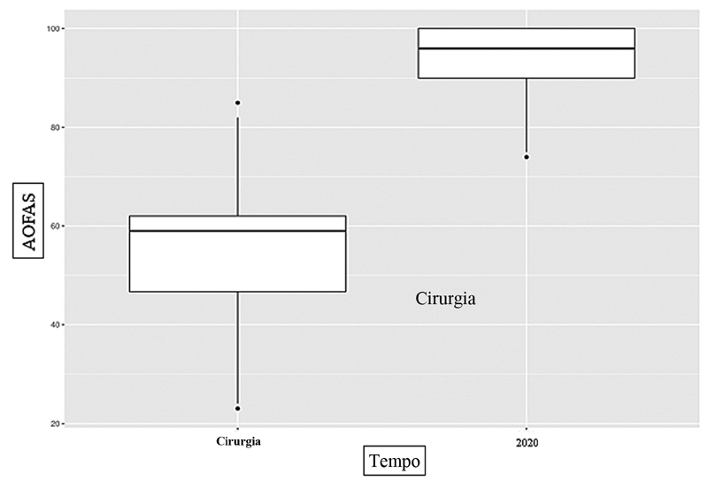
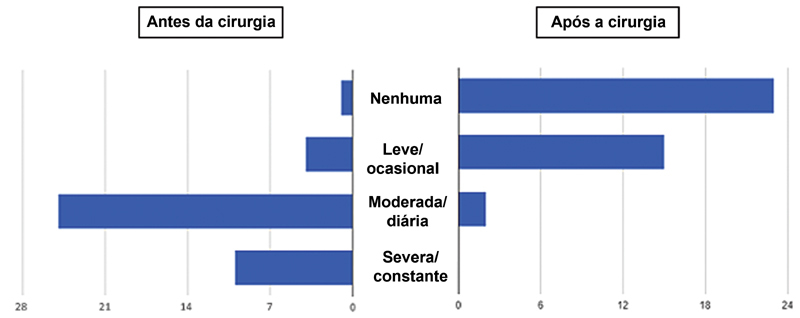
Objective The present study assesses the results of a minimally invasive surgical technique for acute and chronic ankle instability management. Methods The present case series study retrospectively evaluated 40 patients undergoing arthroscopic-assisted percutaneous ankle ligament reconstruction from 2013 to 2019. Results The present study included 17 males and 23 females with an average age of 38.3 years old. Postintervention follow-up using American Orthopaedic Foot and Ankle Society (AOFAS) Ankle-Hindfoot scores identified improvement of > 30 points in function and pain control. The most frequently occurring associated injuries were osteochondral (35%). No patient required reintervention or had infection during follow-up. Conclusion The technique in the present study is easy and achieves satisfactory results for function and pain control. Level of Evidence IV.
Keywords: ankle joint; arthroscopy; joint instability; ligaments, articular; subtalar joint; tendons.
Sociedade Brasileira de Ortopedia e Traumatologia. This is an open access article published by Thieme under the terms of the Creative Commons Attribution-NonDerivative-NonCommercial License, permitting copying and reproduction so long as the original work is given appropriate credit. Contents may not be used for commecial purposes, or adapted, remixed, transformed or built upon. ( https://creativecommons.org/licenses/by-nc-nd/4.0/ ).
Conflict of interest statement
Conflito de Interesses Os autores declaram não haver conflito de interesses.
Figures


Similar articles
-
Arthroscopic-assisted Broström-Gould for chronic ankle instability: a long-term follow-up.Am J Sports Med. 2011 Nov;39(11):2381-8. doi: 10.1177/0363546511416069. Epub 2011 Jul 29. Am J Sports Med. 2011. PMID: 21803979
-
Minimally invasive reconstruction of the lateral ankle ligaments using semitendinosus autograft or tendon allograft.Foot Ankle Int. 2014 Oct;35(10):1015-21. doi: 10.1177/1071100714540145. Epub 2014 Jun 20. Foot Ankle Int. 2014. PMID: 24951483
-
A modified all-inside arthroscopic remnant-preserving technique of lateral ankle ligament reconstruction: medium-term clinical and radiologic results comparable with open reconstruction.Int Orthop. 2020 Oct;44(10):2155-2165. doi: 10.1007/s00264-020-04773-w. Epub 2020 Aug 15. Int Orthop. 2020. PMID: 32803356
-
[Arthroscopic repair of chronic lateral ankle instability].Oper Orthop Traumatol. 2019 Jun;31(3):201-210. doi: 10.1007/s00064-019-0595-7. Epub 2019 Mar 27. Oper Orthop Traumatol. 2019. PMID: 30918997 Review. German.
-
Arthroscopic Repair of Lateral Ankle Ligament for Chronic Lateral Ankle Instability: A Systematic Review.Arthroscopy. 2018 Aug;34(8):2497-2503. doi: 10.1016/j.arthro.2018.02.034. Epub 2018 May 2. Arthroscopy. 2018. PMID: 29730218
Cited by
-
All-inside arthroscopic procedures for chronic lateral ankle instability: evidence-based clinical practice guidelines.Br Med Bull. 2025 Apr 4;154(1):ldaf001. doi: 10.1093/bmb/ldaf001. Br Med Bull. 2025. PMID: 40183802 Free PMC article.
-
Factors Associated with the Clinical Outcomes of Ankle Instability Surgery.Arch Bone Jt Surg. 2025;13(1):30-38. doi: 10.22038/ABJS.2024.74008.3427. Arch Bone Jt Surg. 2025. PMID: 39886343 Free PMC article.
References
-
- Miklovic T M, Donovan L, Protzuk O A, Kang M S, Feger M A. Acute lateral ankle sprain to chronic ankle instability: a pathway of dysfunction. Phys Sportsmed. 2018;46(01):116–122. - PubMed
-
- Guillo S, Bauer T, Lee J W.Consensus in chronic ankle instability: aetiology, assessment, surgical indications and place for arthroscopy Orthop Traumatol Surg Res 201399(8, Suppl)S411–S419. - PubMed
-
- Rodriguez-Merchan E C. Chronic ankle instability: diagnosis and treatment. Arch Orthop Trauma Surg. 2012;132(02):211–219. - PubMed
LinkOut - more resources
Full Text Sources