

Ectopic fat is associated with cardiac remodeling-A comprehensive assessment of regional fat depots in type 2 diabetes using multi-parametric MRI
- PMID: 35966535
- PMCID: PMC9366177
- DOI: 10.3389/fcvm.2022.813427
Ectopic fat is associated with cardiac remodeling-A comprehensive assessment of regional fat depots in type 2 diabetes using multi-parametric MRI
Abstract
Background: Different regional depots of fat have distinct metabolic properties and may relate differently to adverse cardiac remodeling. We sought to quantify regional depots of body fat and to investigate their relationship to cardiac structure and function in Type 2 Diabetes (T2D) and controls.
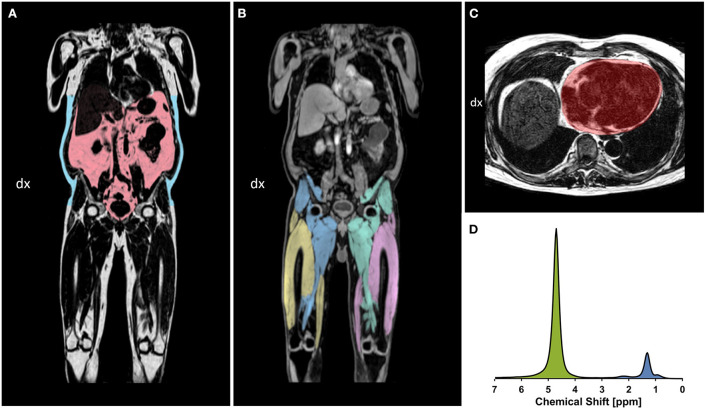
Methods: From the SCAPIS cohort in Linköping, Sweden, we recruited 92 subjects (35% female, mean age 59.5 ± 4.6 years): 46 with T2D and 46 matched controls. In addition to the core SCAPIS data collection, participants underwent a comprehensive magnetic resonance imaging examination at 1.5 T for assessment of left ventricular (LV) structure and function (end-diastolic volume, mass, concentricity, ejection fraction), as well as regional body composition (liver proton density fat fraction, visceral adipose tissue, abdominal subcutaneous adipose tissue, thigh muscle fat infiltration, fat tissue-free thigh muscle volume and epicardial adipose tissue).
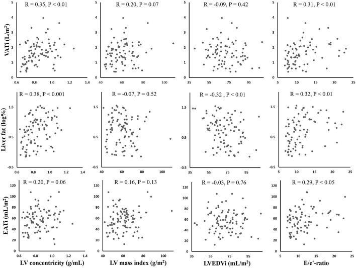
Results: Compared to the control group, the T2D group had increased: visceral adipose tissue volume index (P < 0.001), liver fat percentage (P < 0.001), thigh muscle fat infiltration percentage (P = 0.02), LV concentricity (P < 0.001) and LV E/e'-ratio (P < 0.001). In a multiple linear regression analysis, a negative association between liver fat percentage and LV mass (St Beta -0.23, P < 0.05) as well as LV end-diastolic volume (St Beta -0.27, P < 0.05) was found. Epicardial adipose tissue volume and abdominal subcutaneous adipose tissue volume index were the only parameters of fat associated with LV diastolic dysfunction (E/e'-ratio) (St Beta 0.24, P < 0.05; St Beta 0.34, P < 0.01, respectively). In a multivariate logistic regression analysis, only visceral adipose tissue volume index was significantly associated with T2D, with an odds ratio for T2D of 3.01 (95% CI 1.28-7.05, P < 0.05) per L/m2 increase in visceral adipose tissue volume.
Conclusions: Ectopic fat is predominantly associated with cardiac remodeling, independently of type 2 diabetes. Intriguingly, liver fat appears to be related to LV structure independently of VAT, while epicardial fat is linked to impaired LV diastolic function. Visceral fat is associated with T2D independently of liver fat and abdominal subcutaneous adipose tissue.
Keywords: cardiac remodeling; ectopic fat; left ventricular diastolic function; left ventricular structure; magnetic resonance imaging; type 2 diabetes; visceral fat.
Copyright © 2022 Edin, Ekstedt, Scheffel, Karlsson, Swahn, Östgren, Engvall, Ebbers, Leinhard, Lundberg and Carlhäll.
Conflict of interest statement
Authors MK and OL are employees of AMRA Medical AB. Authors OL and PL are stockholders in AMRA Medical AB. Author ME reports personal fees from Advisory Board AMRA Medical AB. The remaining authors declare that the research was conducted in the absence of any commercial or financial relationships that could be construed as a potential conflict of interest.
Figures
References
-
- Hiuge-Shimizu A, Kishida K, Funahashi T, Ishizaka Y, Oka R, Okada M, et al. Absolute value of visceral fat area measured on computed tomography scans and obesity-related cardiovascular risk factors in large-scale Japanese general population (the VACATION-J study). Ann Med. (2012) 44:82–92. 10.3109/07853890.2010.526138 - DOI - PubMed
-
- Schlett CL, Lorbeer R, Arndt C, Auweter S, Machann J, Hetterich H, et al. Association between abdominal adiposity and subclinical measures of left-ventricular remodeling in diabetics, prediabetics and normal controls without history of cardiovascular disease as measured by magnetic resonance imaging: Results from the KORA-FF4 S. Cardiovasc Diabetol. (2018) 17:1–12. 10.1186/s12933-018-0721-0 - DOI - PMC - PubMed