. 2022 Jul 28:13:915721.
doi: 10.3389/fmicb.2022.915721. eCollection 2022.

The ABCT31 Transporter Regulates the Export System of Phenylacetic Acid as a Side-Chain Precursor of Penicillin G in Monascus ruber M7

Affiliations

The ABCT31 Transporter Regulates the Export System of Phenylacetic Acid as a Side-Chain Precursor of Penicillin G in Monascus ruber M7

Rabia Ramzan et al. Front Microbiol. .

Abstract

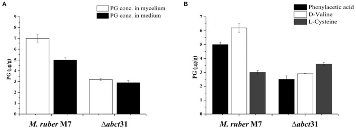
The biosynthesis of penicillin G (PG) is compartmentalized, and the transportation of the end and intermediate products, and substrates (precursors) such as L-cysteine (L-Cys), L-valine (L-Val) and phenylacetic acid (PAA) requires traversing membrane barriers. However, the transportation system of PAA as a side chain of PG are unclear yet. To discover ABC transporters (ABCTs) involved in the transportation of PAA, the expression levels of 38 ABCT genes in the genome of Monascus ruber M7, culturing with and without PAA, were examined, and found that one abct gene, namely abct31, was considerably up-regulated with PAA, indicating that abct31 may be relative with PAA transportation. Furthermore the disruption of abct31 was carried out, and the effects of two PG substrate's amino acids (L-Cys and L-Val), PAA and some other weak acids on the morphologies and production of secondary metabolites (SMs) of Δabct31 and M. ruber M7, were performed through feeding experiments. The results revealed that L-Cys, L-Val and PAA substantially impacted the morphologies and SMs production of Δabct31 and M. ruber M7. The UPLC-MS/MS analysis findings demonstrated that Δabct31 did not interrupt the synthesis of PG in M. ruber M7. According to the results, it suggests that abct31 is involved in the resistance and detoxification of the weak acids, including the PAA in M. ruber M7.

Keywords: ABC transporter; amino acid; phenylacetic acid; secondary metabolites; weak acid.


Conflict of interest statement

The authors declare that the research was conducted in the absence of any commercial or financial relationships that could be construed as a potential conflict of interest.

Figures

Figure 1
Figure 1
Quantitative real-time PCR analysis of the expression of all ABC transporter genes in the M. ruber M7 toward PAA. (A) The comparative expression of all ABC transporter genes in M. ruber M7 without PAA; (B) A comparative expression of all ABC transporter genes in M. ruber M7 with PAA. M. ruber M7 was cultivated and incubated at 28°C for 7 d in the presence (black bar) and absence (gray bar) of PAA at 10 mM. The values were represented as average values ± sd.
Figure 2
Figure 2
Domain analysis of abct31 analyzed by CDD program.
Figure 3
Figure 3
Homology model and checking results for ABCT31 protein. Verification of homology modeling (A) Homology model of ABCT31 protein TMD, Transmembrane domain; NBD, Nucleotide-binding domains; (B) Plot displaying the model's normalized Q mean score; (C) Plot displaying the model's local quality; (D) Z score value of the model developed; (E) Ramachandran plot validating the energy-minimized model's backbone dihedral angles.
Figure 4
Figure 4
Structural features and location analysis of the ABCT31 protein. (A) Features loop and walker of the abct31 protein; (B) Topology analysis of the ABCT31 protein; (C) Location of abct31 gene in M. ruber M7.
Figure 5
Figure 5
ABC transporter carries the substrate.
Figure 6
Figure 6
Construction and verification of Δabct31 strain. (A) Pictorial presentation for the homologous recombination approach to create abct31 disruption strains. (B) M: Trans 2k II Marker; Lane1: PCR product of 5' franking region (770 bp); Lane 2: PCR product of 3' franking (434 bp). (C) M: Trans 2k plus II Marker; Lane 1: hph (2,137 bp). (D) M: Trans 2k plus II Marker; Lane 1: PCR product of recombinant fragment of 5'UTR, hph and 3'UTR (3341 bp). (E) M: Trans 2k plus II Marker; Lane1: Restriction enzyme digestion analysis of vector pC-abct31 (KpnI/HindIII). (F) Validation of abct31 homologous recombination events. M: Trans 2k plus II Marker; Lane 1, the abct31 strain; Lane 2, the wild-type strain (M7). Different distinct bands were obtained by PCR amplification for selected pair of primer. (G) Southern hybridization analysis. Lane 1, Xba1 digested genomic DNA of Δabct31; lane 2, Xba1 digested genomic DNA of M7 respectively; M: λDNA/HindIII marker; Probe 1: abct31 gene; Probe 2: hph gene.
Figure 7
Figure 7
Morphological comparison of M. ruber M7and Δabct31. (A) Colony morphology of M. ruber M7 and Δabct31 on PDA, CYA, G25N, MA plates, and cultured at 28°C for 15 days; (B) Cleistothecia and conidia development in M. ruber M7and Δabct31 inoculated on PDA, CYA, G25N, MA plates, and cultured at 28°C for 8 days.
Figure 8
Figure 8
Morphological comparison of M. ruber M7and Δabct31 to evaluate the sensitivity toward pathway amino acid supplementation. (A) Colony morphology of M. ruber M7 and Δabct31 inoculated on PDA plates supplemented with D-valine, phenylacetic acid, and L-cysteine at 2 mM concentrations for each cultured at 28°C for 15 days. (B) Cleistothecia and conidia development in M. ruber M7 and Δabct31 cultured on PDA plates which supplemented with D-valine, phenylacetic acid, and L-cysteine at 2 mM concentrations for each and cultured at 28°C for 10 days.
Figure 9
Figure 9
Comparison of biomass (dry cell weight) of the M. ruber M7 and Δabct31 against pathway amino acid. (A) PDB medium without amino acid (control); (B) PDB medium with D-valine; (C) PDB medium with phenylacetic acid; (D) PDB medium with L-cysteine, at 2 mM concentrations and incubated at 28°C without agitation. The bar representing the mean of triplicate values and error bars show standard deviation.
Figure 10
Figure 10
Comparison of PG concentration of the wild-type strain and the Δabct31 transformants on the 7th day. (A) PG concentration in mycelium and medium; (B) Comparison of PG concentration affected by supplementation of D-valine, phenylacetic acid, and L-cysteine at 2 mM on 7th day. The bar representing the mean of triplicate values and error bars show standard deviation.
Figure 11
Figure 11
Comparison of eluted products of M. ruber M7 and Δabct31 transformant for solid-state fermentation on rice. (A) Metabolic profile of M. ruber M7 at 250 nm; (B) Metabolic profile of Δabct31 at 250 nm; (C) UV-Vis spectrum of benzyl penicillin (PG) which is shown by the arrows in metabolic profiles.
Figure 12
Figure 12
Detection of β-metabolites detected M. ruber M7 and Δabct31 transformants by UPLC-MS/MS. UPLC-MS/MS spectrogram of the M. ruber M7 and Δabct31 for solid-state fermented on rice displays that the atomic mass of ion (m/z 335.336) matched well with the standard penicillin, the molecular weight for isopenicillin N is 359.3 g/mol, in spectrogram ion m/z 359.00 representing the isopenicillin N peak. (A) Spectra in A penicillin G (PG) standard with the mass 335.336; (B) A pattern of fragmentation of M. ruber M7; (C) A pattern of fragmentation of Δabct31.
Figure 13
Figure 13
Colony morphology of M. ruber M7 and Δabct31 for the feeding of weak acids. The morphologies of M. ruber M7 (1st vertical row) and Δabct31 (2nd vertical row) colonies on PDA plates supplemented with weak acid at 28°C for 12d. (A) Colony morphology of M. ruber M7 and Δabct31 for the feeding of sorbic acids at 0, 1, 2.5, 5, 7.5, and 10 mM concentration, respectively; (B) Colony morphology of M. ruber M7 and Δabct31 for the feeding of acetic acids at 0, 0.5, 0.75, 1, 1.25, 1.5 mM concentration, respectively; (C) Colony morphology of M. ruber M7 and Δabct31 for the feeding of adipic acids at 0, 1, 2, and 3 mM concentration, respectively.
Figure 14
Figure 14
Expression of abct31 in M. ruber M7 to weak acid supplementation with or without PAA. NC was presented in the control group in which M. ruber M7 was cultivated on PDA without sorbic acid. SA presented the sorbic acid group in which M. ruber M7 was cultivated on a PDA with (A) Sorbic acid at 7.5 Mm; (B) Acetic acid at 1.5 Mm; (C) Adipic acid at 3 mM. Both groups were incubated at 28°C for 7d in the presence (black vertical bar) and absence (empty vertical bar) of PAA at 10 mM.

Similar articles

Cited by

References

    1. Aguilera A., Gómez-González B. (2008). Genome instability: a mechanistic view of its causes and consequences. Nat. Rev. Genet. 9, 204–217. 10.1038/nrg2268 - DOI - PubMed
    1. Al Shawi A., Rasul A., Khan M., Iqbal F., Tonghui M. (2011). Eupatilin: a flavonoid compound isolated from the artemisia plant, induces apoptosis and G2/M phase cell cycle arrest in human melanoma A375 cells. African J. Pharm. Pharmacol. 5, 582–588. 10.5897/AJPP11.079 - DOI
    1. Ávalos J., Díaz-Sánchez V., García-Martínez, Jorge Castrillo M., Ruger-Herreros M., Limón M. C. (2014). Biosynthesis and molecular genetics of fungal secondary metabolites. Fungal Biol. 2, 67–79. 10.1007/978-1-4939-1191-2 - DOI
    1. Barreiro C., García-Estrada C. (2019). Proteomics and Penicillium chrysogenum: unveiling the secrets behind penicillin production. J. Proteomics. 198, 119–131. 10.1016/j.jprot.2018.11.006 - DOI - PubMed
    1. Buchan D. W. A., Minneci F., Nugent T. C. O., Bryson K., Jones D. T. (2013). Scalable web services for the PSIPRED protein analysis workbench. Nucleic Acids Res. 41, 349–357. 10.1093/nar/gkt381 - DOI - PMC - PubMed

LinkOut - more resources