. 2022 Jul 27:13:946703.
doi: 10.3389/fmicb.2022.946703. eCollection 2022.

Molecular and biological characterization of hepatitis B virus subgenotype F1b clusters: Unraveling its role in hepatocarcinogenesis

Affiliations

Molecular and biological characterization of hepatitis B virus subgenotype F1b clusters: Unraveling its role in hepatocarcinogenesis

María Mercedes Elizalde et al. Front Microbiol. .

Abstract

Hepatitis B virus (HBV) subgenotype F1b infection has been associated with the early occurrence of hepatocellular carcinoma in chronically infected patients from Alaska and Peru. In Argentina, however, despite the high prevalence of subgenotype F1b infection, this relationship has not been described. To unravel the observed differences in the progression of the infection, an in-depth molecular and biological characterization of the subgenotype F1b was performed. Phylogenetic analysis of subgenotype F1b full-length genomes revealed the existence of two highly supported clusters. One of the clusters, designated as gtF1b Basal included sequences mostly from Alaska, Peru and Chile, while the other, called gtF1b Cosmopolitan, contained samples mainly from Argentina and Chile. The clusters were characterized by a differential signature pattern of eight nucleotides distributed throughout the genome. In vitro characterization of representative clones from each cluster revealed major differences in viral RNA levels, virion secretion, antigen expression levels, as well as in the localization of the antigens. Interestingly, a differential regulation in the expression of genes associated with tumorigenesis was also identified. In conclusion, this study provides new insights into the molecular and biological characteristics of the subgenotype F1b clusters and contributes to unravel the different clinical outcomes of subgenotype F1b chronic infections.

Keywords: characterization; clusters; hepatitis B virus; hepatocarcinogenesis; subgenotype F1b.


Conflict of interest statement

The authors declare that the research was conducted in the absence of any commercial or financial relationships that could be construed as a potential conflict of interest.

Figures
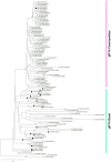
Figure 1
Figure 1
Phylogenetic analysis of subgenotype F1b sequences. A maximum likelihood tree was constructed on subgenotype F1b complete genome sequences derived from chronically infected patients (n = 99), using subgenotype F1a as an outgroup (n = 2, ○). The numbers at each node correspond to bootstrap values (greater than 90%) obtained with 1,000 replicates. The scale bar indicates the genetic distances. Sequences are name by the country of origin, followed by the year of the sample collection and the accession number. ALA, Alaska; ARG, Argentina; BRZ, Brazil; CHL, Chile; CRC, Costa Rica; IRL, Ireland; JPN, Japan; MEX. Mexico; PER, Peru; SAL, El Salvador; URU, Uruguay; USA, United states; VEN, Venezuela. ■ Nucleotide sequences associated with HCC cases. □ Nucleotide sequences non-associated with HCC cases. ∆ Data unknown.
Figure 2
Figure 2
Differential nucleotide signature pattern between subgenotype F1b clusters. Schematic representation of HBV genome including enhancers I and II, promoters (white boxes) and proteins (gray boxes). Polymorphisms in nucleotide positions: 843, 1688, 1966, 2125, 2200, 2480, 2633, 2771 are shown.
Figure 3
Figure 3
Analysis of HBV replicative capacity between gtF1b Cosmopolitan and gtF1b Basal variants. HuH-7 cells were transfected with linear full-length HBV genomes of gtF1b Cosmopolitan and gtF1b Basal variants and three days’ post-transfection cells and supernatants were harvested. (A) DNA was isolated after Hirt extraction and subjected to Southern blot analysis. (B) Total DNA was extracted, treated with T5 exonuclease to remove non-supercoiled dsDNA, and cccDNA levels were assessed by qPCR. Results were normalized to mitochondrial DNA. (C) Total RNA was extracted, and HBV RNA was detected by Northern blot. GAPDH RNA served as loading control. (D) Relative intensity of the Precore/pgRNA band was quantified using ImageJ software. (E) Cytoplasmic DNA was extracted HBV DNA replicative intermediates were assessed by Southern blot. (F) Extracellular HBV DNA was quantified by qPCR. PF-rcDNA: protein free HBV relaxed circular DNA; PF-dslDNA: protein free HBV double-stranded linear DNA. rcDNA: HBV relaxed circular DNA; dsLDNA: HBV double-stranded linear DNA; SS: HBV single-stranded DNA. Shown values represent the mean ± standard deviation of three independent experiments. ***p < 0.0001; **p < 0.005; ns: no statistical differences.
Figure 4
Figure 4
Analysis of intracellular and secreted HBsAg and HBeAg levels of gtF1b Cosmopolitan and gtF1b Basal variants. HuH-7 cells were transfected with linear full-length HBV genomes of gtF1b Cosmopolitan and gtF1b Basal variants. Three days’ post-transfection, cells and culture supernatants were harvested. Intracellular and extracellular levels of HBsAg and HBeAg were determined by ECLIA (A,C). Extracellular/intracellular HBsAg and HBeAg ratio (B,D). Values shown represent the mean ± standard deviation of three independent experiments. ***p < 0.0001; *p < 0.05; ns: no statistical differences.
Figure 5
Figure 5
Relative composition of HBsAg proteins of gtF1b Cosmopolitan and gtF1b Basal variants. HuH-7 cells were transfected with linear full-length HBV genomes of gtF1b Cosmopolitan and gtF1b Basal variants. Three days’ post-transfection, cellular lysates (A) and the supernatants (C) were analyzed by Western Blot, using a SHBs-specific monoclonal (HB01) antibody. SHBs and LHBs occur in unglycosylated (LHBs and SHBs) and glycosylated (gLHBs and gSHBs) forms, while MHBs occurs in glycosylated (gMHBs) and double glycosylated (ggMHBs) forms. (B) Intracellular and (D) extracellular LHBs/MHBs/SHBs ratio was determined. Ct: cell transfected with pUC19 empty vector (control).
Figure 6
Figure 6
Analysis of HBV promoter activities of gtF1b Cosmopolitan and gtF1b Basal variants. HuH-7 cells were transfected with luciferase reporter constructs containing SPI (A), SPII (B), Core promoter with Precore TATA box-like mutated sequence (C), and Core promoter with pgRNA TATA box-like mutated sequence (D) of gtF1b Cosmopolitan and gtF1b Basal variants. Three days’ post-transfection, cells were harvested, and luciferase activity was detected. Values shown represent the mean ± standard deviation of three independent experiments. The pGL4.13[luc2/SV40] and pGL4.10[luc2] vectors were included as positive and negative controls, respectively. *p < 0.05; ***p < 0.0001; ns: no statistical differences.
Figure 7
Figure 7
Expression analysis of HCC-related genes in gtF1b Cosmopolitan and gtF1b Basal variants. HuH-7 cells were transfected with pUC19 empty vector (control), or linear full-length HBV genomes of gtF1b Cosmopolitan and gtF1b Basal variants. Three days’ post-transfection, cells were harvested, total RNA was extracted, and mRNA levels of HCC-related genes were analyzed by RT-qPCR. Relative expression was calculated using the method of 2−ΔΔCt. Values shown represent the mean ± standard deviation of three independent experiments. *p < 0.05; **p < 0.005; ***p < 0.0001.

References

    1. Allweiss L., Dandri M. (2016). Experimental in vitro and in vivo models for the study of human hepatitis B virus infection. J. Hepatol. 64, S17–S31. doi: 10.1016/j.jhep.2016.02.012 - DOI - PubMed
    1. Araujo N. M., Teles S. A., Spitz N. (2020). Comprehensive analysis of clinically significant hepatitis B virus mutations in relation to genotype, subgenotype and geographic region. Front. Microbiol. 11:616023. doi: 10.3389/fmicb.2020.616023, PMID: - DOI - PMC - PubMed
    1. Bertani S., Pineau P., Loli S., Moura J., Zimic M., Deharo E., et al. . (2013). An atypical age-specific pattern of hepatocellular carcinoma in Peru: a threat for Andean populations. PLoS One 8, e67756. doi: 10.1371/journal.pone.0067756, PMID: - DOI - PMC - PubMed
    1. Bouchard M. J., Puro R. J., Wang L., Schneider R. J. (2003). Activation and inhibition of cellular calcium and tyrosine kinase signaling pathways identify targets of the HBx protein involved in hepatitis B virus replication. J. Virol. 77, 7713–7719. doi: 10.1128/jvi.77.14.7713-7719.2003, PMID: - DOI - PMC - PubMed
    1. Chakraborty U., Dinh T. A., Alani E. (2018). Genomic instability promoted by overexpression of mismatch repair factors in yeast: a model for understanding cancer progression. Genetics 209, 439–456. doi: 10.1534/genetics.118.300923, PMID: - DOI - PMC - PubMed

LinkOut - more resources