

Enlarged perivascular spaces and white matter hyperintensities in patients with frontotemporal lobar degeneration syndromes
- PMID: 35966773
- PMCID: PMC9366845
- DOI: 10.3389/fnagi.2022.923193
Enlarged perivascular spaces and white matter hyperintensities in patients with frontotemporal lobar degeneration syndromes
Abstract
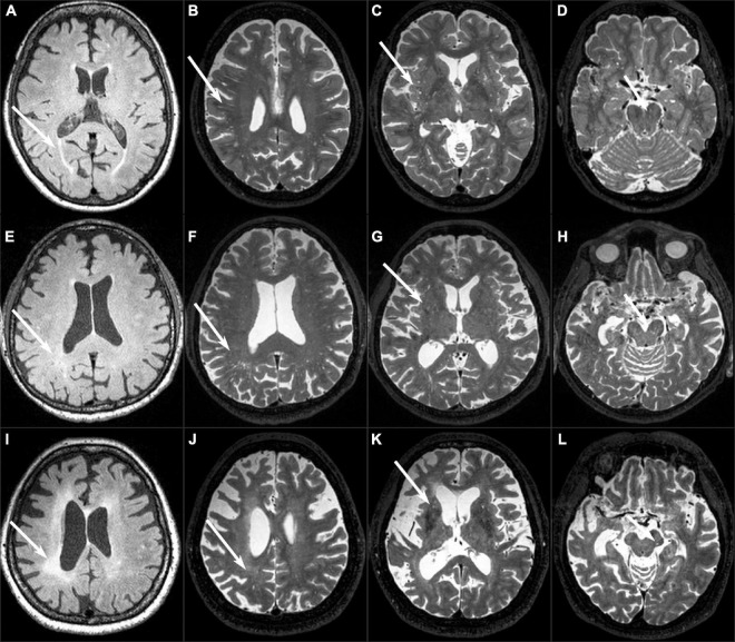
Objective: The aim of this study was to investigate the distribution characteristics of enlarged perivascular spaces (EPVS) and white matter hyperintensities (WMH) and their associations with disease severity across the frontotemporal lobar degeneration (FTLD) syndromes spectrum.
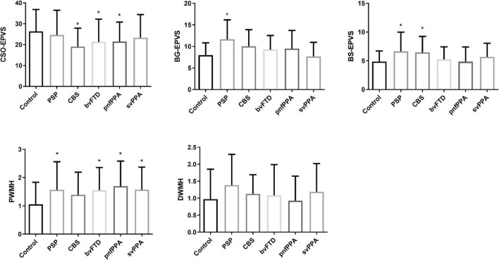
Methods: This study included 73 controls, 39 progressive supranuclear palsy Richardson's syndrome (PSP-RS), 31 corticobasal syndrome (CBS), 47 behavioral variant frontotemporal dementia (bvFTD), 36 non-fluent variant primary progressive aphasia (nfvPPA), and 50 semantic variant primary progressive aphasia (svPPA). All subjects had brain magnetic resonance imaging (MRI) and neuropsychological tests, including progressive supranuclear palsy rating scale (PSPRS) and FTLD modified clinical dementia rating sum of boxes (FTLD-CDR). EPVS number and grade were rated on MRI in the centrum semiovale (CSO-EPVS), basal ganglia (BG-EPVS), and brain stem (BS-EPVS). Periventricular (PWMH) and deep (DWMH) were also graded on MRI. The distribution characteristics of EPVS and WMH were compared between control and disease groups. Multivariable linear regression analysis was performed to evaluate the association of EPVS and WMH with disease severity.
Results: Compared with control subjects, PSP-RS and CBS had more BS-EPVS; CBS, bvFTD, and nfvPPA had less CSO-EPVS; all disease groups except CBS had higher PWMH (p < 0.05). BS-EPVS was associated with PSPRS in PSP-RS (β = 2.395, 95% CI 0.888-3.901) and CBS (β = 3.115, 95% CI 1.584-4.647). PWMH was associated with FTLD-CDR in bvFTD (β = 1.823, 95% CI 0.752-2.895), nfvPPA (β = 0.971, 95% CI 0.030-1.912), and svPPA (OR: 1.330, 95% CI 0.457-2.204).
Conclusion: BS-EPVS could be a promising indicator of disease severity in PSP-RS and CBS, while PWMH could reflect the severity of bvFTD, nfvPPA, and svPPA.
Keywords: CBS; PSP-RS; bvFTD; enlarged perivascular spaces; nfvPPA; svPPA; white matter hyperintensities.
Copyright © 2022 Wang, Sun, Li, Zou, Li, Wu and Li.
Conflict of interest statement
The authors declare that the research was conducted in the absence of any commercial or financial relationships that could be construed as a potential conflict of interest.
Figures
References
LinkOut - more resources
Full Text Sources
Miscellaneous