

Identification and immunological characterization of cuproptosis-related molecular clusters in Alzheimer's disease
- PMID: 35966780
- PMCID: PMC9366224
- DOI: 10.3389/fnagi.2022.932676
Identification and immunological characterization of cuproptosis-related molecular clusters in Alzheimer's disease
Abstract
Introduction: Alzheimer's disease is the most common dementia with clinical and pathological heterogeneity. Cuproptosis is a recently reported form of cell death, which appears to result in the progression of various diseases. Therefore, our study aimed to explore cuproptosis-related molecular clusters in Alzheimer's disease and construct a prediction model.
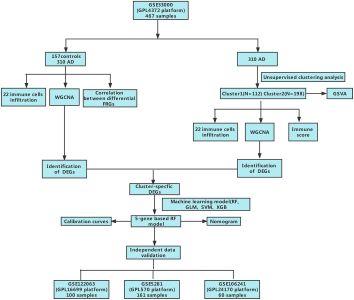
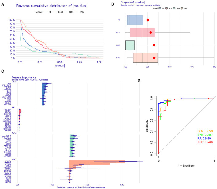
Methods: Based on the GSE33000 dataset, we analyzed the expression profiles of cuproptosis regulators and immune characteristics in Alzheimer's disease. Using 310 Alzheimer's disease samples, we explored the molecular clusters based on cuproptosis-related genes, along with the related immune cell infiltration. Cluster-specific differentially expressed genes were identified using the WGCNA algorithm. Subsequently, the optimal machine model was chosen by comparing the performance of the random forest model, support vector machine model, generalized linear model, and eXtreme Gradient Boosting. Nomogram, calibration curve, decision curve analysis, and three external datasets were applied for validating the predictive efficiency.
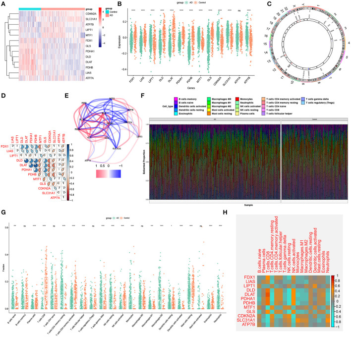
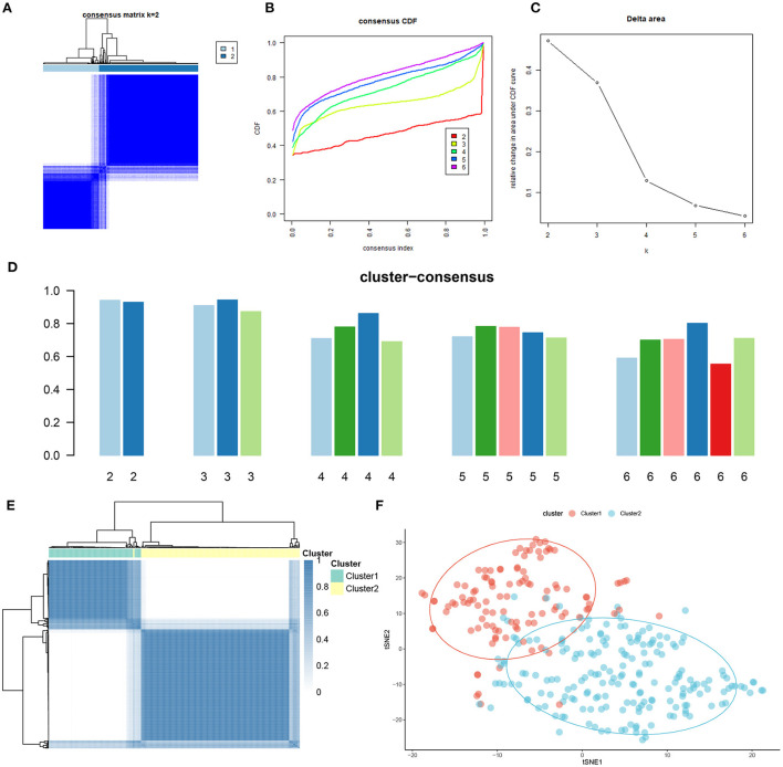
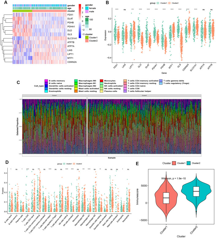
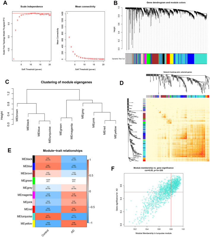
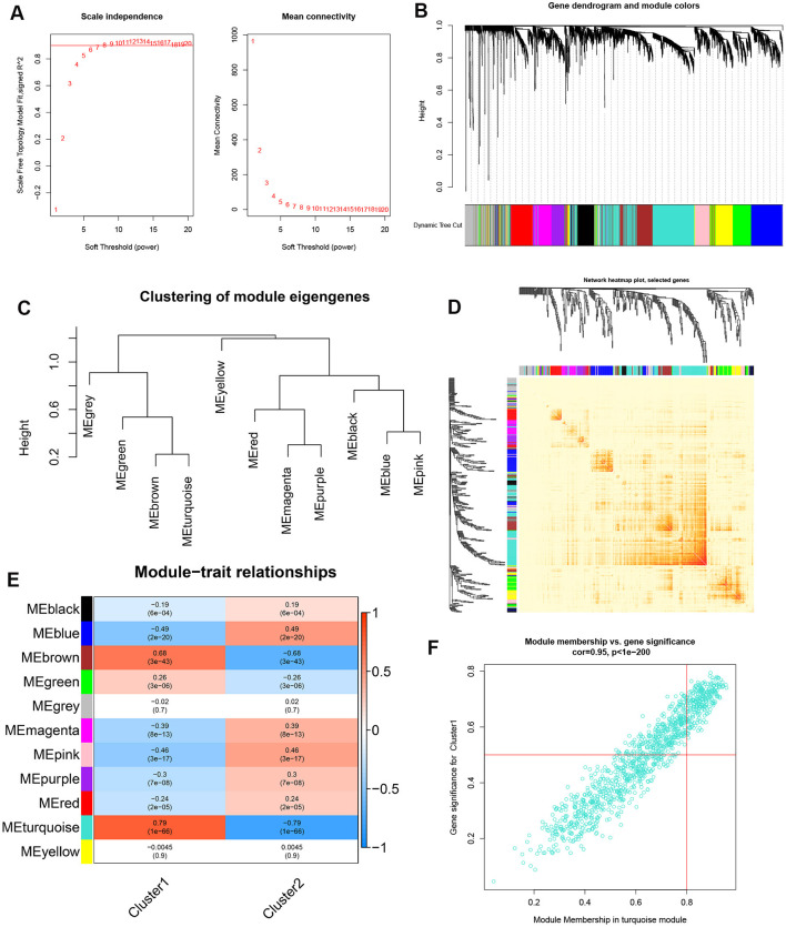
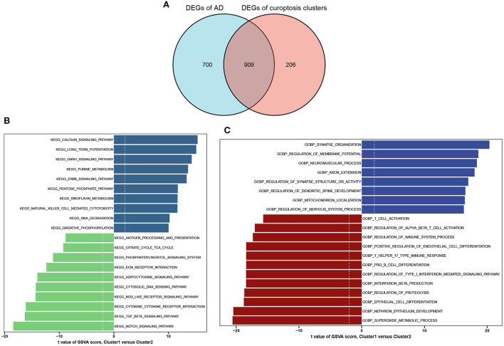
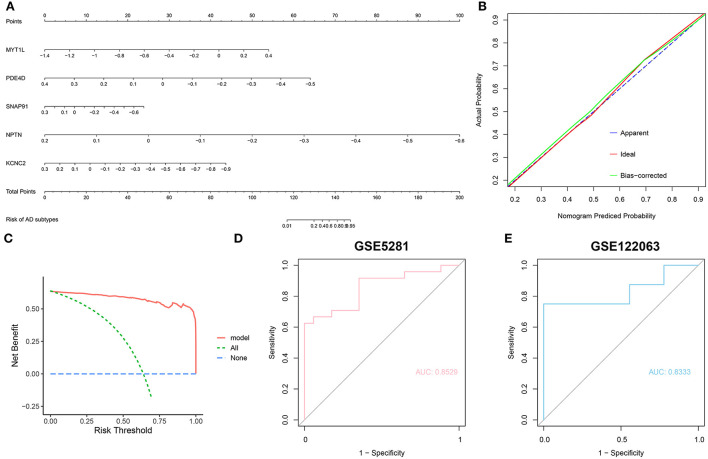
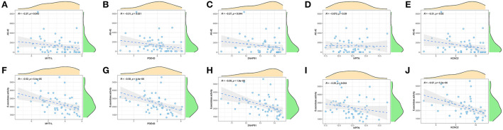
Results: The dysregulated cuproptosis-related genes and activated immune responses were determined between Alzheimer's disease and non-Alzheimer's disease controls. Two cuproptosis-related molecular clusters were defined in Alzheimer's disease. Analysis of immune infiltration suggested the significant heterogeneity of immunity between distinct clusters. Cluster2 was characterized by elevated immune scores and relatively higher levels of immune infiltration. Functional analysis showed that cluster-specific differentially expressed genes in Cluster2 were closely related to various immune responses. The Random forest machine model presented the best discriminative performance with relatively lower residual and root mean square error, and a higher area under the curve (AUC = 0.9829). A final 5-gene-based random forest model was constructed, exhibiting satisfactory performance in two external validation datasets (AUC = 0.8529 and 0.8333). The nomogram, calibration curve, and decision curve analysis also demonstrated the accuracy to predict Alzheimer's disease subtypes. Further analysis revealed that these five model-related genes were significantly associated with the Aβ-42 levels and β-secretase activity.
Conclusion: Our study systematically illustrated the complicated relationship between cuproptosis and Alzheimer's disease, and developed a promising prediction model to evaluate the risk of cuproptosis subtypes and the pathological outcome of Alzheimer's disease patients.
Keywords: Alzheimer's disease; cuproptosis; immune infiltration; machine learning; molecular clusters; prediction model.
Copyright © 2022 Lai, Lin, Lin, Wu, Zhao and Lin.
Conflict of interest statement
The authors declare that the research was conducted in the absence of any commercial or financial relationships that could be construed as a potential conflict of interest.
Figures
Similar articles
-
Identification and immune features of cuproptosis-related molecular clusters in polycystic ovary syndrome.Sci Rep. 2023 Jan 18;13(1):980. doi: 10.1038/s41598-022-27326-0. Sci Rep. 2023. PMID: 36653385 Free PMC article.
-
Identification of immune infiltration and cuproptosis-related molecular clusters in tuberculosis.Front Immunol. 2023 Jul 11;14:1205741. doi: 10.3389/fimmu.2023.1205741. eCollection 2023. Front Immunol. 2023. PMID: 37497230 Free PMC article.
-
Identification and immunological role of cuproptosis in osteoporosis.Heliyon. 2024 Feb 20;10(5):e26759. doi: 10.1016/j.heliyon.2024.e26759. eCollection 2024 Mar 15. Heliyon. 2024. PMID: 38455534 Free PMC article.
-
Ferroptosis, Pyroptosis, and Cuproptosis in Alzheimer's Disease.ACS Chem Neurosci. 2023 Oct 4;14(19):3564-3587. doi: 10.1021/acschemneuro.3c00343. Epub 2023 Sep 13. ACS Chem Neurosci. 2023. PMID: 37703318 Review.
-
Machine learning for the life-time risk prediction of Alzheimer's disease: a systematic review.Brain Commun. 2021 Oct 21;3(4):fcab246. doi: 10.1093/braincomms/fcab246. eCollection 2021. Brain Commun. 2021. PMID: 34805994 Free PMC article. Review.
Cited by
-
Cuproptosis-related genes prediction feature and immune microenvironment in major depressive disorder.Heliyon. 2023 Jul 26;9(8):e18497. doi: 10.1016/j.heliyon.2023.e18497. eCollection 2023 Aug. Heliyon. 2023. PMID: 37576193 Free PMC article.
-
Identification and experimental validation of cuproptosis regulatory program in a sepsis immune microenvironment through a combination of single-cell and bulk RNA sequencing.Front Immunol. 2024 Jun 14;15:1336839. doi: 10.3389/fimmu.2024.1336839. eCollection 2024. Front Immunol. 2024. PMID: 38947313 Free PMC article.
-
A cuproptosis score model and prognostic score model can evaluate clinical characteristics and immune microenvironment in NSCLC.Cancer Cell Int. 2024 Feb 10;24(1):68. doi: 10.1186/s12935-024-03267-8. Cancer Cell Int. 2024. PMID: 38341588 Free PMC article.
-
A glance through the effects of CD4+ T cells, CD8+ T cells, and cytokines on Alzheimer's disease.Comput Struct Biotechnol J. 2023 Nov 8;21:5662-5675. doi: 10.1016/j.csbj.2023.10.058. eCollection 2023. Comput Struct Biotechnol J. 2023. PMID: 38053545 Free PMC article. Review.
-
Identification of copper death-associated molecular clusters and immunological profiles for lumbar disc herniation based on the machine learning.Sci Rep. 2024 Aug 20;14(1):19294. doi: 10.1038/s41598-024-69700-0. Sci Rep. 2024. PMID: 39164344 Free PMC article.
References
LinkOut - more resources
Full Text Sources