. 2022 Jul 29:9:901187.
doi: 10.3389/fmed.2022.901187. eCollection 2022.

Selective endothelial removal: A case series of a phase I/II surgical trial with long-term follow up

Affiliations

Selective endothelial removal: A case series of a phase I/II surgical trial with long-term follow up

Yu-Chi Liu et al. Front Med (Lausanne). .

Abstract

Peters anomaly is a congenital condition which results in a central corneal opacity from birth. Selective Endothelial Removal (SER) is a novel surgical technique and a form of regenerative therapy, which encourages clearance of the central corneal opacity by the patient's own corneal endothelial cells, and it may potentially be beneficial for the treatment of Peters anomaly. We have performed a phase I/II surgical trial, evaluating the safety of SER in four eyes (three patients) with Peters Anomaly. These patients underwent SER at between 9 and 39 months of age, each demonstrating clearance of central corneal opacities and improvements in vision post-operatively. No complications occurred in any of these eyes, at a minimal post-operative follow-up duration of 48 months. We conclude that SER for Peters anomaly is a safe surgical procedure. While encouraging efficacy outcomes have been observed, these findings should be further evaluated in a larger scale Phase II/III surgical trial.

Keywords: Peters anomaly; cornea; endothelium; keratoplasty; regenerative medicine.


Conflict of interest statement

The authors declare that the research was conducted in the absence of any commercial or financial relationships that could be construed as a potential conflict of interest.

Figures

FIGURE 1
FIGURE 1
Intra-operative video screenshots of patient 2’s right eye. (A) Selective endothelial removal resulted in the intentional creation of a central endothelial defect underlying the central corneal opacity, evidenced by positive staining with trypan blue. (B) Following trypan blue staining, a full intracameral air bubble was injected. The absence of a reflective ring around the central region of trypan blue staining indicated that the Descemet’s membrane was intact despite selective endothelial removal.
FIGURE 2
FIGURE 2
Picture collage of eyes which underwent selective endothelial removal for the treatment of Peters anomaly. The last reviews for patients 1 and 3 were at the 60th and 48th post-operative month, while the last review with photos being taken for patient 2 was at the 24th post-operative month. Gradual post-operative improvements in the central corneal opacities were seen in all eyes. Pre-op, pre-operative; POM, post-operative month.
FIGURE 3
FIGURE 3
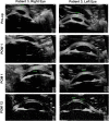
Ultrasound biomicroscopy (UBM) images for both eyes of patient 3. Pre-operatively, thick paracentral bands of iridocorneal adhesions with shallow anterior chambers may be seen in both eyes. Serial post-operative UBM images demonstrate that these bands have been lysed, with normalization of anterior chamber depths. Pre-op, pre-operative; POW, post-operative week; POM, post-operative month.
FIGURE 4
FIGURE 4
Anterior segment optical coherence tomography scans of patient 2. (A) Right eye with type 1 Peters anomaly. (B) Left eye which is normal. There is a posterior corneal defect in the central cornea of the right eye, evidenced by a central corneal thickness of 468 μm in contrast to a central corneal thickness of 562 μm in the normal fellow eye.

Similar articles

Cited by

References

    1. Kurilec JM, Zaidman GW. Incidence of Peters anomaly and congenital corneal opacities interfering with vision in the United States. Cornea. (2014) 33:848–50. 10.1097/ICO.0000000000000182 - DOI - PubMed
    1. Kuper C, Kuwabara T, Stark WJ. The histopathology of Peters’ anomaly. Am J Ophthalmol. (1975) 80:653–60. 10.1016/0002-9394(75)90397-9 - DOI - PubMed
    1. Ni W, Wang W, Hong J, Zhang P, Liu C. A novel histopathologic finding in the Descemet’s membrane of a patient with Peters Anomaly: a case-report and literature review. BMC Ophthalmol. (2015) 15:139. 10.1186/s12886-015-0131-y - DOI - PMC - PubMed
    1. Sowden JC. Molecular and developmental mechanisms of anterior segment dysgenesis. Eye (Lond). (2007) 21:1310–8. 10.1038/sj.eye.6702852 - DOI - PubMed
    1. Elbaz U, Ali A, Strungaru H, Mireskandari K. Phenotypic spectrum of peters anomaly: implications for management. Cornea. (2022) 41:192–200. 10.1097/ICO.0000000000002768 - DOI - PubMed

LinkOut - more resources