. 2022 Aug 4:2022:8675096.
doi: 10.1155/2022/8675096. eCollection 2022.

Linderalactone Suppresses Pancreatic Cancer Development In Vitro and In Vivo via Negatively Regulating PI3K/AKT Signaling Pathway

Affiliations

Linderalactone Suppresses Pancreatic Cancer Development In Vitro and In Vivo via Negatively Regulating PI3K/AKT Signaling Pathway

Dongchao Xu et al. J Oncol. .

Abstract

Linderalactone is one of the main extracts of Linderae Radix, which is widely used in traditional Chinese medicine. There have been few studies on the antitumor effect of linderalactone in the past. In this study, we explored the anti-pancreatic cancer activity of linderalactone in vitro and in vivo. The results showed that linderalactone inhibited the proliferation of pancreatic cancer cells in a time- and dose-dependent manner. Cell migration and invasion were significantly inhibited by linderalactone. The cell cycle was arrested in the G2/M phase, and the expression levels of cell cycle-associated proteins changed significantly with linderalactone treatment. In addition, linderalactone induced cell apoptosis and altered the expression of apoptotic markers, such as caspase 3 and PARP1. Mechanistically, linderalactone suppressed the PI3K/AKT signaling pathway by downregulating the phosphorylation of PI3K and AKT. The xenograft study results were consistent with the in vitro results, and there was no obvious chemical toxicity. Thus, our research demonstrated that linderalactone exhibits antitumor activity against pancreatic cancer and may be developed as a potential anti-pancreatic cancer agent in the future.


Conflict of interest statement

The authors declare no conflicts of interest.

Figures

Figure 1
Figure 1
Linderalactone inhibits the viability of pancreatic cancer cells. (a) Morphological changes of pancreatic cancer cells exposed to LL. (b) The chemical structure of LL. (c) CCK8 assay detected the viability of pancreatic cancer cells under varying concentrations and time of LL treatment. Scale bar = 200 μm.
Figure 2
Figure 2
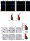
Linderalactone inhibits the proliferation of pancreatic cancer cells. (a, b) EdU staining assay detected the proliferation of BXPC-3 and CFPAC-1 cell lines under varying concentrations of LL treatment after 24 hours. (c, d) Quantification of EdU stain: the percentage of EdU positive cells to the total number of cells. (e) Clone formation assay to detect the effect of LL on BXPC-3 and CFPAC-1 cell lines. (f) Quantification of clone formation. Scale bar = 200 μm. p < 0.05; ∗∗p < 0.01.
Figure 3
Figure 3
Linderalactone inhibits the migration and invasion of pancreatic cancer cells. (a) Wound-healing assay detects the migration ability of BXPC-3 under varying concentrations of LL after 24 hours. (b) Quantification of wound-healing assay: the area of cell migration in 24 hours accounts for the percentage of the wound area in 0 hours. (c) The transwell assay detected the migration ability of cells under varying concentrations of LL treatment after 24 hours. (d) Quantification of transwell assay: percentage of the compound treatment group in the control group. (e) The transwell assay with Matrigel to detect cell invasion ability under varying concentrations of LL treatment after 24 hours. (f) Quantification of transwell assay: percentage of the compound treatment group in the control group. Scale bar = 200 μm. p < 0.05; ∗∗p < 0.01.
Figure 4
Figure 4
Linderalactone blocks the cell cycle and induces apoptosis. (a) Flow cytometry using PI staining to detect the cycle distribution of BXPC-3 and CFPAC-1 cell lines treated with varying concentrations of LL for 24 hours. (b) The percentage of cell cycle distribution for BXPC-3 and CFPAC-1. (c) Annexin V/PI staining to detect the apoptotic death of BXPC-3 and CFPAC-1. (d) The percentage of cell apoptosis for BXPC-3 and CFPAC-1. (e, f) Western blot analysis of key cell cycle and cell apoptosis markers. p < 0.05; ∗∗p < 0.01.
Figure 5
Figure 5
Linderalactone exhibits antitumor activity by inhibiting the PI3K/AKT signaling pathway. (a, b) Western blot analysis of phosphorylated PI3K and phosphorylated AKT, total PI3K, and total AKT expression in BXPC-3 and CFPAC-1 cells after treatment with varying concentrations of LL. (c, d) After LL or YS-49 treatment, the expression of phosphorylated PI3K and phosphorylated AKT and total PI3K and total AKT in BXPC-3 and CFPAC-1 cells. (e) After LL or YS-49 treatment, the clone formation assay detects the proliferation ability of BXPC-3 and CFPAC-1. (f) Quantification of clone formation. p < 0.05; ∗∗p < 0.01.
Figure 6
Figure 6
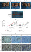
Linderalactone inhibits tumor progression in vivo. (a) Images of the control group and low-dose (LL 25 mg/kg) and high-dose (LL 50 mg/kg) tumor-bearing mice. (b) Quantitative volume of the tumor progression: the tumor volume was measured every three days for three weeks after the injection of LL. (c) Quantitative mice weight: the body weight of the mice was measured every three days for three weeks after the injection of LL. (d) Immunohistochemical detection of PCNA and Ki-67 expression level in tumor cells. p < 0.05; ∗∗p < 0.01.

Similar articles

Cited by

References

    1. Siegel R. L., Miller K. D., Fuchs H. E., Jemal A. Cancer statistics, 2021. CA: A Cancer Journal for Clinicians . 2021;71(1):7–33. doi: 10.3322/caac.21654. - DOI - PubMed
    1. Sung H., Ferlay J., Siegel R. L., et al. Global cancer statistics 2020: GLOBOCAN estimates of incidence and mortality worldwide for 36 cancers in 185 countries. CA: A Cancer Journal for Clinicians . 2021;71(3):209–249. doi: 10.3322/caac.21660. - DOI - PubMed
    1. Kindler H. L. A glimmer of hope for pancreatic cancer. New England Journal of Medicine . 2018;379(25):2463–2464. doi: 10.1056/nejme1813684. - DOI - PubMed
    1. Mizrahi J. D., Surana R., Valle J. W., Shroff R. T. Pancreatic cancer. The Lancet . 2020;395(10242):2008–2020. doi: 10.1016/s0140-6736(20)30974-0. - DOI - PubMed
    1. Okusaka T., Furuse J. Recent advances in chemotherapy for pancreatic cancer: evidence from Japan and recommendations in guidelines. Journal of Gastroenterology . 2020;55(4):369–382. doi: 10.1007/s00535-020-01666-y. - DOI - PMC - PubMed