The Development of the Mesenteric Model of Abdominal Anatomy
- PMID: 35966981
- PMCID: PMC9365479
- DOI: 10.1055/s-0042-1743585
The Development of the Mesenteric Model of Abdominal Anatomy
Abstract
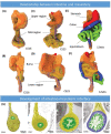
Recent advances in mesenteric anatomy have clarified the shape of the mesentery in adulthood. A key finding is the recognition of mesenteric continuity, which extends from the oesophagogastric junction to the mesorectal level. All abdominal digestive organs develop within, or on, the mesentery and in adulthood remain directly connected to the mesentery. Identification of mesenteric continuity has enabled division of the abdomen into two separate compartments. These are the mesenteric domain (upon which the abdominal digestive system is centered) and the non-mesenteric domain, which comprises the urogenital system, musculoskeletal frame, and great vessels. Given this anatomical endpoint differs significantly from conventional descriptions, a reappraisal of mesenteric developmental anatomy was recently performed. The following narrative review summarizes recent advances in abdominal embryology and mesenteric morphogenesis. It also examines the developmental basis for compartmentalizing the abdomen into two separate domains along mesenteric lines.
Keywords: abdomen; anatomy; embryology; mesentery; peritoneum.
Thieme. All rights reserved.
Figures


Similar articles
-
The development and structure of the mesentery.Commun Biol. 2021 Aug 18;4(1):982. doi: 10.1038/s42003-021-02496-1. Commun Biol. 2021. PMID: 34408242 Free PMC article.
-
Mesentery - a 'New' organ.Emerg Top Life Sci. 2020 Sep 8;4(2):191-206. doi: 10.1042/ETLS20200006. Emerg Top Life Sci. 2020. PMID: 32539112 Review.
-
Development of mesenteric tissues.Semin Cell Dev Biol. 2019 Aug;92:55-62. doi: 10.1016/j.semcdb.2018.10.005. Epub 2018 Oct 25. Semin Cell Dev Biol. 2019. PMID: 30347243 Review.
-
Development of a Novel Technique to Dissect the Mesentery That Preserves Mesenteric Continuity and Enables Characterization of the ex vivo Mesentery.Front Surg. 2020 Jan 23;6:80. doi: 10.3389/fsurg.2019.00080. eCollection 2019. Front Surg. 2020. PMID: 32039231 Free PMC article.
-
Update on the mesentery: structure, function, and role in disease.Lancet Gastroenterol Hepatol. 2022 Jan;7(1):96-106. doi: 10.1016/S2468-1253(21)00179-5. Epub 2021 Nov 22. Lancet Gastroenterol Hepatol. 2022. PMID: 34822760 Review.
Cited by
-
Peritoneal Adhesions in Osteopathic Medicine: Theory, Part 1.Cureus. 2023 Jul 26;15(7):e42472. doi: 10.7759/cureus.42472. eCollection 2023 Jul. Cureus. 2023. PMID: 37502471 Free PMC article. Review.
-
Fascial Nomenclature: Update 2024.Cureus. 2024 Feb 11;16(2):e53995. doi: 10.7759/cureus.53995. eCollection 2024 Feb. Cureus. 2024. PMID: 38343702 Free PMC article. Review.
References
-
- Gray H. Rochester, Kent: Grange; 1858. Gray's Anatomy: A Facsimile.
-
- Moore K L, Agur A MR, Dalley A F, Moore K L.Essential Clinical Anatomy 5 th ed. Philadelphia, PA: Wolters Kluwer Health; 2015
-
- Coffey J C, Culligan K, Walsh L G. An appraisal of the computed axial tomographic appearance of the human mesentery based on mesenteric contiguity from the duodenojejunal flexure to the mesorectal level. Eur Radiol. 2016;26(03):714–721. - PubMed

