

Case Reports
doi: 10.7759/cureus.26772.
eCollection 2022 Jul.
Colonic Polypoid Vascular Ectasia in a Patient With Rectal Prolapse
Affiliations
- PMID: 35967181
- PMCID: PMC9366030
- DOI: 10.7759/cureus.26772
Item in Clipboard
Case Reports
Colonic Polypoid Vascular Ectasia in a Patient With Rectal Prolapse
Cureus.
.
Abstract
Vascular ectasia is a common cause of lower gastrointestinal (GI) bleeding in older patients. They typically present as flat or slightly raised fern-like bright red lesions. We report a rare case of a vascular ectasia presenting as a pedunculated polypoid lesion in a young patient with rectal prolapse. The pedunculated polyp was removed using hot snare polypectomy. This case highlights a unique presentation of a rare lesion and endoscopic management of these lesions.
Keywords: colonoscopy; pedunculated polyp; polypectomy; rectal prolapse; vascular ectasia.
Copyright © 2022, Meader et al.
Conflict of interest statement
The authors have declared that no competing interests exist.
Figures
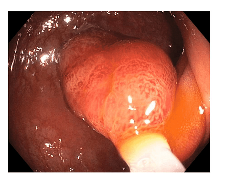
This image demonstrates a 20 mm-pedunculated polyp with hyperemic mucosa and a long stalk.
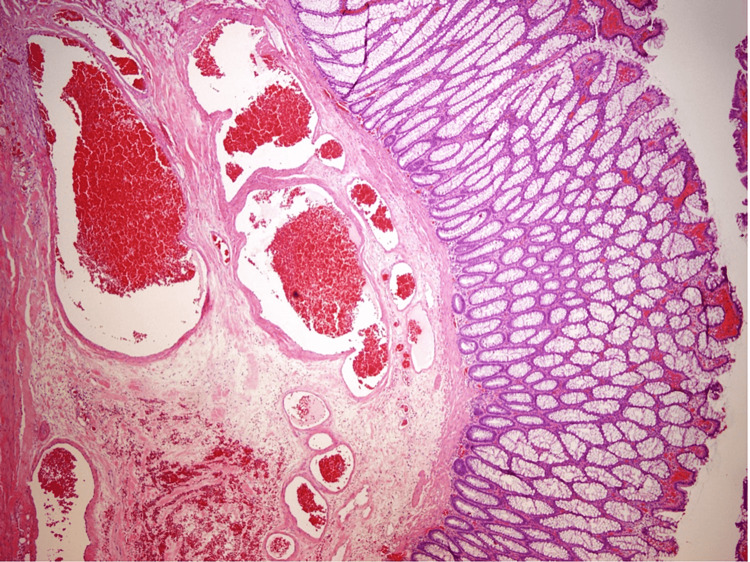
Low-power photograph showing colonic type mucosa with surface erosion, reactive changes, and underlying submucosal dilated vascular spaces (vascular ectasia) in addition to a large thrombosed vascular space. 1X magnification.
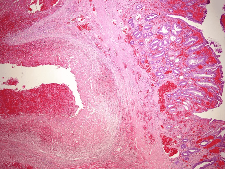
Low-power photograph showing colonic type mucosa with underlying submucosal abnormally dilated congested vascular spaces consistent with vascular ectasia. 1X magnification.
Similar articles
-
Colonic pedunculated polypoid vascular ectasia mimicking ileocolic intussusception: a rare case report.Ann Med Surg (Lond). 2023 May 26;85(7):3674-3678. doi: 10.1097/MS9.0000000000000913. eCollection 2023 Jul. Ann Med Surg (Lond). 2023. PMID: 37427223 Free PMC article.
-
A large polypoid vascular ectasia removed by using a polypectomy with a detachable snare in an asymptomatic patient.Ann Coloproctol. 2013 Feb;29(1):31-3. doi: 10.3393/ac.2013.29.1.31. Epub 2013 Feb 28. Ann Coloproctol. 2013. PMID: 23586013 Free PMC article.
-
Can endoscopic submucosal dissection technique be an alternative treatment option for a difficult giant (≥ 30 mm) pedunculated colorectal polyp?Dis Colon Rectum. 2013 May;56(5):660-6. doi: 10.1097/DCR.0b013e318276d2b9. Dis Colon Rectum. 2013. PMID: 23575407
-
Endoscopic resection of asymptomatic, colonic, polypoid arteriovenous malformations: Two case reports and a literature review.Saudi J Gastroenterol. 2017 Jan-Feb;23(1):67-70. doi: 10.4103/1319-3767.199111. Saudi J Gastroenterol. 2017. PMID: 28139503 Free PMC article. Review.
-
Experience in the endoscopic management of large colonic polyps.ANZ J Surg. 2003 Dec;73(12):988-95. doi: 10.1046/j.1445-2197.2003.t01-23-.x. ANZ J Surg. 2003. PMID: 14632888 Review.
Cited by
-
Colonic pedunculated polypoid vascular ectasia mimicking ileocolic intussusception: a rare case report.Ann Med Surg (Lond). 2023 May 26;85(7):3674-3678. doi: 10.1097/MS9.0000000000000913. eCollection 2023 Jul. Ann Med Surg (Lond). 2023. PMID: 37427223 Free PMC article.
References
-
- Angiodysplasia of the colon: an expression of occlusive vascular disease. Heer M, Sulser H, Hany A. https://pubmed.ncbi.nlm.nih.gov/2440788/ Hepatogastroenterology. 1987;34:127–131. - PubMed
-
- Gastrointestinal angiodysplasia associated with aortic valve disease: part of a spectrum of angiodysplasia of the gut. Weaver GA, Alpern HD, Davis JS, Ramsey WH, Reichelderfer M. https://www.gastrojournal.org/article/S0016-5085(79)80002-5/fulltext. Gastroenterology. 1979;77:1–11. - PubMed
-
- On the nature and etiology of vascular ectasias of the colon. Degenerative lesions of aging. Boley SJ, Sammartano R, Adams A, DiBiase A, Kleinhaus S, Sprayregen S. https://pubmed.ncbi.nlm.nih.gov/300063/ Gastroenterology. 1977;72:650–660. - PubMed
-
- Review article: gastrointestinal angiodysplasia - pathogenesis, diagnosis and management. Sami SS, Al-Araji SA, Ragunath K. https://pubmed.ncbi.nlm.nih.gov/24138285/ Aliment Pharmacol Ther. 2014;39:15–34. - PubMed
-
- Vascular lesions of the gastrointestinal tract. Regula J, Wronska E, Pachlewski J. https://doi.org/10.1016/j.bpg.2007.10.026. Best Pract Res Clin Gastroenterol. 2008;22:313–328. - PubMed
Publication types
LinkOut - more resources
Full Text Sources