

Preclinical studies of the triazolo[1,5- a]pyrimidine derivative WS-716 as a highly potent, specific and orally active P-glycoprotein (P-gp) inhibitor
- PMID: 35967279
- PMCID: PMC9366537
- DOI: 10.1016/j.apsb.2022.03.023
Preclinical studies of the triazolo[1,5- a]pyrimidine derivative WS-716 as a highly potent, specific and orally active P-glycoprotein (P-gp) inhibitor
Abstract
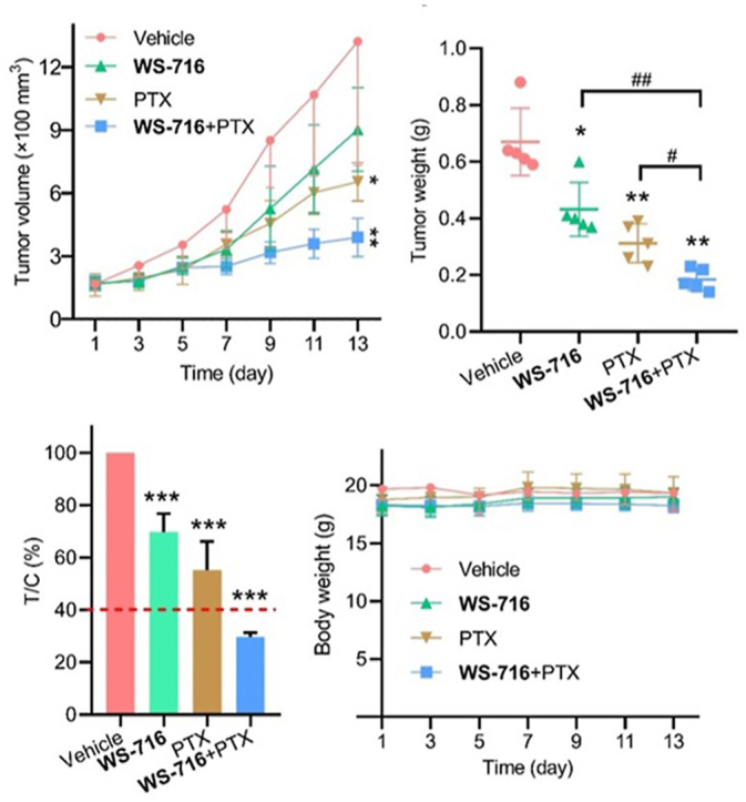
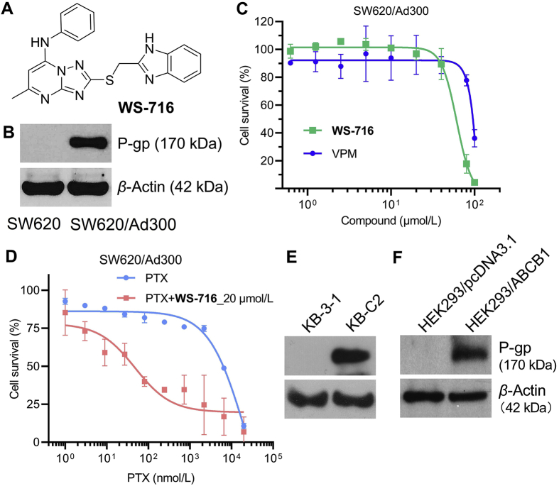
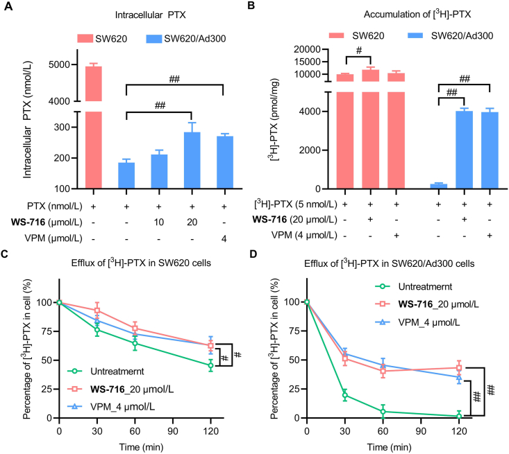
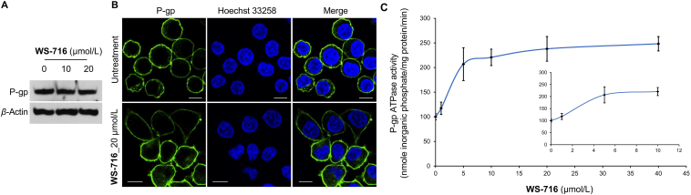
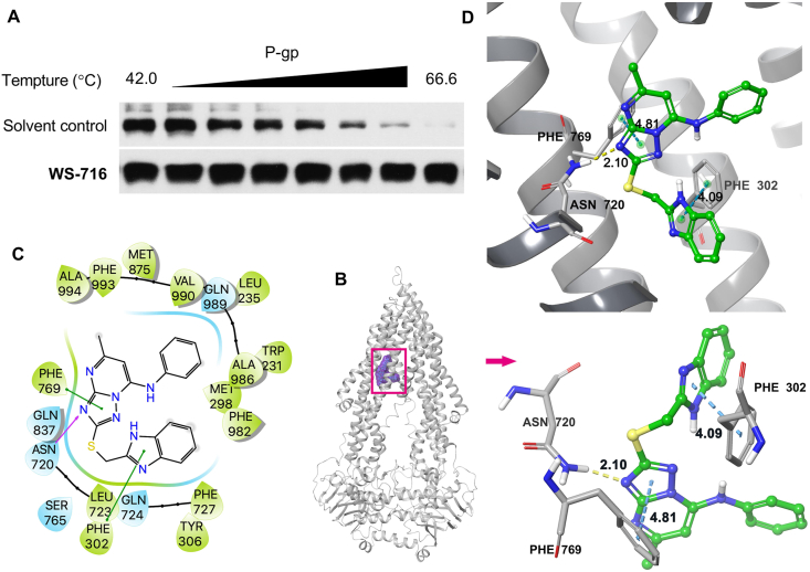
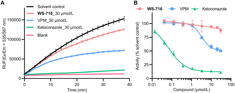
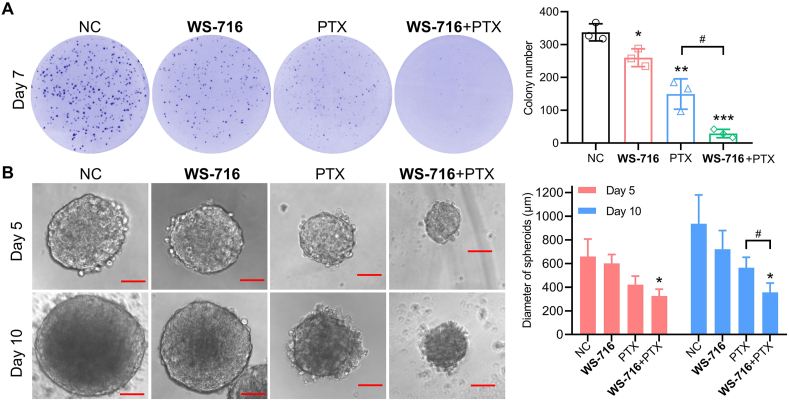
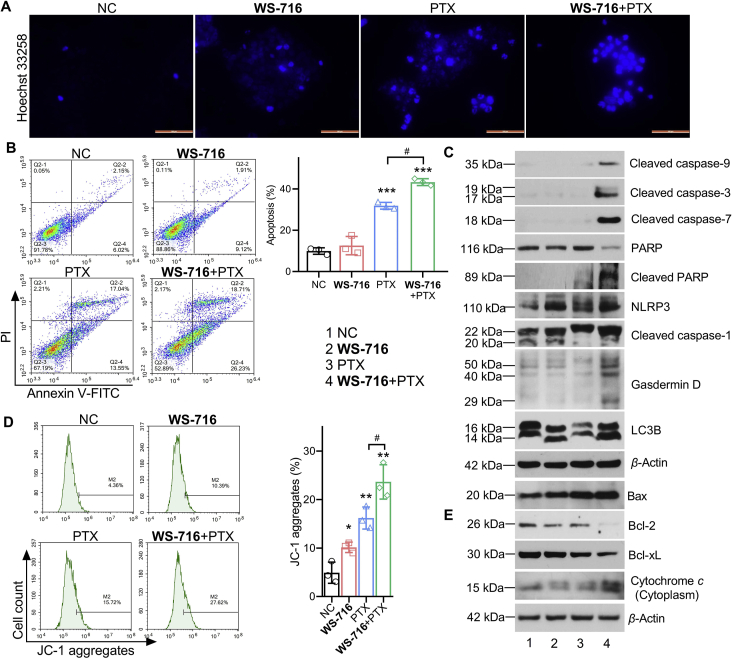
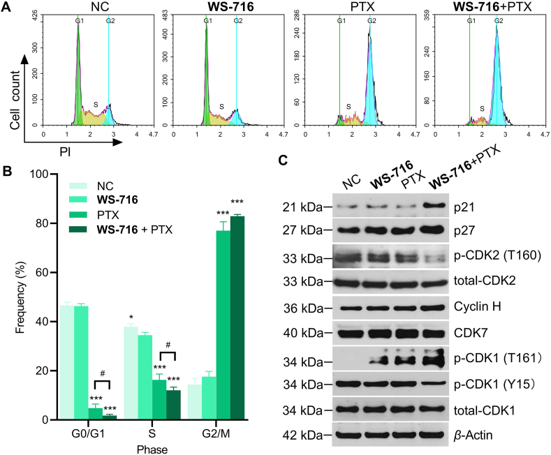
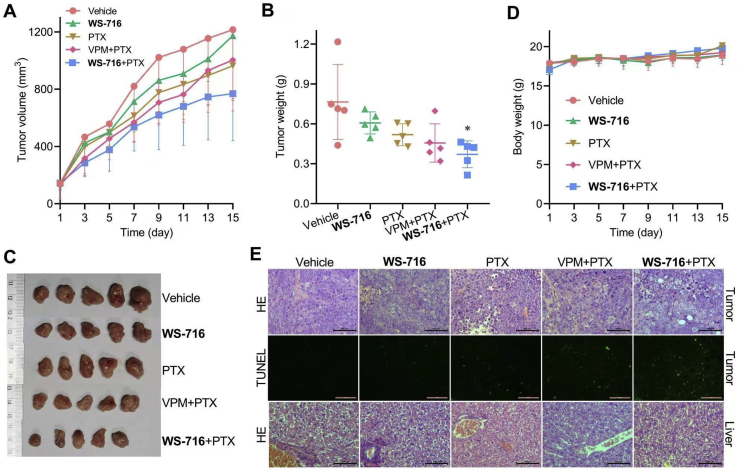
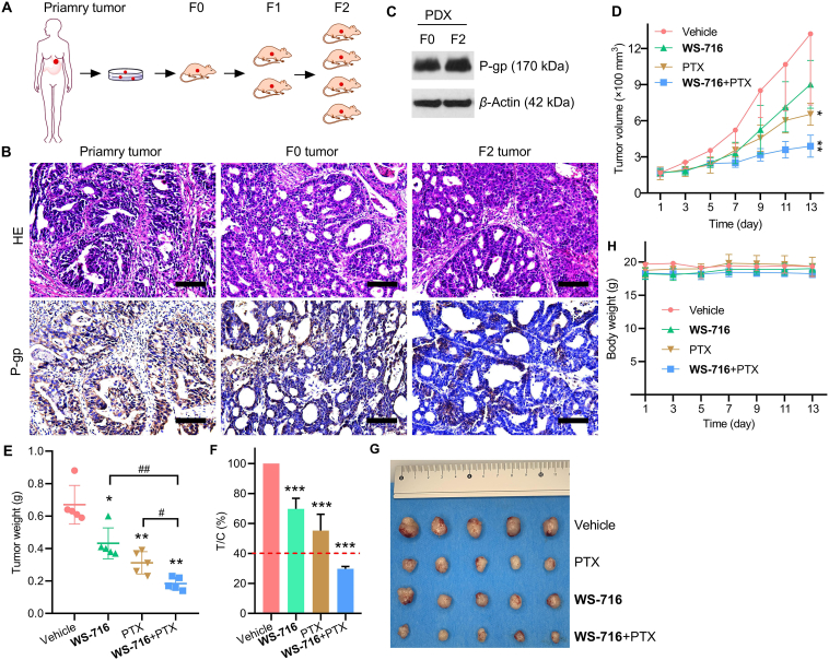
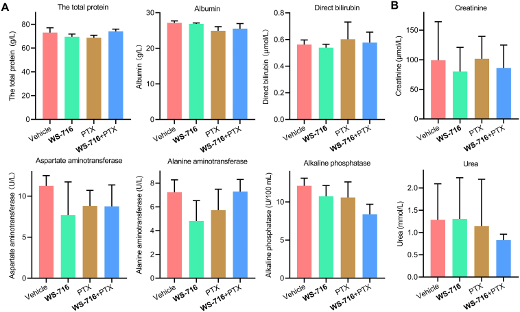
Multidrug resistance (MDR) is the main cause of clinical treatment failure and poor prognosis in cancer. Targeting P-glycoprotein (P-gp) has been regarded as an effective strategy to overcome MDR. In this work, we reported our preclinical studies of the triazolo[1,5-a]pyrimidine-based compound WS-716 as a highly potent, specific, and orally active P-gp inhibitor. Through direct binding to P-gp, WS-716 inhibited efflux function of P-gp and specifically reversed P-gp-mediated MDR to paclitaxel (PTX) in multiple resistant cell lines, without changing its expression or subcellular localization. WS-716 and PTX synergistically inhibited formation of colony and 3D spheroid, induced apoptosis and cell cycle arrest at G2/M phase in resistant SW620/Ad300 cells. In addition, WS-716 displayed minimal effect on the drug-metabolizing enzyme cytochrome P4503A4 (CYP3A4). Importantly, WS-716 increased sensitivity of both pre-clinically and clinically derived MDR tumors to PTX in vivo with the T/C value of 29.7% in patient-derived xenograft (PDX) models. Relative to PTX treatment alone, combination of WS-716 and PTX caused no obvious adverse reactions. Taken together, our preclinical studies revealed therapeutic promise of WS-716 against MDR cancer, the promising data warrant its further development for cancer therapy.
Keywords: ATP-Binding cassette; Cancer therapy; Drug combination; Multidrug resistance (MDR); P-gp inhibitors; Preclinical studies; Triazolo[1,5-a]pyrimidine.
© 2022 Chinese Pharmaceutical Association and Institute of Materia Medica, Chinese Academy of Medical Sciences. Production and hosting by Elsevier B.V.
Conflict of interest statement
The authors declare no conflicts of interest.
Figures
Similar articles
-
Discovery of the Triazolo[1,5-a]Pyrimidine-Based Derivative WS-898 as a Highly Efficacious and Orally Bioavailable ABCB1 Inhibitor Capable of Overcoming Multidrug Resistance.J Med Chem. 2021 Nov 11;64(21):16187-16204. doi: 10.1021/acs.jmedchem.1c01498. Epub 2021 Nov 1. J Med Chem. 2021. PMID: 34723530
-
Structure-Based Design, Synthesis, and Biological Evaluation of New Triazolo[1,5-a]Pyrimidine Derivatives as Highly Potent and Orally Active ABCB1 Modulators.J Med Chem. 2020 Dec 24;63(24):15979-15996. doi: 10.1021/acs.jmedchem.0c01741. Epub 2020 Dec 5. J Med Chem. 2020. PMID: 33280384
-
An ester derivative of tenacigenin B from Marsdenia tenacissima (Roxb.) Wight et Arn reversed paclitaxel-induced MDR in vitro and in vivo by inhibiting both P-gp and MRP2.J Ethnopharmacol. 2022 Aug 10;294:115353. doi: 10.1016/j.jep.2022.115353. Epub 2022 May 6. J Ethnopharmacol. 2022. PMID: 35533911
-
Therapeutic strategies to overcome taxane resistance in cancer.Drug Resist Updat. 2021 Mar;55:100754. doi: 10.1016/j.drup.2021.100754. Epub 2021 Feb 27. Drug Resist Updat. 2021. PMID: 33691261 Review.
-
Improvement of conventional anti-cancer drugs as new tools against multidrug resistant tumors.Drug Resist Updat. 2020 May;50:100682. doi: 10.1016/j.drup.2020.100682. Epub 2020 Feb 7. Drug Resist Updat. 2020. PMID: 32087558
Cited by
-
Mechanism of multidrug resistance to chemotherapy mediated by P‑glycoprotein (Review).Int J Oncol. 2023 Nov;63(5):119. doi: 10.3892/ijo.2023.5567. Epub 2023 Sep 1. Int J Oncol. 2023. PMID: 37654171 Free PMC article. Review.
-
Preclinical studies of the falnidamol as a highly potent and specific active ABCB1 transporter inhibitor.BMC Cancer. 2025 Jan 7;25(1):24. doi: 10.1186/s12885-024-13371-7. BMC Cancer. 2025. PMID: 39773145 Free PMC article.
-
P-Glycoprotein as a Therapeutic Target in Hematological Malignancies: A Challenge to Overcome.Int J Mol Sci. 2025 May 14;26(10):4701. doi: 10.3390/ijms26104701. Int J Mol Sci. 2025. PMID: 40429842 Free PMC article. Review.
-
Targeting epigenetic regulators to overcome drug resistance in cancers.Signal Transduct Target Ther. 2023 Feb 17;8(1):69. doi: 10.1038/s41392-023-01341-7. Signal Transduct Target Ther. 2023. PMID: 36797239 Free PMC article. Review.
-
Self-assembled nanoformulations of paclitaxel for enhanced cancer theranostics.Acta Pharm Sin B. 2023 Aug;13(8):3252-3276. doi: 10.1016/j.apsb.2023.02.021. Epub 2023 Mar 5. Acta Pharm Sin B. 2023. PMID: 37655323 Free PMC article. Review.
References
-
- Fuchs D.A., Johnson R.K. Cytologic evidence that taxol, an antineoplastic agent from Taxus brevifolia, acts as a mitotic spindle poison. Cancer Treat Rep. 1978;62:1219–1222. - PubMed
-
- Gottesman M.M. Mechanisms of cancer drug resistance. Annu Rev Med. 2002;53:615–627. - PubMed
-
- Rosenberg M.F., Callaghan R., Ford R.C., Higgins C.F. Structure of the multidrug resistance P-glycoprotein to 2.5 nm resolution determined by electron microscopy and image analysis. J Biol Chem. 1997;272:10685–10694. - PubMed
-
- Gottesman M.M., Pastan I. Biochemistry of multidrug resistance mediated by the multidrug transporter. Annu Rev Biochem. 1993;62:385–427. - PubMed
LinkOut - more resources
Full Text Sources
Miscellaneous
