. 2022 Jul 29:13:919237.
doi: 10.3389/fimmu.2022.919237. eCollection 2022.

Radiosensitivity in patients affected by ARPC1B deficiency: a new disease trait?

Affiliations

Radiosensitivity in patients affected by ARPC1B deficiency: a new disease trait?

Maria Chiriaco et al. Front Immunol. .

Abstract

Actin-related protein 2/3 complex subunit 1B (ARPC1B) deficiency is a recently described inborn error of immunity (IEI) presenting with combined immunodeficiency and characterized by recurrent infections and thrombocytopenia. Manifestations of immune dysregulation, including colitis, vasculitis, and severe dermatitis, associated with eosinophilia, hyper-IgA, and hyper-IgE are also described in ARPC1B-deficient patients. To date, hematopoietic stem cell transplantation seems to be the only curative option for patients. ARPC1B is part of the actin-related protein 2/3 complex (Arp2/3) and cooperates with the Wiskott-Aldrich syndrome protein (WASp) in the regulation of the actin cytoskeleton remodeling and in driving double-strand break clustering for homology-directed repair. In this study, we aimed to investigate radiosensitivity (RS) in ARPC1B-deficient patients to assess whether it can be considered an additional disease trait. First, we performed trio-based next-generation-sequencing studies to obtain the ARPC1B molecular diagnosis in our index case characterized by increased RS, and then we confirmed, using three different methods, an increment of radiosensitivity in all enrolled ARPC1B-deficient patients. In particular, higher levels of chromatid-type aberrations and γH2AX foci, with an increased number of cells arrested in the G2/M-phase of the cell cycle, were found in patients' cells after ionizing radiation exposition and radiomimetic bleomycin treatment. Overall, our data suggest increased radiosensitivity as an additional trait in ARPC1B deficiency and support the necessity to investigate this feature in ARPC1B patients as well as in other IEI with cytoskeleton defects to address specific clinical follow-up and optimize therapeutic interventions.

Keywords: ARPC1B; DNA damage response (DDR); combined immunodeficiency; immune dysregulation; radiosensitivity.


Conflict of interest statement

The authors declare that the research was conducted in the absence of any commercial or financial relationships that could be construed as a potential conflict of interest. The reviewer VB declared a shared affiliation with the author J-PV to the handling editor at the time of review.

Figures
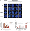
Figure 1
Figure 1
DNA DSB repair kinetics in healthy donors (HDs), PtII-1, Pt1-ARPC1B, and Pt3-ARPC1B. (A) Representative images of γH2AX staining in primary fibroblasts from HDs and patients irradiated with 1Gy X-rays and fixed after 0.5, 2, 4, and 24 h. (B) Histograms showing the number of γH2AX foci/cells in fibroblasts reported as mean ± SEM (n = 3; HDs = 3) at different time points after irradiation. (C) Frequency (mean ± SEM; n = 3; HDs = 6) of chromatid-type aberrations on irradiated EBVB cells in the G2-cell cycle phase. Statistical analysis was performed with a two-way ANOVA test and Tukey posttest. * p < 0.05; ** p < 0.01; *** p < 0.001; **** p < 0.0001.
Figure 2
Figure 2
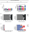
Radiosensitivity evaluation in ARPC1B-deficient cells after 2 h of bleomycin treatment. (A) FACS analysis of γH2AX expression in EBVB cells from HDs, Pt-II1, Pt1-3 ARPC1B, and Pt-WAS with or w/o BLM treatment (9 µM/1 h). Column bar graphs show the mean ± SEM (n > 3; HDs = 15). (B) Distribution of cells into the cell cycle phases before and after 2 h of BLM treatment (FACS). Each bar represents the mean of cells determined in at least three independent experiments. (C) Quantification of the γH2AX+ EBVB cells in each cell cycle phase (FACS). The percentage of cells in G0/G1, S, and G2/M cell cycle phases (γH2AX+ with respect to singlet cells) was represented as mean ± SEM (n > 3). Statistical significance was calculated with one-way ANOVA and Bonferroni’s posttest: ** p < 0.01.
Figure 3
Figure 3
Repair-time kinetics of the cell survival fraction after BLM treatment. Kinetic experiment on untreated or BLM-treated EBVB cells from PtII-1, Pt1-3 ARPC1B patients, two WAS patients, and four HDs, investigated during 24 h of recovery time culture. (A) The graph shows the percentage of γH2AX+ EBVB cells. (B) Cell survival fractions were reported as the percentage of 7AAD EBVB cells. (C) Amount of γH2AX+ EBVB cells in each cell cycle phase: G0/G1 (left panel), S (middle panel), and G2/M (right panel). Black line indicates the mean ± SEM of the four HDs. (D) Percentage of γH2AX+ EBVB cells were reported as mean ± SEM calculated for each disease group (three ARPC1B patients and two WAS patients) and PtII-1 against HD group. Statistical significance was calculated with two-way ANOVA and Bonferroni’s posttest: * p < 0.05, ** p < 0.01, *** p < 0.001, and **** p < 0.0001.
Figure 4
Figure 4
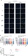
γH2AX expression in ARPC1B-deficient EBVB BLM-treated cells by immunofluorescence. (A) Representative images of γH2AX (green) and nuclei counterstaining (blue) investigated on cells from PtII-1, Pt1-3 ARPC1B, Pt1-WAS, and HD before and after 4 h of BLM treatment (9 µM/1 h). Each left column shows ×20 magnification, and each right column shows ×60 magnification. Bar scale represented 75 µm (×20) and 25 µm (×60). (B) Histograms show the γH2AX-integrated intensity evaluated in the patient’s EBVB cells compared to HDs (n = 4; HDs = 4). Each point represents the integrated intensity of a single cell. The line indicates the mean ± SEM (n > 2). (C) The graph shows a between-group statistical comparison of the γH2AX-integrated intensity detected in PtII-1, ARPC1B-, and WAS-disease group of patients against the HD group. The statistical analyses were performed with two-way ANOVA and Bonferroni’s posttest: * p < 0.05, ** p < 0.01, *** p < 0.001, and **** p < 0.0001.

References

    1. Kahr WH, Pluthero FG, Elkadri A, Warner N, Drobac M, Chen CH, et al. . Loss of the Arp2/3 complex component ARPC1B causes platelet abnormalities and predisposes to inflammatory disease. Nat Commun (2017) 8:14816. doi: 10.1038/ncomms14816 - DOI - PMC - PubMed
    1. Kuijpers TW, Tool ATJ, van der Bijl I, de Boer M, van Houdt M, de Cuyper IM, et al. . Combined immunodeficiency with severe inflammation and allergy caused by ARPC1B deficiency. J Allergy Clin Immunol (2017) 140(1):273–7. doi: 10.1016/j.jaci.2016.09.061 - DOI - PubMed
    1. Somech R, Lev A, Lee YN, Simon AJ, Barel O, Schiby G, et al. . Disruption of thrombocyte and T lymphocyte development by a mutation in ARPC1B. J Immunol (2017) 199(12):4036–45. doi: 10.4049/jimmunol.1700460 - DOI - PMC - PubMed
    1. Brigida I, Zoccolillo M, Cicalese MP, Pfajfer L, Barzaghi F, Scala S, et al. . T-Cell defects in patients with ARPC1B germline mutations account for combined immunodeficiency. Blood (2018) 132(22):2362–74. doi: 10.1182/blood-2018-07-863431 - DOI - PMC - PubMed
    1. Volpi S, Cicalese MP, Tuijnenburg P, Tool ATJ, Cuadrado E, Abu-Halaweh M, et al. . A combined immunodeficiency with severe infections, inflammation, and allergy caused by ARPC1B deficiency. J Allergy Clin Immunol (2019) 143(6):2296–9. doi: 10.1016/j.jaci.2019.02.003 - DOI - PMC - PubMed

Substances

LinkOut - more resources