. 2022 Jul 28:13:928300.
doi: 10.3389/fimmu.2022.928300. eCollection 2022.

Cystic fibrosis transmembrane regulator correction attenuates heart failure-induced lung inflammation

Affiliations

Cystic fibrosis transmembrane regulator correction attenuates heart failure-induced lung inflammation

Franziska E Uhl et al. Front Immunol. .

Abstract

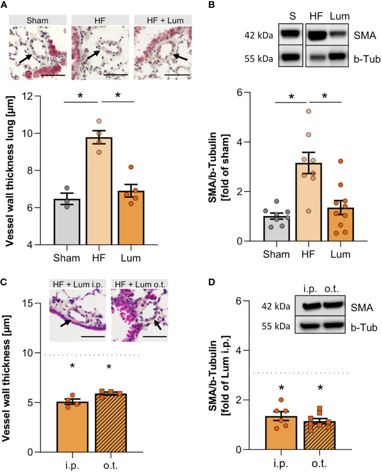
Heart failure (HF) affects 64 million people worldwide. Despite advancements in prevention and therapy, quality of life remains poor for many HF patients due to associated target organ damage. Pulmonary manifestations of HF are well-established. However, difficulties in the treatment of HF patients with chronic lung phenotypes remain as the underlying patho-mechanistic links are still incompletely understood. Here, we aim to investigate the cystic fibrosis transmembrane regulator (CFTR) involvement in lung inflammation during HF, a concept that may provide new mechanism-based therapies for HF patients with pulmonary complications. In a mouse model of HF, pharmacological CFTR corrector therapy (Lumacaftor (Lum)) was applied systemically or lung-specifically for 2 weeks, and the lungs were analyzed using histology, flow cytometry, western blotting, and qPCR. Experimental HF associated with an apparent lung phenotype characterized by vascular inflammation and remodeling, pronounced tissue inflammation as evidenced by infiltration of pro-inflammatory monocytes, and a reduction of pulmonary CFTR+ cells. Moreover, the elevation of a classically-activated phenotype of non-alveolar macrophages coincided with a cell-specific reduction of CFTR expression. Pharmacological correction of CFTR with Lum mitigated the HF-induced downregulation of pulmonary CFTR expression and increased the proportion of CFTR+ cells in the lung. Lum treatment diminished the HF-associated elevation of classically-activated non-alveolar macrophages, while promoting an alternatively-activated macrophage phenotype within the lungs. Collectively, our data suggest that downregulation of CFTR in the HF lung extends to non-alveolar macrophages with consequences for tissue inflammation and vascular structure. Pharmacological CFTR correction possesses the capacity to alleviate HF-associated lung inflammation.

Keywords: cystic fibrosis transmembrane regulator; heart failure; inflammation; lung; macrophages.


Conflict of interest statement

The authors declare that the research was conducted in the absence of any commercial or financial relationships that could be construed as a potential conflict of interest.

Figures

Figure 1
Figure 1
Heart failure-associated structural changes in the lung are confined to blood vessels. (A) Representative Haematoxylin and Eosin (H&E) staining of lungs from sham and heart failure (HF) mice (arrows indicate vessel walls; scale bar 20 µm) and quantification of wall thickness of small vessels in H&E-stained lung slices. N=7 per group. (B) Representative western blot and quantification of the smooth muscle actin (SMA) protein expression in lung tissue from sham and HF mice. N=8 per group. (C) Masson Trichrome (MTC) staining of lungs from sham and HF mice (arrows indicate collagen, stained in blue; scale bar 20 µm) and qualitative quantification of collagen in MTC-stained lung sections. N=8 for Sham, N=10 for HF. (D) Quantification of hydroxyproline content of lung tissue from sham and HF mice. N=7 for Sham, N=6 for HF. N denotes the number of mice. Data expressed as mean ± SEM. * denotes p ≤ 0.05 after unpaired t-test.
Figure 2
Figure 2
Heart failure associates with lung infiltration of CD80+ pro-inflammatory macrophages. (A) Representative images of lung sections from sham and heart failure (HF) mice that were stained for monocyte/macrophages (MOMA) in red, smooth muscle actin (SMA) in green, DAPI stained nuclei in blue. Arrows indicate vessel wall-associated MOMA positivity; scale bar 20 µm. Quantification of the percentage of MOMA positive cells in lung vessel walls. N=3 (n=8) for Sham, N=3 (n=7) for HF. (B) Flow cytometry results representing the number of CD45hi Ly6C+ SiglecF- and (C) CD45hi Ly6Chi SiglecF- macrophages. N=8 for Sham, N=10 for HF each. Representative dot blots of Ly6C and SiglecF expression of F4/80+ macrophages in the lung of sham and HF mice. (D) Flow cytometric assessment of F4/80+ CD80+ classically activated macrophages, (E) F4/80+ CD80+ SiglecF- classically-activated non-alveolar macrophages, and (F) F4/80+ CD80+ SiglecF+ classically-activated alveolar macrophages in lung tissue of sham and HF mice. Representative dot blots of SiglecF and CD80 expression of F4/80+ macrophages in the lung of sham and HF mice. N=8 for Sham, N=10 for HF each. N denotes the number of mice; n denotes the number of independent measures. Data expressed as mean ± SEM. * denotes p ≤ 0.05 after unpaired t-test.
Figure 3
Figure 3
Pulmonary tumour necrosis factor alpha increase is accompanied by decreased cystic fibrosis transmembrane regulator expression in the heart failure lung. (A) Representative western blot and quantification of tumour necrosis factor alpha (TNF-α) expression in the lungs of sham and heart failure (HF) mice. N=7 per group. (B) Representative western blot and quantification of CFTR protein expression in lungs of sham and HF mice. N=7 for Sham, N=8 for HF. Flow cytometric assessment of proportion of (C) CFTR+ F4/80+ SiglecF- non-alveolar macrophages and (D) CFTR+ F4/80+ SiglecF+ alveolar macrophages in lung tissue from naïve and HF mice. N=5 for naïve, N=8 for HF each. Representative histograms of CFTR+ SiglecF- and CFTR+ SiglecF+ cells from naïve (grey) and HF (coral) mice. N denotes the number of mice. Data expressed as mean ± SEM. * denotes p ≤ 0.05 after unpaired t-test.
Figure 4
Figure 4
Systemic application of cystic fibrosis transmembrane regulator (CFTR) correctors increases pulmonary CFTR expression. (A) Percentage of CFTR+ cells in lungs of sham, heart failure (HF), and Lumacaftor (Lum) treated (intraperitoneally (i.p.)) HF mice and representative dot plots. N=8 for Sham, N=8 for HF + vehicle, N=10 for HF + Lum. (B) Representative western blot and quantification of the CFTR expression in the lungs of HF mice treated with Lumacaftor either i.p. or o.t. N=6 for HF + Lum i.p., N=8 for HF + Lum o.t. (C) Median fluorescence intensity and representative histograms of CFTR+ cells in the lungs of HF mice treated with Lumacaftor either i.p. (coral) or o.t. (orange). N=6 for HF + Lum i.p., N=8 for HF + Lum o.t. N denotes the number of mice. Data expressed as mean ± SEM. In (A), * denotes p ≤ 0.05 relative to HF after one-way ANOVA with Dunnett’s post-hoc testing; in (B, C), $ denotes p ≤ 0.05 after unpaired t-test.
Figure 5
Figure 5
Cystic fibrosis transmembrane regulator correction mitigates heart failure-associated alteration of pulmonary vascular structure. (A) Quantification of the vessel wall thickness of smaller vessels in the lungs of sham, heart failure (HF), and Lumacaftor (Lum) treated HF mice. N=3 for Sham, N=4 for HF + vehicle, N=5 for HF + Lum. Insets showing representative images of H&E-stained lung sections; scale bars 50 µm; arrows indicating vessel wall. (B) Representative western blot and quantification of the smooth muscle actin (SMA) expression in lung tissue from sham, HF, and Lum treated HF mice. N=8 for Sham, N=8 for HF + vehicle, N=10 for HF + Lum. (C) Quantification of the vessel wall thickness of smaller vessels in the lungs of Lum treated (intraperitoneally (i.p.) or orotracheally (o.t.)) HF mice. N=4 for HF + Lum i.p., N=4 for HF + Lum o.t. The dotted line indicates the level of HF mice. Insets showing representative images of H&E-stained lung sections; scale bars 50µm; arrows indicating vessel wall. (D) Representative western blot and quantification of SMA expression in lung tissue from Lum treated (i.p. and o.t.) HF mice. N=6 for HF + Lum i.p., N=8 for HF + Lum o.t. The dotted line indicates the level of HF mice. N denotes the number of mice. Data expressed as mean ± SEM. * denotes p ≤ 0.05 relative to HF after one-way ANOVA with Dunnett’s post-hoc testing.
Figure 6
Figure 6
Cystic fibrosis transmembrane regulator correction normalizes levels of non-alveolar macrophages and increases CD206+ alveolar macrophages. Proportion of pulmonary F4/80+-macrophages in sham, heart failure (HF), and Lumacaftor (Lum) treated ((intraperitoneally (i.p.) or orotracheally (o.t.)) HF mice positive for (A) CD80 and (D) CD206+. N=8 for Sham, N=10 for HF + vehicle, N=6 for HF + Lum i.p., N=8 for HF + Lum o.t. each. Percentage of pulmonary non-alveolar F4/80+ and SiglecF- macrophages in sham, HF, and Lum treated (i.p. and o.t.) HF mice positive for (B) CD80+ and (E) CD206+. N=8 for Sham, N=10 for HF + vehicle, N=6 for HF + Lum i.p., N=8 for HF + Lum o.t. each. Percentage of pulmonary alveolar F4/80+ and SiglecF+ macrophages in sham, HF, and Lum treated (i.p. and o.t.) HF mice positive for (C) CD80+ and (F) CD206+. N=8 for Sham, N=10 for HF + vehicle, N=6 for HF + Lum i.p., N=8 for HF + Lum o.t. each. N denotes the number of mice. In (A, B, D, F), data expressed as mean ± SEM; * denotes p ≤ 0.05 relative to HF for multiple comparisons with Dunnett’s post-hoc testing. In (C, E), data expressed as median ± IQR; * denotes p ≤ 0.05 relative to HF for multiple comparisons with Dunn’s post-hoc testing.

Similar articles

Cited by

References

    1. Bueno H, Moura B, Lancellotti P, Bauersachs J. The year in cardiovascular medicine 2020: Heart failure and cardiomyopathies. Eur Heart J (2021) 42(6):657–70. doi: 10.1093/eurheartj/ehaa1061 - DOI - PubMed
    1. Savarese G, Lund LH. Global public health burden of heart failure. Card Fail Rev (2017) 3(1):7–11. doi: 10.15420/cfr.2016:25:2 - DOI - PMC - PubMed
    1. Moradi M, Daneshi F, Behzadmehr R, Rafiemanesh H, Bouya S, Raeisi M. Quality of life of chronic heart failure patients: A systematic review and meta-analysis. Heart Fail Rev (2020) 25(6):993–1006. doi: 10.1007/s10741-019-09890-2 - DOI - PubMed
    1. Harjola VP, Mullens W, Banaszewski M, Bauersachs J, Brunner-La Rocca HP, Chioncel O, et al. . Organ dysfunction, injury and failure in acute heart failure: From pathophysiology to diagnosis and management. a review on behalf of the acute heart failure committee of the heart failure association (Hfa) of the European society of cardiology (Esc). Eur J Heart Fail (2017) 19(7):821–36. doi: 10.1002/ejhf.872 - DOI - PMC - PubMed
    1. Meissner A, Yang J, Kroetsch JT, Sauve M, Dax H, Momen A, et al. . Tumor necrosis factor-Alpha-Mediated downregulation of the cystic fibrosis transmembrane conductance regulator drives pathological sphingosine-1-Phosphate signaling in a mouse model of heart failure. Circulation (2012) 125(22):2739–50. doi: 10.1161/CIRCULATIONAHA.111.047316 - DOI - PubMed

Publication types

Substances