. 2022 Jul 29:13:939106.
doi: 10.3389/fimmu.2022.939106. eCollection 2022.

Aspirin eugenol ester alleviates lipopolysaccharide-induced acute lung injury in rats while stabilizing serum metabolites levels

Affiliations

Aspirin eugenol ester alleviates lipopolysaccharide-induced acute lung injury in rats while stabilizing serum metabolites levels

Qi Tao et al. Front Immunol. .

Abstract

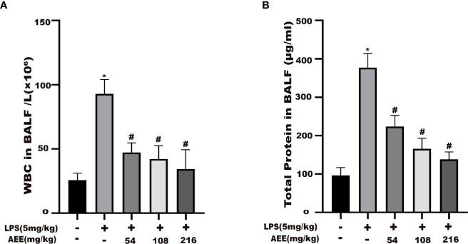
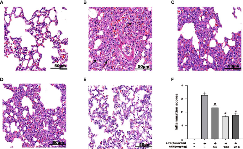
Aspirin eugenol ester (AEE) was a novel drug compound with aspirin and eugenol esterified. AEE had various pharmacological activities, such as anti-inflammatory, antipyretic, analgesic, anti-oxidative stress and so on. In this study, it was aimed to investigate the effect of AEE on the acute lung injury (ALI) induced by lipopolysaccharide (LPS) in rats. In vitro experiments evaluated the protective effect of AEE on the LPS-induced A549 cells. The tumor necrosis factor-α (TNF-α), interleukin-6 (IL-6), and interleukin-1β (IL-1β) were measured in the cell supernatant. The Wistar rats were randomly divided into five groups (n = 8): control group, model group (LPS group), LPS + AEE group (AEE, 54 mg·kg-1), LPS + AEE group (AEE, 108 mg·kg-1), LPS + AEE group (AEE, 216 mg·kg-1). The lung wet-to-dry weight (W/D) ratio and immune organ index were calculated. WBCs were counted in bronchoalveolar lavage fluid (BALF) and total protein concentration was measured. Hematoxylin-Eosin (HE) staining of lung tissue was performed. Glutathione (GSH), glutathione peroxidase (GPx), catalase (CAT), antioxidant superoxide dismutase (SOD), total antioxidant capacity (T-AOC), lactate dehydrogenase (LDH), C-reactive protein (CRP), myeloperoxidase (MPO), malondialdehyde (MDA), macrophage mobility inhibitory factor (MIF), TNF-α, IL-6, and IL-1β activity were measured. The metabolomic analysis of rat serum was performed by UPLC-QTOF-MS/MS. From the results, compared with LPS group, AEE improved histopathological changes, reduced MDA, CRP, MPO, MDA, and MIF production, decreased WBC count and total protein content in BALF, pro-inflammatory cytokine levels, immune organ index and lung wet-dry weight (W/D), increased antioxidant enzyme activity, in a dose-dependent manner. The results of serum metabolomic analysis showed that the LPS-induced ALI caused metabolic disorders and oxidative stress in rats, while AEE could ameliorate it to some extent. Therefore, AEE could alleviate LPS-induced ALI in rats by regulating abnormal inflammatory responses, slowing down oxidative stress, and modulating energy metabolism.

Keywords: acute lung injury; aspirin eugenol ester (AEE); inflammation; metabolites; oxidative stress.


Conflict of interest statement

The authors declare that the research was conducted in the absence of any commercial or financial relationships that could be construed as a potential conflict of interest.

Figures

Figure 1
Figure 1
The effects of AEE and LPS with different concentrations on A549 cell viability, respectively. (A) AEE, (B) LPS. Values are presented as the means ± SD where applicable (n = 6). *P < 0.05 compared to control group.
Figure 2
Figure 2
Effect of AEE on LPS-induced inflammatory cytokines. (A) TNF-α, (B) IL-6 and (C) IL-1β in the supernatant of A549 cells. (D) TNF-α, (E) IL-6, and (F) IL-1β in the serum of LPS-induced ALI rats. Values are presented as the means ± SD where applicable (n = 6). *P < 0.05 compared to control group, #P < 0.05 compared to LPS group.
Figure 3
Figure 3
Effect of AEE on the number of WBC in BALF and total protein level in BALF of LPS-induced ALI rats. (A) WBC in BALF, (B) Total protein in BALF. Values are presented as the means ± SD where applicable (n = 8). *P < 0.05 compared to control group, #P < 0.05 compared to LPS group.
Figure 4
Figure 4
Effect of AEE on splenic index in LPS-induced ALI rats. Values are presented as the means ± SD where applicable (n = 8). *P < 0.05 compared to control group, #P < 0.05 compared to LPS group.
Figure 5
Figure 5
Effect of AEE on lung wet to dry weight ratio (W/D). Values are presented as the means ± SD where applicable (n = 8). *P < 0.05 compared to control group, #P < 0.05 compared to LPS group.
Figure 6
Figure 6
Effect of AEE on concentrations of CRP (A), MIF (B) in serum, MPO (C) in lung tissue. Values are presented as the means ± SD where applicable (n = 8). *P < 0.05 compared to control group, #P < 0.05 compared to LPS group.
Figure 7
Figure 7
Effect of AEE on the level of oxidative stress in LPS-induced ALI rats. (A) GSH, (B) GPx, (C) CAT, (D) T-AOC, (E) SOD, (F) LDH, (G) MDA. Values are presented as the means ± SD where applicable (n = 8). *P < 0.05 compared to control group, #P < 0.05 compared to LPS group.
Figure 8
Figure 8
Effect of AEE on histopathological changes in the lungs of LPS-induced ALI rats. (A) Control group, (B) LPS group, (C) LPS + AEE group (AEE, 54 mg kg-1), (D) LPS + AEE group (AEE, 108 mg kg-1), (E) LPS + AEE group (AEE, 216 mg kg-1), (F) Inflammatory change score chart. *P < 0.05 compared to control group, # P < 0.05 compared to LPS group.
Figure 9
Figure 9
Metabolomic analysis of the effect of AEE on LPS-induced ALI in rats. (A, G) PCA score plots based on rat sera obtained from control, LPS and AEE groups in positive and negative modes, ESI+: R2 = 0.878, ESI-: R2 = 0.829. (B, H) The loading plots of AEE and LPS groups in positive and negative modes. (C, I) OPLS-DA score plots for AEE and LPS groups in positive and negative models, ESI+: R2 X = 0.807, R2 Y = 0.926, Q2 = 0.768; ESI-: R2 X = 0.646, R2 Y = 0.998, Q2 = 0.958. (D, J) Permutation test for the OPLS-DA model, ESI+: intercepts of R2 = 0.626 and Q2 = -0.502 and ESI-: intercepts of R2 = 0.946 and Q2 = -0.245. (E, K) OPLS-DA score plots for control and LPS groups in positive and negative modes, ESI+: R2 X = 0.977, R2 Y = 1, Q2 = 0.992; ESI-: R2 X = 0.887, R2 Y = 1, Q2 = 0.951. (F, L) Permutation test for the OPLS-DA model, ESI+: intercepts of R2 = 0.999 and Q2 = -0.16, ESI-: intercepts of R2 = 0.991 and Q2 = -0.243. Values are presented as the means ± SD where applicable (n = 6).
Figure 10
Figure 10
Results of ploidy enrichment and pathway analysis of potential metabolites in serum.

Similar articles

Cited by

References

    1. Kim YY, Lee S, Kim MJ, Kang BC, Dhakal H, Choi YA, et al. . Tyrosol attenuates lipopolysaccharide-induced acute lung injury by inhibiting the inflammatory response and maintaining the alveolar capillary barrier. Food Chem Toxicol (2017) 109:526–33. doi: 10.1016/j.fct.2017.09.053 - DOI - PubMed
    1. Niu X, Zang L, Li W, Xiao X, Yu J, Yao Q, et al. . Anti-inflammatory effect of yam glycoprotein on lipopolysaccharide-induced acute lung injury via the NLRP3 and NF-κB/TLR4 signaling pathway. Int Immunopharmacol (2020) 81:106024. doi: 10.1016/j.intimp.2019.106024 - DOI - PubMed
    1. Aggarwal NR, King LS, D'Alessio FR. Diverse macrophage populations mediate acute lung inflammation and resolution. Am J Physiol Lung Cell Mol Physiol (2014) 306(8):L709–25. doi: 10.1152/ajplung.00341.2013 - DOI - PMC - PubMed
    1. Zhu T, Zhang W, Wang D-X. Insulin up-regulates epithelial sodium channel in LPS-induced acute lung injury model in rats by SGK1 activation. Injury (2012) 43(8):1277–83. doi: 10.1016/j.injury.2012.04.004 - DOI - PubMed
    1. Wang Y, Huang C, Bian E, Lei T, Lv X, Li J. NLRC5 negatively regulates inflammatory responses in LPS-induced acute lung injury through NF-κB and p38 MAPK signal pathways. Toxicol Appl Pharmacol (2020) 403:115150. doi: 10.1016/j.taap.2020.115150 - DOI - PubMed

MeSH terms