Review
. 2022 Jul 28:13:928173.
doi: 10.3389/fimmu.2022.928173. eCollection 2022.

Current status of xenotransplantation research and the strategies for preventing xenograft rejection

Affiliations
Review

Current status of xenotransplantation research and the strategies for preventing xenograft rejection

Qiao Zhou et al. Front Immunol. .

Abstract

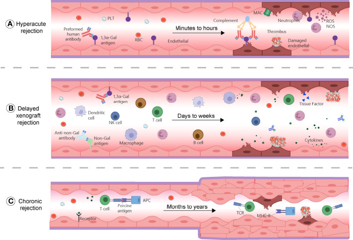
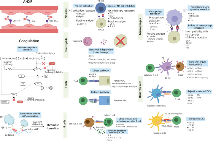
Transplantation is often the last resort for end-stage organ failures, e.g., kidney, liver, heart, lung, and pancreas. The shortage of donor organs is the main limiting factor for successful transplantation in humans. Except living donations, other alternatives are needed, e.g., xenotransplantation of pig organs. However, immune rejection remains the major challenge to overcome in xenotransplantation. There are three different xenogeneic types of rejections, based on the responses and mechanisms involved. It includes hyperacute rejection (HAR), delayed xenograft rejection (DXR) and chronic rejection. DXR, sometimes involves acute humoral xenograft rejection (AHR) and cellular xenograft rejection (CXR), which cannot be strictly distinguished from each other in pathological process. In this review, we comprehensively discussed the mechanism of these immunological rejections and summarized the strategies for preventing them, such as generation of gene knock out donors by different genome editing tools and the use of immunosuppressive regimens. We also addressed organ-specific barriers and challenges needed to pave the way for clinical xenotransplantation. Taken together, this information will benefit the current immunological research in the field of xenotransplantation.

Keywords: Xenotransplantation; chronic rejection; delayed xenograft rejection; glucocorticoids; hyperacute rejection; immunosuppressants.


Conflict of interest statement

The authors declare that the research was conducted in the absence of any commercial or financial relationships that could be construed as a potential conflict of interest.

Figures

Figure 1
Figure 1
Mechanisms of rejections during xenotransplantation. (A) Hyperacute rejection occurs within minutes to hours and is caused by the binding of the host’s pre-existing antibodies to α-Gal antigens on the graft, which results in complement activation and membrane attacking complex (MAC) formation. This reaction causes endothelial cells lysis, fibrinoid occlusion, and vasculature destruction. Neutrophils, through the production of ROS and NOS also contribute to this process. (B) Delayed xenograft rejection (DXR) occurs within days to weeks and include acute humoral xenograft rejection (AHXR), cellular xenograft rejection, and coagulation dysregulation. AHXR is antibody-mediated and involve non-Gal antibodies and α-Gal antibodies reactivity against non-Gal epitopes and α-Gal of the graft. Various innate and adaptive immune cells, proinflammatory cytokines, and coagulation dysregulation contribute to rejection, resulting in massive deposition of immunoglobins, fibrin, endothelial cell lysis, and interstitial bleeding. (C) Chronic rejection occurs within months to years. Xenoantigens are surveyed by host APCs and presented to T cells, leading to their activation and triggering inflammatory cascades, characterized by thrombotic microangiopathy, proliferation of the graft vascular endothelial cells, vessel narrowing, and interstitial fibrosis. APC, antigen presenting cell; MAC, membrane attacking complex; MHC-II, major histocompatibility complex class II; NK cell, natural killer cell; NOS, nitric oxide species; PLT, platelet; RBC, red blood cells; ROS, reactive oxygen species; TCR, T cell receptor.
Figure 2
Figure 2
Delayed xenograft rejection: AHXR, cellular xenograft rejection and coagulation dysregulation. The antibodies involved in AHXR are mainly directed against 1,3α-Gal epitopes and non-Gal epitopes such as Neu-5GC and SDa blood group. During cellular responses, ligands on the graft cells activate recipients’ NK cells and macrophages via different activating receptors. Meanwhile, graft cells fail to deliver inhibitory signals to the activated cells and enhance their proinflammatory and cytotoxic properties. Neutrophils can generate ROS, tissue-digesting enzyme, and NETs upon activation by graft cells, which causes tissue damage. Three types of DCs are involved in graft rejection. IRI-related DCs activate both cytotoxic T cells and B cells, thus triggering both cell- and antibody-mediated rejection. Rejection-related DCs promote acute and chronic rejection by activating T cells. Tolerogenic DCs suppress immune rejection by inducing Tregs. The different surface markers characterizing the different immune cells are listed in the right-hand side of the cells. T cells are involved in rejection through both direct and indirect pathways. In the direct pathway, porcine APCs present antigens and activate host’s T cells. In the indirect pathway, the graft antigens are presented by the host’s APCs. B cells are activated by T helper cells and secreted cytokines. Antibodies released by activated B cells that have differentiated into plasma cells contribute to graft rejection. AHXR and cellular xenograft rejection are both accompanied with coagulation dysregulation, where porcine TFPI cannot fully inhibit factor Xa and fails to inactivate TF. Porcine TBM also fails to regulate protein C. Porcine vWF can aggregate spontaneously and activate the host’s platelets through GP1b receptors. Altogether these reactions lead to the formation of thrombus in the graft vessels. AHXR, acute humoral xenograft rejection; Ag, antigen; aPC, activate protein C; DC, dendritic cell; IRI: ischemia-reperfusion injury; NET, nuclear extracellular traps; TBM, thrombomodulin; TF, tissue factor; Tregs, T regulatory cells; vWF, von Willebrand Factor.
Figure 3
Figure 3
Mechanism of action of the immunosuppressants commonly used in xenotransplantation. GCs exert anti-inflammatory and immunosuppressive effects by inhibiting macrophages, eosinophils, T cells, and to a lesser extent, B cells, by binding to GC receptors in cytoplasm. Cyclosporin binds to cyclophilin, then this drug-immunophilin complex binds to calcineurin, which subsequently prevents Th cell activation and IL-2 production, which eventually inhibits T cell clonal proliferation. Tacrolimus inhibits T cells proliferation by binding to FKBP, which inhibits several transcription factors involved in the production of proinflammatory cytokines. Cyclophosphamide blocks DNA alkylation in various cell types, leading to programmed cell death induction and preventing cell division. Leflunomide inhibits the synthesis of pyrimidines, thus arresting cell cycle in S phase. Mycophenolate mofetil prevents T and B cell proliferation by specifically inhibiting a purine pathway required for lymphocyte division. Polyclonal anti-thymocyte globulins are mainly directed against T cells. However, other immune cells sharing common surface antigens with T cells can also be affected to a lesser extent. Monoclonal antibodies target specific cytokine pathways (e.g., IL-6Rα) or cell surface markers, such as CD3, for anti-C3 IT, or CD20, for rituximab. The IL-6 receptor inhibitor tocilizumab reduces systemic inflammation and inhibits of CD8+ T cell and B cell differentiation. Anti-CD3 IT can deplete CD3+ T cells transiently and reduces the number of T cells in circulation and in lymph nodes. Rituximab is a B cell-depleting drug that targets CD20. Rapamycin exerts immunosuppressive and anti-proliferative effects of T cells by inhibiting the activation of S6K1 and PI3 kinase signalling. CTLA-4Ig and anti-CD40mAb target the costimulatory pathways CD80/86:CD28 and CD154:CD40, respectively, thereby dampening T cell activation. The compstatin analogue Cp40 and Tesidolumab inhibits complement C3 and C5 respectively, thereby reducing complement activities. FKBP, FK506 binding protein; GCs, glucocorticoids; IT, immunotoxin; mAb, monoclonal antibody; MMF, mycophenolate mofetil, pECs, porcine endothelial cells.

Similar articles

Cited by

References

    1. Jawoniyi O, Gormley K, McGleenan E, Noble HR. Organ donation and transplantation: Awareness and roles of healthcare professionals-a systematic literature review. J Clin Nurs (2018) 27(5-6):e726–38. doi: 10.1111/jocn.14154 - DOI - PubMed
    1. Pedersen PB, Henriksen DP, Brabrand M, Lassen AT. Prevalence of organ failure and mortality among patients in the emergency department: a population-based cohort study. BMJ Open (2019) 9(10):e032692. doi: 10.1136/bmjopen-2019-032692 - DOI - PMC - PubMed
    1. Lone NI, Walsh TS. Impact of intensive care unit organ failures on mortality during the five years after a critical illness. Am J Respir Crit Care Med (2012) 186(7):640–7. doi: 10.1164/rccm.201201-0059OC - DOI - PubMed
    1. Jones, Ben. Keeping kidneys. Bull World Health Organisation (2012) 90(10):718–9. doi: 10.2471/BLT.12.021012 - DOI - PMC - PubMed
    1. D. Working Group on Incentives for Living. Matas AJ, Satel S, Munn S, Richards JR, Tan-Alora A, et al. . Danguilan: Incentives for organ donation: proposed standards for an internationally acceptable system. Am J Transplant (2012) 12(2):306–12. doi: 10.1111/j.1600-6143.2011.03881.x - DOI - PMC - PubMed

Publication types