Does hand modulate the reshaping of the attentional system during rightward prism adaptation? An fMRI study
- PMID: 35967619
- PMCID: PMC9363778
- DOI: 10.3389/fpsyg.2022.909815
Does hand modulate the reshaping of the attentional system during rightward prism adaptation? An fMRI study
Abstract
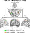
Adaptation to right-deviating prisms (R-PA), that is, learning to point with the right hand to targets perceived through prisms, has been shown to change spatial topography within the inferior parietal lobule (IPL) by increasing responses to left, central, and right targets on the left hemisphere and decreasing responses to right and central targets on the right hemisphere. As pointed out previously, this corresponds to a switch of the dominance of the ventral attentional network from the right to the left hemisphere. Since the encoding of hand movements in pointing paradigms is side-dependent, the choice of right vs. left hand for pointing during R-PA may influence the visuomotor adaptation process and hence the reshaping of the attentional system. We have tested this hypothesis in normal subjects by comparing activation patterns to visual targets in left, central, and right fields elicited before and after adaptation to rightward-deviating prisms using the right hand (RWRH) with those in two control groups. The first control group underwent adaptation to rightward-deviating prisms using the left hand, whereas the second control group underwent adaptation to leftward-deviating prisms using the right hand. The present study confirmed the previously described enhancement of left and central visual field representation within left IPL following R-PA. It further showed that the use of right vs. left hand during adaptation modulates this enhancement in some but not all parts of the left IPL. Interestingly, in some clusters identified in this study, L-PA with right hand mimics partially the effect of R-PA by enhancing activation elicited by left stimuli in the left IPL and by decreasing activation elicited by right stimuli in the right IPL. Thus, the use of right vs. left hand modulates the R-PA-induced reshaping of the ventral attentional system. Whether the choice of hand during R-PA affects also the reshaping of the dorsal attentional system remains to be determined as well as possible clinical applications of this approach. Depending on the patients' conditions, using the right or the left hand during PA might potentiate the beneficial effects of this intervention.
Keywords: attention; fMRI; functional reshaping; hand; inferior parietal lobule; prism adaptation.
Copyright © 2022 Farron, Clarke and Crottaz-Herbette.
Conflict of interest statement
The authors declare that the research was conducted in the absence of any commercial or financial relationships that could be construed as a potential conflict of interest.
Figures


Similar articles
-
Choosing Sides: Impact of Prismatic Adaptation on the Lateralization of the Attentional System.Front Psychol. 2022 Jun 23;13:909686. doi: 10.3389/fpsyg.2022.909686. eCollection 2022. Front Psychol. 2022. PMID: 35814089 Free PMC article. Review.
-
A Brief Exposure to Leftward Prismatic Adaptation Enhances the Representation of the Ipsilateral, Right Visual Field in the Right Inferior Parietal Lobule.eNeuro. 2017 Sep 27;4(5):ENEURO.0310-17.2017. doi: 10.1523/ENEURO.0310-17.2017. eCollection 2017 Sep-Oct. eNeuro. 2017. PMID: 28955725 Free PMC article.
-
Supramodal effect of rightward prismatic adaptation on spatial representations within the ventral attentional system.Brain Struct Funct. 2018 Apr;223(3):1459-1471. doi: 10.1007/s00429-017-1572-2. Epub 2017 Nov 18. Brain Struct Funct. 2018. PMID: 29151115
-
Prism adaptation enhances decoupling between the default mode network and the attentional networks.Neuroimage. 2019 Oct 15;200:210-220. doi: 10.1016/j.neuroimage.2019.06.050. Epub 2019 Jun 22. Neuroimage. 2019. PMID: 31233909
-
Modulation of visual attention by prismatic adaptation.Neuropsychologia. 2016 Nov;92:31-41. doi: 10.1016/j.neuropsychologia.2016.06.022. Epub 2016 Jun 21. Neuropsychologia. 2016. PMID: 27342255 Review.
Cited by
-
Choosing Sides: Impact of Prismatic Adaptation on the Lateralization of the Attentional System.Front Psychol. 2022 Jun 23;13:909686. doi: 10.3389/fpsyg.2022.909686. eCollection 2022. Front Psychol. 2022. PMID: 35814089 Free PMC article. Review.
-
Direction of visual shift and hand congruency enhance spatial realignment during visuomotor adaptation.Exp Brain Res. 2023 Oct;241(10):2475-2486. doi: 10.1007/s00221-023-06697-4. Epub 2023 Sep 1. Exp Brain Res. 2023. PMID: 37658176
References
-
- Brett M., Anton J.-L., Valabregue R., Poline J. -B. (2002). “Region of interest analysis using an SPM toolbox,” in Presented at the 8th International Conference on Functional Mapping of the Human Brain, Vol. 16 (Sendai: ).
LinkOut - more resources
Full Text Sources
Research Materials
Miscellaneous

