

Exosomes Derived from Secretome Human Umbilical Vein Endothelial Cells (Exo-HUVEC) Ameliorate the Photo-Aging of Skin Fibroblast
- PMID: 35967916
- PMCID: PMC9374532
- DOI: 10.2147/CCID.S371330
Exosomes Derived from Secretome Human Umbilical Vein Endothelial Cells (Exo-HUVEC) Ameliorate the Photo-Aging of Skin Fibroblast
Abstract
Purpose: This is an in-vitro experimental study to analyze the effect of Exo-HUVEC on endothelial cell (CD31), cell proliferation, matrix metalloproteinase 1 (MMP-1) and collagen type 1 on irradiated fibroblast with UVB as photo-aging model.
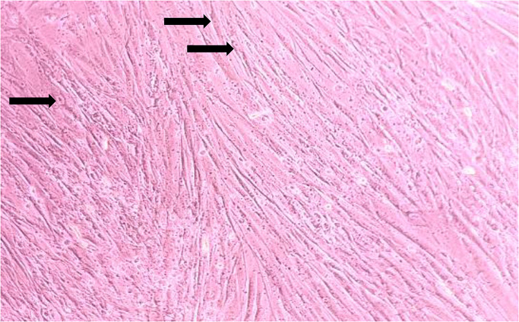
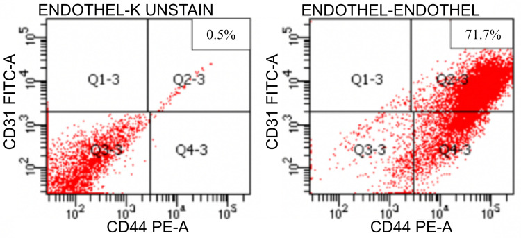
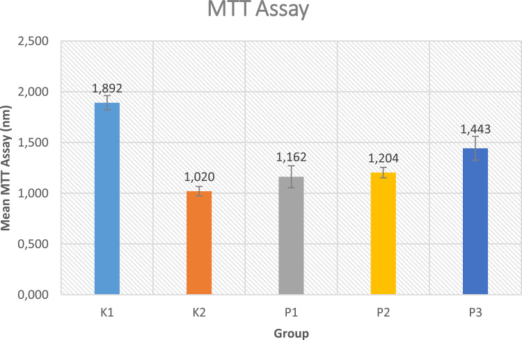
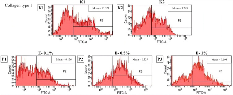
Patients and methods: Fibroblast cultures were divided into 5 groups, namely without UVB exposure, UVB exposure 600mJ/cm2 for 80 seconds as photo-aging model, and UVB exposure +Exo-HUVEC exposure 0.1%, 0.5% and 1%. The endothelial cell was stained with a CD31 marker, MMP-1 were examined with ELISA, cell proliferation is detected using an MTT assay; meanwhile, collagen type 1 deposition and endothelial cell were measured using flowcytometry.
Results: This study found positive endothelial cell marker CD31. Significant difference was found in cell proliferation, MMP-1 and collagen type 1 level between the control group with UVB irradiation and the treatment group with Exo-HUVEC (p < 0.05).
Conclusion: Exo-HUVEC significantly increases cell proliferation and collagen type 1 level, while decrease MMP-1 levels on irradiated fibroblast; therefore, Exo-HUVEC ameliorate the photo-aging of skin fibroblast.
Keywords: Exo-HUVEC; MMP-1; cell proliferation; collagen type 1; endothelial cell; photo-aging.
© 2022 Ellistasari et al.
Conflict of interest statement
The authors report no conflicts of interest in this work.
Figures


Similar articles
-
Exosomes Derived from Human Induced Pluripotent Stem Cells Ameliorate the Aging of Skin Fibroblasts.Int J Mol Sci. 2018 Jun 9;19(6):1715. doi: 10.3390/ijms19061715. Int J Mol Sci. 2018. PMID: 29890746 Free PMC article.
-
Achatina fulica mucous improves cell viability and increases collagen deposition in UVB-irradiated human fibroblast culture.J Stem Cells Regen Med. 2020 May 27;16(1):26-31. doi: 10.46582/jsrm.1601005. eCollection 2020. J Stem Cells Regen Med. 2020. PMID: 32536768 Free PMC article.
-
Exosomes Derived from Human Umbilical Cord Mesenchymal Stem Cells Accelerate Diabetic Wound Healing via Promoting M2 Macrophage Polarization, Angiogenesis, and Collagen Deposition.Int J Mol Sci. 2022 Sep 9;23(18):10421. doi: 10.3390/ijms231810421. Int J Mol Sci. 2022. PMID: 36142334 Free PMC article.
-
Bone marrow mesenchymal stem cell-derived exosome miR-29b-3p alleviates UV irradiation-induced photoaging in skin fibroblast.Photodermatol Photoimmunol Photomed. 2023 May;39(3):235-245. doi: 10.1111/phpp.12827. Epub 2022 Aug 23. Photodermatol Photoimmunol Photomed. 2023. PMID: 35950642
-
Oleracone C from Portulaca oleracea attenuates UVB-induced changes in matrix metalloproteinase and type I procollagen production via MAPK and TGF-β/Smad pathways in human keratinocytes.Int J Cosmet Sci. 2023 Apr;45(2):166-176. doi: 10.1111/ics.12828. Epub 2023 Jan 12. Int J Cosmet Sci. 2023. PMID: 36415152
Cited by
-
Exosomal Lnc NEAT1 from endothelial cells promote bone regeneration by regulating macrophage polarization via DDX3X/NLRP3 axis.J Nanobiotechnology. 2023 Mar 20;21(1):98. doi: 10.1186/s12951-023-01855-w. J Nanobiotechnology. 2023. PMID: 36941678 Free PMC article.
-
Exosomes: The emerging mechanisms and potential clinical applications in dermatology.Int J Biol Sci. 2024 Feb 25;20(5):1778-1795. doi: 10.7150/ijbs.92897. eCollection 2024. Int J Biol Sci. 2024. PMID: 38481799 Free PMC article. Review.
-
A temperature-sensitive chitosan hydrogels loaded with nano-zinc oxide and exosomes from human umbilical vein endothelial cells accelerates wound healing.Regen Ther. 2025 May 17;30:63-74. doi: 10.1016/j.reth.2025.04.020. eCollection 2025 Dec. Regen Ther. 2025. PMID: 40491560 Free PMC article.
-
Endothelial cells-derived exosomes-based hydrogel improved tendinous repair via anti-inflammatory and tissue regeneration-promoting properties.J Nanobiotechnology. 2024 Jul 9;22(1):401. doi: 10.1186/s12951-024-02607-0. J Nanobiotechnology. 2024. PMID: 38982446 Free PMC article.
-
Exosomes in skin photoaging: biological functions and therapeutic opportunity.Cell Commun Signal. 2024 Jan 12;22(1):32. doi: 10.1186/s12964-023-01451-3. Cell Commun Signal. 2024. PMID: 38217034 Free PMC article. Review.
References
LinkOut - more resources
Full Text Sources
