Temporal analysis of Lassa virus infection and transmission in experimentally infected Mastomys natalensis
- PMID: 35967978
- PMCID: PMC9364215
- DOI: 10.1093/pnasnexus/pgac114
Temporal analysis of Lassa virus infection and transmission in experimentally infected Mastomys natalensis
Abstract
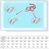
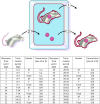
Little is known about the temporal patterns of infection and transmission of Lassa virus (LASV) within its natural reservoir (Mastomys natalensis). Here, we characterize infection dynamics and transmissibility of a LASV isolate (Soromba-R) in adult lab-reared M. natalensis originating from Mali. The lab-reared M. natalenesis proved to be highly susceptible to LASV isolates from geographically distinct regions of West Africa via multiple routes of exposure, with 50% infectious doses of < 1 TCID50. Postinoculation, LASV Soromba-R established a systemic infection with no signs of clinical disease. Viral RNA was detected in all nine tissues examined with peak concentrations detected between days 7 and 14 postinfection within most organs. There was an overall trend toward clearance of virus within 40 days of infection in most organs. The exception is lung specimens, which retained positivity throughout the course of the 85-day study. Direct (contact) and indirect (fomite) transmission experiments demonstrated 40% of experimentally infected M. natalensis were capable of transmitting LASV to naïve animals, with peak transmissibility occurring between 28 and 42 days post-inoculation. No differences in patterns of infection or transmission were noted between male and female experimentally infected rodents. Adult lab-reared M. natalensis are highly susceptible to genetically distinct LASV strains developing a temporary asymptomatic infection associated with virus shedding resulting in contact and fomite transmission within a cohort.
Published by Oxford University Press on behalf of National Academy of Sciences 2022.
Figures


Similar articles
-
Inoculation route-dependent Lassa virus dissemination and shedding dynamics in the natural reservoir - Mastomys natalensis.Emerg Microbes Infect. 2021 Dec;10(1):2313-2325. doi: 10.1080/22221751.2021.2008773. Emerg Microbes Infect. 2021. PMID: 34792436 Free PMC article.
-
Establishment of a Genetically Confirmed Breeding Colony of Mastomys natalensis from Wild-Caught Founders from West Africa.Viruses. 2021 Mar 31;13(4):590. doi: 10.3390/v13040590. Viruses. 2021. PMID: 33807214 Free PMC article.
-
Assessing the Potential of Sexual Transmission of Lassa Virus in a Natural Rodent Reservoir.J Infect Dis. 2025 Jun 24:jiaf314. doi: 10.1093/infdis/jiaf314. Online ahead of print. J Infect Dis. 2025. PMID: 40581828
-
Vaccine platforms for the prevention of Lassa fever.Immunol Lett. 2019 Nov;215:1-11. doi: 10.1016/j.imlet.2019.03.008. Epub 2019 Apr 23. Immunol Lett. 2019. PMID: 31026485 Free PMC article. Review.
-
Lassa virus diversity and feasibility for universal prophylactic vaccine.F1000Res. 2019 Jan 31;8:F1000 Faculty Rev-134. doi: 10.12688/f1000research.16989.1. eCollection 2019. F1000Res. 2019. PMID: 30774934 Free PMC article. Review.
Cited by
-
Modelling Lassa virus dynamics in West African Mastomys natalensis and the impact of human activities.J R Soc Interface. 2024 Jul;21(216):20240106. doi: 10.1098/rsif.2024.0106. Epub 2024 Jul 24. J R Soc Interface. 2024. PMID: 39045680 Free PMC article.
-
Evidence of multiple bacterial, viral, and parasitic infectious disease agents in Mastomys natalensis rodents in riverine areas in selected parts of Zambia.Infect Ecol Epidemiol. 2024 Dec 31;15(1):2441537. doi: 10.1080/20008686.2024.2441537. eCollection 2025. Infect Ecol Epidemiol. 2024. PMID: 39764202 Free PMC article.
-
The first imported case of Lassa fever in China: a case description.Quant Imaging Med Surg. 2025 Jan 2;15(1):1066-1072. doi: 10.21037/qims-24-1784. Epub 2024 Nov 22. Quant Imaging Med Surg. 2025. PMID: 39839037 Free PMC article. No abstract available.
-
Luna Virus and Helminths in Wild Mastomys natalensis in Two Contrasting Habitats in Zambia: Risk Factors and Evidence of Virus Dissemination in Semen.Pathogens. 2022 Nov 14;11(11):1345. doi: 10.3390/pathogens11111345. Pathogens. 2022. PMID: 36422597 Free PMC article.
-
Infection with mpox virus via the genital mucosae increases shedding and transmission in the multimammate rat (Mastomys natalensis).Nat Microbiol. 2024 May;9(5):1231-1243. doi: 10.1038/s41564-024-01666-1. Epub 2024 Apr 22. Nat Microbiol. 2024. PMID: 38649413
References
-
- Safronetz D, Feldmann H, Falzarano D.. 2012. Arenaviruses and filoviruses: viral haemorrhagic fevers. In: Medical microbiology. 18th edn. Galveston (TX): University of Texas Medical Branch. DOI: 10.1016/B978-0-7020-4089-4.00068-8.
-
- Maes P, et al. 2018. Taxonomy of the family Arenaviridae and the order Bunyavirales: update 2018. Arch Virol. 163:2295–2310. - PubMed
-
- Muckenfuss RS, Armstrong C, Webster LT.. 1934. Etiology of the 1933 epidemic of encephalitis. JAMA. 103:731–733.
LinkOut - more resources
Full Text Sources
Miscellaneous

