

Lipoid proteinosis: Review of Indian cases
- PMID: 35968171
- PMCID: PMC9364649
- DOI: 10.4103/jomfp.jomfp_249_21
Lipoid proteinosis: Review of Indian cases
Abstract
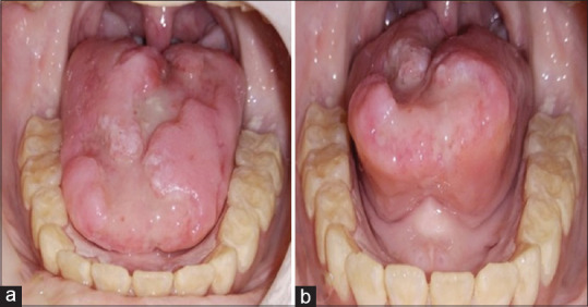
Lipoid proteinosis (LP) is a rare autosomal recessive disorder characterized by the deposition of amorphous hyaline material in the dermis and submucosal connective tissue. To date <500 cases of LP have been described and oral manifestations described in a very few reports. Indian cases are much less reported and reviewed. Hence, here review of 51 Indian LP cases along with a case of histologically proven LP in 12-year-old male patient with typical skin, ocular, laryngeal, oral and radiographic features is done. Cases from 1969 to 2021 were collected using keyword LP on google and google scholar and Indian cases were analyzed afterward. Review with case presentation regarding oral manifestations will help the oral physician to diagnose LP in early stage.
Keywords: Indian cases; lipoid proteinosis; oral manifestations; oral physician; review.
Copyright: © 2022 Journal of Oral and Maxillofacial Pathology.
Conflict of interest statement
There are no conflicts of interest.
Figures


References
-
- Hamada T, McLean WH, Ramsay M, Ashton GH, Nanda A, Jenkins T, et al. Lipoid proteinosis maps to 1q21 and is caused by mutations in the extracellular matrix protein 1 gene (ECM1) Hum Mol Genet. 2002;11:833–40. - PubMed
-
- Lee KC, Peters SM, Ko YC, Kunkle TC, Perrino MA, Yoon AJ, et al. Oral manifestations of lipoid proteinosis in a 10-year-old female: A case report and literature update. Oral Surg Oral Med Oral Pathol Oral Radiol. 2018;126:e228–32. - PubMed
Publication types
LinkOut - more resources
Full Text Sources