

Predictors of ischemic events in patients with unilateral extracranial vertebral artery dissection: A single-center exploratory study
- PMID: 35968293
- PMCID: PMC9366300
- DOI: 10.3389/fneur.2022.939001
Predictors of ischemic events in patients with unilateral extracranial vertebral artery dissection: A single-center exploratory study
Abstract
Objective: Extracranial vertebral artery dissection (EVAD) is one of the main causes of stroke in young and middle-aged patients. However, the diagnosis is challenging. This study aimed to identify the characteristics of EVAD on color duplex ultrasonography (CDU) and high-resolution magnetic resonance imaging (hrMRI), hoping to improve the accuracy and determine the relative contribution of vessel findings and clinical factors to acute ischemic events.
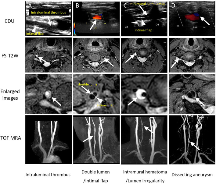
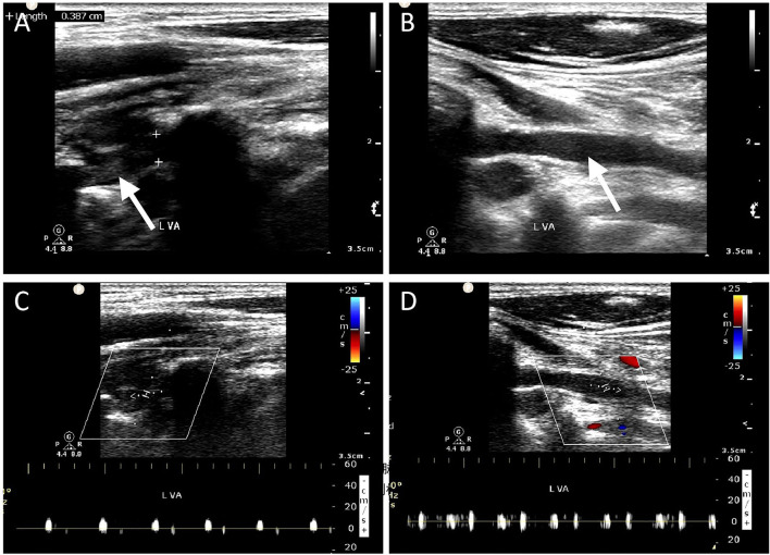
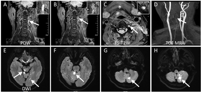
Methods: Patients with unilateral EVAD were recruited and divided into ischemia and non-ischemia groups. Clinical features of patients and the lesion location; a variety of signs which indicate dissection, including the presence of an intimal flap, double lumen, intramural hematoma, dissecting aneurysm, intraluminal thrombus, and irregular lumen; and other quantitative parameters of each dissected segment on CDU and hrMRI were reviewed, respectively. Multiple logistic regression was performed to explore the association between clinical, imaging characteristics, and ischemic events in patients with unilateral EVAD.
Results: Ninety-six patients with unilateral EVAD who met the inclusion criteria were enrolled during a six-year period. Overall, 41 cases (42.7%) were confirmed as ischemic stroke (n = 40) or transient ischemic attack (n = 1) during the 48 h after the onset of symptoms. Men, infections during the last week, and smoking were more common in the ischemia group. Intraluminal thrombus and occlusion on CDU were more prevalent in patients with cerebral ischemia than in those without (36.6 vs. 5.5%; p < 0.001, and 39.0 vs. 9.1%; p = 0.001, respectively). On hrMRI, intraluminal thrombus and occlusion were also more frequent in the ischemia group than in the non-ischemia group (34.1 vs. 5.5%; p < 0.001, and 34.1 vs. 9.1%; p = 0.003, respectively). In addition, lesion length on hrMRI was significantly longer for patients with ischemia (81.5 ± 41.7 vs. 64.7 ± 30.8 mm; p = 0.025). In multivariable logistic regression analysis, male gender, infections during the last week, and the presence of intraluminal thrombus on CDU and hrMRI were independently associated with acute ischemic events.
Conclusion: Male sex, infections during the last week, and the presence of intraluminal thrombus due to dissection are associated with an increased risk of ischemic events in patients with unilateral EVAD.
Keywords: color duplex ultrasonography; high resolution magnetic resonance imaging; intraluminal thrombus; stroke; vertebral artery dissection.
Copyright © 2022 Yan, Lu, Ding, Pu, Hu, Teng and Hui.
Conflict of interest statement
The authors declare that the research was conducted in the absence of any commercial or financial relationships that could be construed as a potential conflict of interest. The handling editor J-CB declared a shared affiliation with the author ZT at the time of review.
Figures
References
-
- Biller J, Sacco RL, Albuquerque FC, Demaerschalk BM, Fayad P, Long PH, et al. Cervical arterial dissections and association with cervical manipulative therapy: a statement for healthcare professionals from the American heart association/American stroke association. Stroke. (2014) 45:3155–74. 10.1161/STR.0000000000000016 - DOI - PubMed
LinkOut - more resources
Full Text Sources
Miscellaneous