

The clinicopathological features and treatments of odontogenic keratocysts
- PMID: 35968329
- PMCID: PMC9360231
The clinicopathological features and treatments of odontogenic keratocysts
Abstract
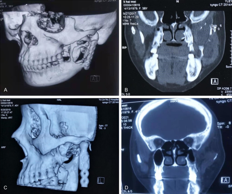
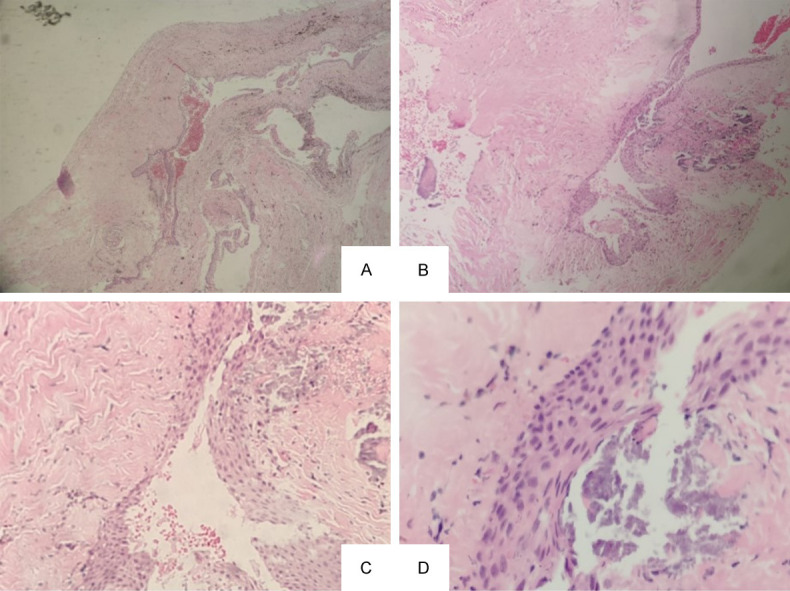
An odontogenic keratocyst (OKC) is a benign but aggressive intraosseous tumor derived from the remains of the original tooth germ or dental lamina. It has a marked ability to recur and become cancerous. However, patients with early-stage OKC often have no symptoms and manifestations. The common clinical manifestation is swelling. Hence, it is critical to precisely diagnose the disease, to use differential diagnosis in combination with auxiliary examination methods, and to select the most appropriate treatment option to reduce the loss of bone tissue and the related damage to patients. In recent years, with the advancement in understanding the molecular basis of this disease and the development of early detection and targeted therapy, the diagnosis and the prognosis of OKC have been improved. The aim of this study was to provide an overview on the clinical features, diagnosis, and treatment of OKC. The molecular and genetic basis of this disease and the characteristics of malignant transformation of OKC were also discussed. Finally, we presented patient cases from our clinical practice to provide some advice on the diagnosis and treatment of OKC.
Keywords: Odontogenic cysts; keratocystic odontogenic tumor; odontogenic keratocyst; odontogenic tumors.
AJCR Copyright © 2022.
Conflict of interest statement
None.
Figures
Similar articles
-
What is Currently Known about Odontogenic Keratocysts?Oral Health Prev Dent. 2022 Jul 22;20:321-330. doi: 10.3290/j.ohpd.b3240829. Oral Health Prev Dent. 2022. PMID: 35866678 Free PMC article. Review.
-
A retrospective study of the malignant change of odontogenic keratocyst.J Stomatol Oral Maxillofac Surg. 2023 Dec;124(6):101466. doi: 10.1016/j.jormas.2023.101466. Epub 2023 Apr 6. J Stomatol Oral Maxillofac Surg. 2023. PMID: 37030439
-
Odontogenic Keratocyst in a 9-Month-Old Patient: A Case Report.J Clin Pediatr Dent. 2021 Jul 1;45(3):199-203. doi: 10.17796/1053-4625-45.3.9. J Clin Pediatr Dent. 2021. PMID: 34192756
-
Peripheral odontogenic keratocyst. A Case report.J Clin Exp Dent. 2023 Feb 1;15(2):e169-e172. doi: 10.4317/jced.60033. eCollection 2023 Feb. J Clin Exp Dent. 2023. PMID: 36911150 Free PMC article.
-
Pediatric odontogenic keratocyst and early diagnosis of Gorlin syndrome: Clinicopathological aids.Saudi Dent J. 2024 Jan;36(1):38-43. doi: 10.1016/j.sdentj.2023.10.012. Epub 2023 Oct 18. Saudi Dent J. 2024. PMID: 38375374 Free PMC article. Review.
Cited by
-
Concurrent Odontogenic Keratocyst and Odontoma: Report of an Unusual and Rare Entity.J Dent (Shiraz). 2023 Dec 1;24(4):438-443. doi: 10.30476/dentjods.2023.98278.2066. eCollection 2023 Dec. J Dent (Shiraz). 2023. PMID: 38149227 Free PMC article.
-
Calcifying odontogenic cyst combined with odontogenic keratocyst: report of a case and review of the literature.Int J Surg Case Rep. 2023 Apr;105:107991. doi: 10.1016/j.ijscr.2023.107991. Epub 2023 Mar 24. Int J Surg Case Rep. 2023. PMID: 37015162 Free PMC article.
-
Multilocular Radiolucent Pathology in the Body and Ramus of the Mandible: A Case Report.Cureus. 2024 Jul 3;16(7):e63722. doi: 10.7759/cureus.63722. eCollection 2024 Jul. Cureus. 2024. PMID: 39100023 Free PMC article.
-
Various Surgical Interventions in Treating Odontogenic Keratocyst: A Radiological Case Report.Healthcare (Basel). 2023 Feb 1;11(3):416. doi: 10.3390/healthcare11030416. Healthcare (Basel). 2023. PMID: 36766990 Free PMC article.
-
Modified Conservative Technique of Odontogenic Keratocyst Treatment With Long-Term Follow-Up: A Case Report.Cureus. 2025 Jan 6;17(1):e77010. doi: 10.7759/cureus.77010. eCollection 2025 Jan. Cureus. 2025. PMID: 39912031 Free PMC article.
References
-
- Barnes L, Eveson JW, Reichart P, Sidransky D. World Health Organization classification of tumours: pathology and genetics of head and neck tumours. Lyon: IARC Press; 2005.
-
- Naggar AK, Chan JKC, Grandis JR, Takata T, Slootweg P. WHO classification of Head and Neck Tumours: odontogenic and maxilofacial bone tumours. 4th edition. Lyon: IARC; 2017. pp. 205–260.
-
- Mehdi A, Saeed B, Narges H, Hooman A, Sepehr A, Zahra A. Review on the most important management of keratocystic odontogenic tumor. Klin Onkol. 2022;35:10–19. - PubMed
-
- Moura BS, Cavalcante MA, Hespanhol W. Keratocystic odontogenic tumor. Rev Col Bras Cir. 2016;43:466–471. - PubMed
LinkOut - more resources
Full Text Sources