

Cases report: MRI findings of asymptomatically familial subependymal heterotopia with filamin A gene abnormality
- PMID: 35968360
- PMCID: PMC9364927
- DOI: 10.3389/fnins.2022.956545
Cases report: MRI findings of asymptomatically familial subependymal heterotopia with filamin A gene abnormality
Abstract
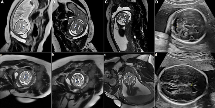
Subependymal heterotopia (SEH) is a rare neuronal migration disorder consisting of gray matter nodules along the lateral ventricular walls and is often associated with other brain malformations. Despite most SEH cases showing epilepsy during their lifetimes, very few patients with asymptomatically familial SEH tend to cause misdiagnosis or missed diagnosis. We present four familial SEH cases without any positive symptoms and medical history, including two fetuses, who were diagnosed by MRI and confirmed by genetic testing with mutation of filamin A. This report emphasizes the role of MRI in the recognition of SEH at an early age of gestation and in asymptomatically familial SEH. MRI provides a fast, repeatable, reliable, and cheap choice for detecting and screening familial SEH.
Keywords: asymptomatic; case report; filamin A; magnetic resonance imaging; subependymal heterotopia.
Copyright © 2022 Lv, Zhou, Zeng, Wang, Zhao, Chen, Li, Song, Xiao, Ding and Cheng.
Conflict of interest statement
The authors declare that the research was conducted in the absence of any commercial or financial relationships that could be construed as a potential conflict of interest. The handling editor JW declared a shared affiliation with the authors BC, ZD, and MX at the time of review.
Figures

Similar articles
-
Bilateral subependymal heterotopia, ventriculomegaly and cerebellar asymmetry: fetal MRI findings of a rare association of brain anomalies.J Radiol Case Rep. 2013 Nov 1;7(11):38-45. doi: 10.3941/jrcr.v7i11.1457. eCollection 2013 Nov. J Radiol Case Rep. 2013. PMID: 24421929 Free PMC article.
-
Antenatal diagnosis of subependymal heterotopia.AJNR Am J Neuroradiol. 2000 Feb;21(2):296-300. AJNR Am J Neuroradiol. 2000. PMID: 10696011 Free PMC article.
-
Mutation in filamin A causes periventricular heterotopia, developmental regression, and West syndrome in males.Epilepsia. 2006 Jan;47(1):211-4. doi: 10.1111/j.1528-1167.2006.00390.x. Epilepsia. 2006. PMID: 16417552
-
Bipolar disorder with Melnick-Needles syndrome and periventricular nodular heterotopia: two case reports and a review of the literature.J Med Case Rep. 2021 Oct 11;15(1):495. doi: 10.1186/s13256-021-03064-1. J Med Case Rep. 2021. PMID: 34629090 Free PMC article. Review.
-
Subependymal heterotopia: a distinct neuronal migration disorder associated with epilepsy.J Neurol Neurosurg Psychiatry. 1994 Oct;57(10):1195-202. doi: 10.1136/jnnp.57.10.1195. J Neurol Neurosurg Psychiatry. 1994. PMID: 7931380 Free PMC article. Review.
Cited by
-
Unveiling the intriguing puzzle: Nodular heterotopia and Mega Cisterna Magna in an adult female.Radiol Case Rep. 2023 Nov 3;19(1):281-284. doi: 10.1016/j.radcr.2023.09.099. eCollection 2024 Jan. Radiol Case Rep. 2023. PMID: 38028293 Free PMC article.
-
Sustained generation of neurons destined for neocortex with oxidative metabolic upregulation upon filamin abrogation.iScience. 2024 Jun 7;27(7):110199. doi: 10.1016/j.isci.2024.110199. eCollection 2024 Jul 19. iScience. 2024. PMID: 38989458 Free PMC article.
References
Publication types
LinkOut - more resources
Full Text Sources