

Abnormal network homogeneity of default-mode network and its relationships with clinical symptoms in antipsychotic-naïve first-diagnosis schizophrenia
- PMID: 35968384
- PMCID: PMC9369006
- DOI: 10.3389/fnins.2022.921547
Abnormal network homogeneity of default-mode network and its relationships with clinical symptoms in antipsychotic-naïve first-diagnosis schizophrenia
Abstract
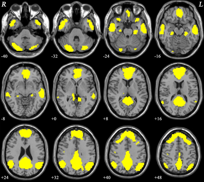
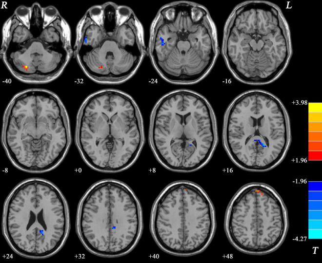
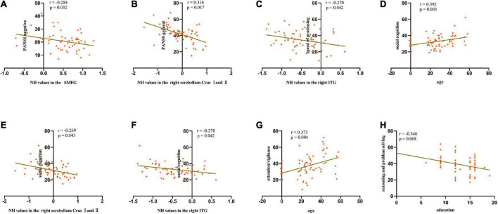
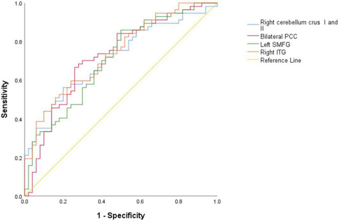
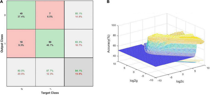
Schizophrenia is a severe mental disorder affecting around 0.5-1% of the global population. A few studies have shown the functional disconnection in the default-mode network (DMN) of schizophrenia patients. However, the findings remain discrepant. In the current study, we compared the intrinsic network organization of DMN of 57 first-diagnosis drug-naïve schizophrenia patients with 50 healthy controls (HCs) using a homogeneity network (NH) and explored the relationships of DMN with clinical characteristics of schizophrenia patients. Receiver operating characteristic (ROC) curves analysis and support vector machine (SVM) analysis were applied to calculate the accuracy of distinguishing schizophrenia patients from HCs. Our results showed that the NH values of patients were significantly higher in the left superior medial frontal gyrus (SMFG) and right cerebellum Crus I/Crus II and significantly lower in the right inferior temporal gyrus (ITG) and bilateral posterior cingulate cortex (PCC) compared to those of HCs. Additionally, negative correlations were shown between aberrant NH values in the right cerebellum Crus I/Crus II and general psychopathology scores, between NH values in the left SMFG and negative symptom scores, and between the NH values in the right ITG and speed of processing. Also, patients' age and the NH values in the right cerebellum Crus I/Crus II and the right ITG were the predictors of performance in the social cognition test. ROC curves analysis and SVM analysis showed that a combination of NH values in the left SMFG, right ITG, and right cerebellum Crus I/Crus II could distinguish schizophrenia patients from HCs with high accuracy. The results emphasized the vital role of DMN in the neuropathological mechanisms underlying schizophrenia.
Keywords: cognitive dysfunction; default-mode network; network homogeneity; resting-state functional magnetic resonance imaging; schizophrenia.
Copyright © 2022 Kong, Chen, Gao, Ni, Ming, Chai, Ling and Xu.
Conflict of interest statement
The authors declare that the research was conducted in the absence of any commercial or financial relationships that could be construed as a potential conflict of interest.
Figures
Similar articles
-
Abnormal default-mode network homogeneity in first-episode, drug-naive schizophrenia at rest.Prog Neuropsychopharmacol Biol Psychiatry. 2014 Mar 3;49:16-20. doi: 10.1016/j.pnpbp.2013.10.021. Epub 2013 Nov 9. Prog Neuropsychopharmacol Biol Psychiatry. 2014. PMID: 24216538
-
Metacognitive Training Modulates Default-Mode Network Homogeneity During 8-Week Olanzapine Treatment in Patients With Schizophrenia.Front Psychiatry. 2020 Mar 27;11:234. doi: 10.3389/fpsyt.2020.00234. eCollection 2020. Front Psychiatry. 2020. PMID: 32292360 Free PMC article.
-
Morinda officinalis oligosaccharides modulate the default-mode network homogeneity in major depressive disorder at rest.Psychiatry Res Neuroimaging. 2024 Sep;343:111847. doi: 10.1016/j.pscychresns.2024.111847. Epub 2024 Jul 4. Psychiatry Res Neuroimaging. 2024. PMID: 38968754
-
Abnormal default-mode network homogeneity and its correlations with neurocognitive deficits in drug-naive first-episode adolescent-onset schizophrenia.Schizophr Res. 2020 Jan;215:140-147. doi: 10.1016/j.schres.2019.10.056. Epub 2019 Nov 26. Schizophr Res. 2020. PMID: 31784338
-
Hyperactivity of the default mode network in schizophrenia and free energy: A dialogue between Freudian theory of psychosis and neuroscience.Front Hum Neurosci. 2022 Dec 14;16:956831. doi: 10.3389/fnhum.2022.956831. eCollection 2022. Front Hum Neurosci. 2022. PMID: 36590059 Free PMC article. Review.
Cited by
-
A comparative machine learning study of schizophrenia biomarkers derived from functional connectivity.Sci Rep. 2025 Jan 22;15(1):2849. doi: 10.1038/s41598-024-84152-2. Sci Rep. 2025. PMID: 39843572 Free PMC article.
References
-
- Anteraper S. A., Collin G., Guell X., Scheinert T., Molokotos E., Henriksen M. T., et al. (2020). Altered resting-state functional connectivity in young children at familial high risk for psychotic illness: a preliminary study. Schizophr. Res. 216 496–503. 10.1016/j.schres.2019.09.006 - DOI - PMC - PubMed
-
- Buckner R. L., Sepulcre J., Talukdar T., Krienen F. M., Liu H., Hedden T., et al. (2009). Cortical hubs revealed by intrinsic functional connectivity: mapping, assessment of stability, and relation to Alzheimer’s disease. J. Neurosci. 29 1860–1873. 10.1523/JNEUROSCI.5062-08.2009 - DOI - PMC - PubMed
-
- de Filippis R., Carbone E. A., Gaetano R., Bruni A., Pugliese V., Segura-Garcia C., et al. (2019). Machine learning techniques in a structural and functional MRI diagnostic approach in schizophrenia: a systematic review. Neuropsychiatr. Dis. Treat. 15 1605–1627. 10.2147/NDT.S202418 - DOI - PMC - PubMed
LinkOut - more resources
Full Text Sources