. 2022 Jul 29:16:968767.
doi: 10.3389/fnins.2022.968767. eCollection 2022.

Modulation of entorhinal cortex-hippocampus connectivity and recognition memory following electroacupuncture on 3×Tg-AD model: Evidence from multimodal MRI and electrophysiological recordings

Affiliations

Modulation of entorhinal cortex-hippocampus connectivity and recognition memory following electroacupuncture on 3×Tg-AD model: Evidence from multimodal MRI and electrophysiological recordings

Bingbing Lin et al. Front Neurosci. .

Abstract

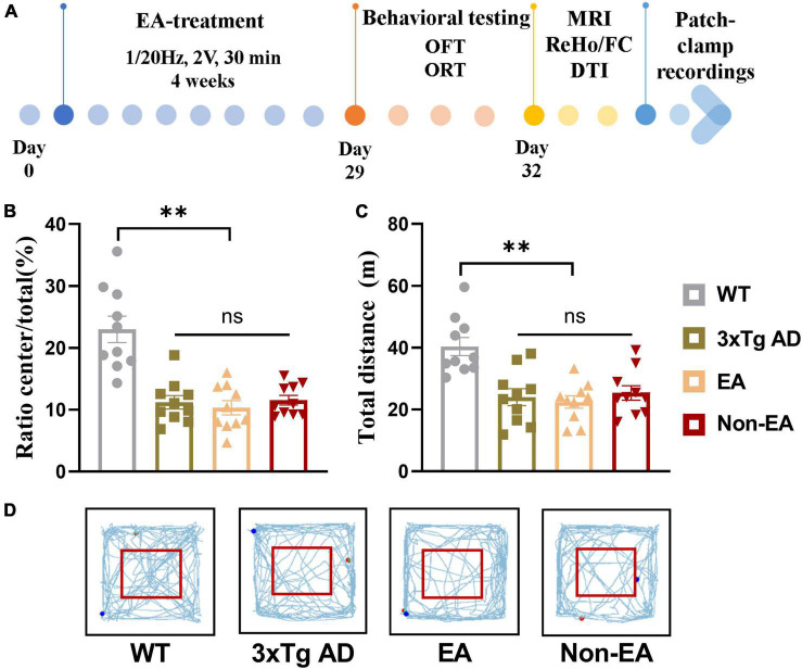
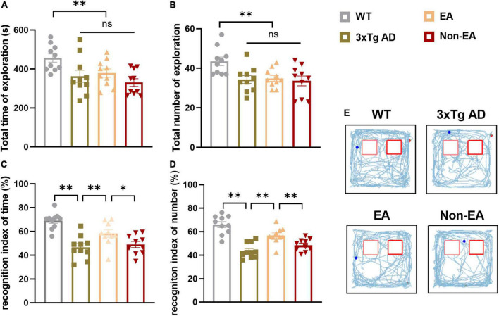
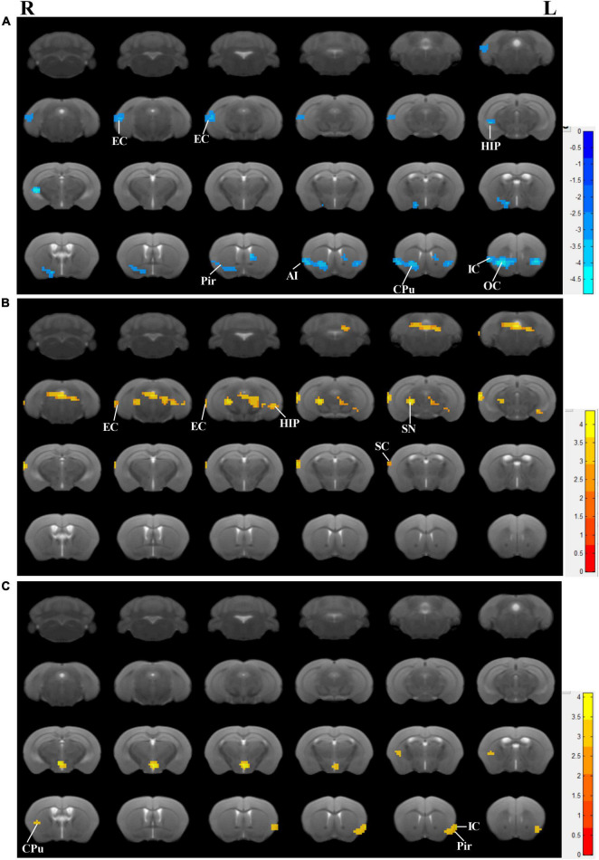
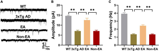
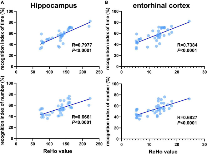
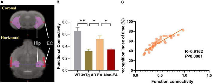
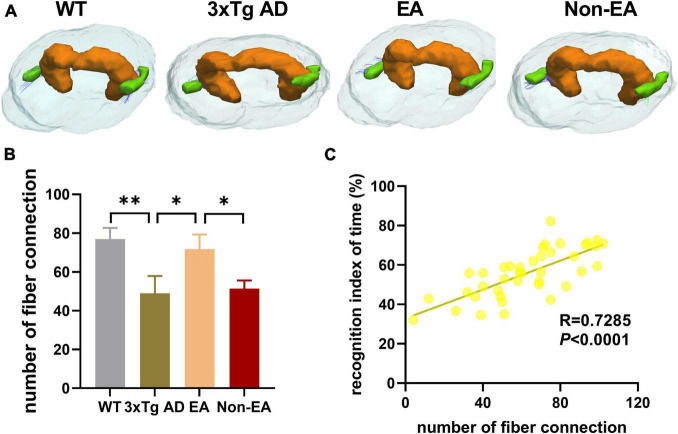
Memory loss and aberrant neuronal network activity are part of the earliest hallmarks of Alzheimer's disease (AD). Electroacupuncture (EA) has been recognized as a cognitive stimulation for its effects on memory disorder, but whether different brain regions or neural circuits contribute to memory recovery in AD remains unknown. Here, we found that memory deficit was ameliorated in 3×Tg-AD mice with EA-treatment, as shown by the increased number of exploring and time spent in the novel object. In addition, reduced locomotor activity was observed in 3×Tg-AD mice, but no significant alteration was seen in the EA-treated mice. Based on the functional magnetic resonance imaging, the regional spontaneous activity alterations of 3×Tg-AD were mainly concentrated in the accumbens nucleus, auditory cortex, caudate putamen, entorhinal cortex (EC), hippocampus, insular cortex, subiculum, temporal cortex, visual cortex, and so on. While EA-treatment prevented the chaos of brain activity in parts of the above regions, such as the auditory cortex, EC, hippocampus, subiculum, and temporal cortex. And then we used the whole-cell voltage-clamp recording to reveal the neurotransmission in the hippocampus, and found that EA-treatment reversed the synaptic spontaneous release. Since the hippocampus receives most of the projections of the EC, the hippocampus-EC circuit is one of the neural circuits related to memory impairment. We further applied diffusion tensor imaging (DTI) tracking and functional connectivity, and found that hypo-connected between the hippocampus and EC with EA-treatment. These data indicate that the hippocampus-EC connectivity is responsible for the recognition memory deficit in the AD mice with EA-treatment, and provide novel insight into potential therapies for memory loss in AD.

Keywords: Alzheimer’s disease; electroacupuncture; electrophysiology; functional connectivity; recognition memory.


Conflict of interest statement

The authors declare that the research was conducted in the absence of any commercial or financial relationships that could be construed as a potential conflict of interest.

Figures

FIGURE 1
FIGURE 1
Effect of EA-treatment on locomotor activity in 3×Tg AD mice. (A) A schematic of the experimental design. Quantification of behavioral results in open field test (n = 10 in each group), involving (B) the percentage of time in the central zone, (C) the total distance traveled throughout the field, (D) and the trajectories of mice in the open field. All data represent the mean ± SEM. **P < 0.01; ns, there was no significant difference.
FIGURE 2
FIGURE 2
Effect of EA-treatment on recognition memory in 3×Tg-AD mice. Quantification of exploratory activity in ORT (n = 10 in each group), involving (A) the total time spent exploring, and (B) the total number of visits to both objects. Quantification of recognition memory, involving (C) the recognition index of time, (D) and the recognition index of number during probe test. (E) The trajectories of mice during probe test in the ORT. All data represent the mean ± SEM. *P < 0.05; **P < 0.01; ns, there was no significant difference.
FIGURE 3
FIGURE 3
Effects of the EA-treatment on regional spontaneous activity. (A) Regions manifesting significant ReHo value differences between 3×Tg-AD mice and wildtype-control mice (blue, 3×Tg-AD < wildtype-control; color bar represents significance of difference), (B) between EA-treated and untreated 3×Tg-AD mice (red, untreated 3×Tg-AD < EA-treated; color bar represents significance of difference), and (C) between Non-EA-treated and untreated 3×Tg-AD mice (red, untreated 3×Tg-AD < Non-EA-treated; color bar represents significance of difference). EC, entorhinal cortex; HIP, hippocampus; Pir, piriform cortex; CPu, caudate putamen; OC, orbital cortex; IC, insular cortex; SC, somatosensory cortex; SN, substantia nigra.
FIGURE 4
FIGURE 4
Effects of the EA-treatment on hippocampal synaptic neurotransmission. (A) Representative sEPSC traces of hippocampal CA1 from all mice by whole-cell voltage-clamp recording (n = 5 in each group). (B) The amplitude of sEPSC, and (C) the frequency of spontaneous firing of hippocampal CA1 were analyzed. Data are presented as the mean ± SEM. **P < 0.01.
FIGURE 5
FIGURE 5
Recognition memory is correlated with aberrant ReHo across the hippocampus and entorhinal cortex. Correlational analysis between the recognition indexes (time/number) and ReHo value obtained from the (A) hippocampus, (B) entorhinal cortex (n = 10 in each group).
FIGURE 6
FIGURE 6
Effects of the EA-treatment on function connectivity between the hippocampus and entorhinal cortex. (A) Representative location image of hippocampus and entorhinal cortex. (B) Quantification of the function connectivity strength between the hippocampus and entorhinal cortex, (C) and the correlational analysis between the recognition index of time and function connectivity (n = 10 in each group). All data represent the mean ± SEM. *P < 0.05; **P < 0.01.
FIGURE 7
FIGURE 7
Effects of the EA-treatment on fiber connection between the hippocampus and entorhinal cortex. (A) Representative image of connected nerve fibers between hippocampus and entorhinal cortex in all group. (B) Quantification of the number of nerve fibers connection between the hippocampus and entorhinal cortex, (C) and the correlational analysis between the recognition index of time and the number of nerve fibers connection (n = 10 in each group). All data represent the mean ± SEM. *P < 0.05; **P < 0.01.

References

    1. Ásgeirsdóttir H. N., Cohen S. J., Stackman R. W., Jr. (2020). Object and place information processing by CA1 hippocampal neurons of C57BL/6J mice. J. Neurophysiol. 123 1247–1264. 10.1152/jn.00278.2019 - DOI - PMC - PubMed
    1. Braak H., Thal D. R., Ghebremedhin E., Del Tredici K. (2011). Stages of the pathologic process in Alzheimer disease: age categories from 1 to 100 years. J. Neuropathol. Exp. Neurol. 70 960–969. 10.1097/NEN.0b013e318232a379 - DOI - PubMed
    1. Cai M., Lee J. H., Yang E. J. (2019). Electroacupuncture attenuates cognition impairment via anti-neuroinflammation in an Alzheimer’s disease animal model. J. Neuroinflam. 16:264. 10.1186/s12974-019-1665-3 - DOI - PMC - PubMed
    1. Cinalli D. A., Cohen S. J., Guthrie K., Stackman R. W., Jr. (2020). Object Recognition Memory: Distinct Yet Complementary Roles of the Mouse CA1 and Perirhinal Cortex. Front. Mol. Neurosci. 13:527543. 10.3389/fnmol.2020.527543 - DOI - PMC - PubMed
    1. Dautricourt S., de Flores R., Landeau B., Poisnel G., Vanhoutte M., Delcroix N., et al. (2021). Longitudinal changes in hippocampal network connectivity in Alzheimer’s Disease. Ann. Neurol. 90 391–406. 10.1002/ana.26168 - DOI - PMC - PubMed

LinkOut - more resources