. 2022 Jul 29:16:864706.
doi: 10.3389/fnins.2022.864706. eCollection 2022.

Stress Affects Central Compensation of Neural Responses to Cochlear Synaptopathy in a cGMP-Dependent Way

Affiliations

Stress Affects Central Compensation of Neural Responses to Cochlear Synaptopathy in a cGMP-Dependent Way

Daria Savitska et al. Front Neurosci. .

Abstract

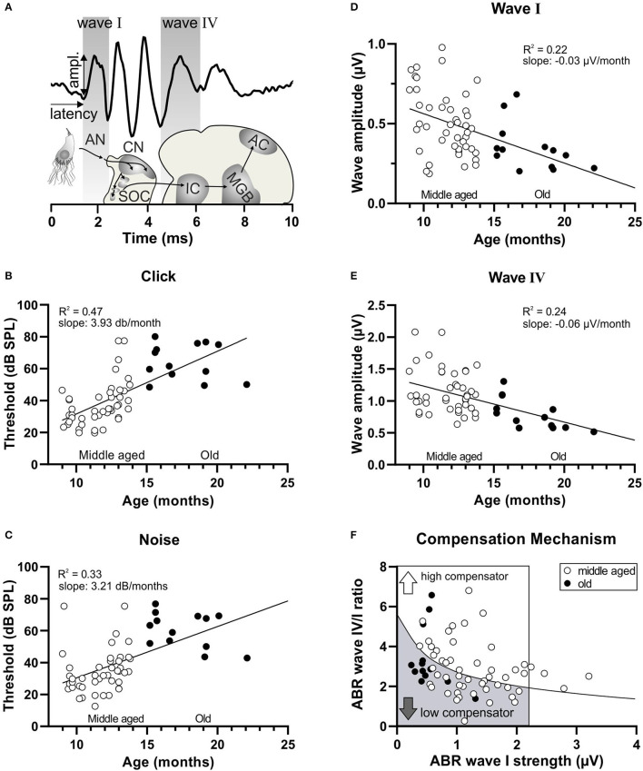
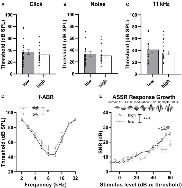
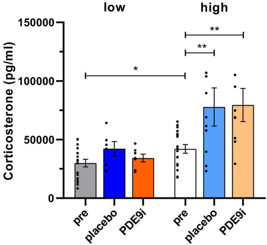
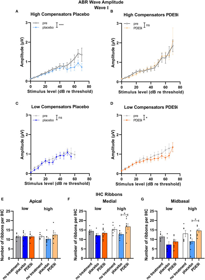
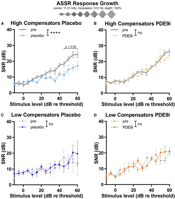
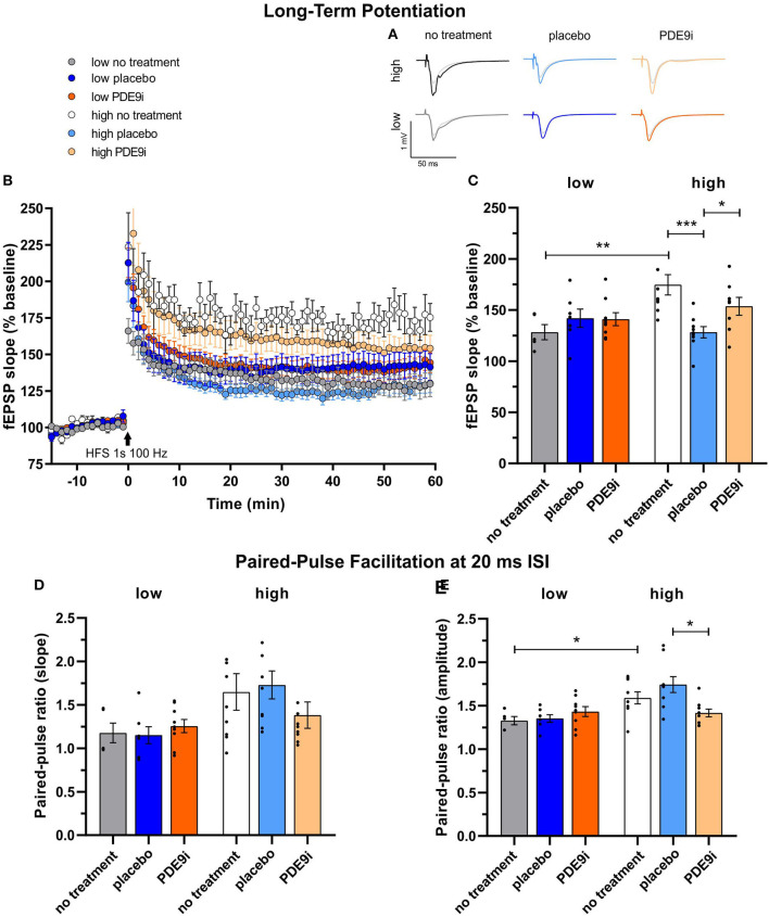
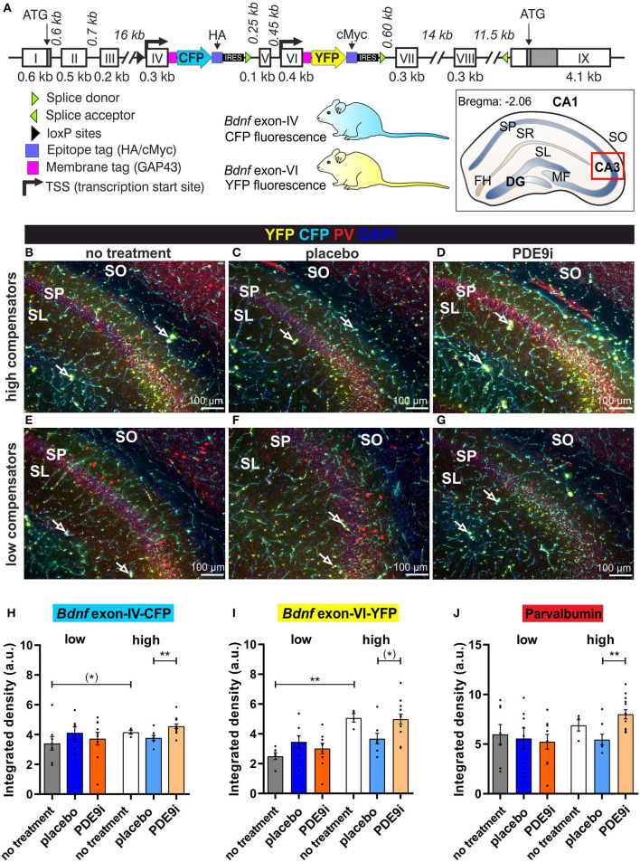
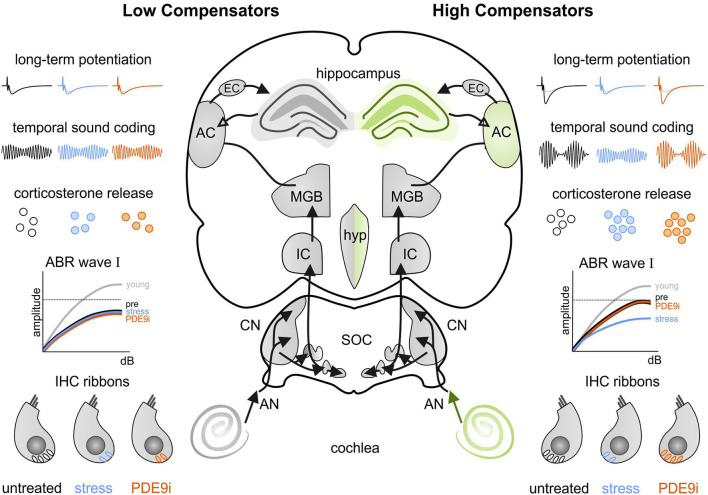
In light of the increasing evidence supporting a link between hearing loss and dementia, it is critical to gain a better understanding of the nature of this relationship. We have previously observed that following cochlear synaptopathy, the temporal auditory processing (e.g., auditory steady state responses, ASSRs), is sustained when reduced auditory input is centrally compensated. This central compensation process was linked to elevated hippocampal long-term potentiation (LTP). We further observed that, independently of age, central responsiveness to cochlear synaptopathy can differ, resulting in either a low or high capacity to compensate for the reduced auditory input. Lower central compensation resulted in poorer temporal auditory processing, reduced hippocampal LTP, and decreased recruitment of activity-dependent brain-derived neurotrophic factor (BDNF) expression in hippocampal regions (low compensators). Higher central compensation capacity resulted in better temporal auditory processing, higher LTP responses, and increased activity-dependent BDNF expression in hippocampal regions. Here, we aimed to identify modifying factors that are potentially responsible for these different central responses. Strikingly, a poorer central compensation capacity was linked to lower corticosterone levels in comparison to those of high compensators. High compensators responded to repeated placebo injections with elevated blood corticosterone levels, reduced auditory brainstem response (ABR) wave I amplitude, reduced inner hair cell (IHC) ribbon number, diminished temporal processing, reduced LTP responses, and decreased activity-dependent hippocampal BDNF expression. In contrast, the same stress exposure through injection did not elevate blood corticosterone levels in low compensators, nor did it reduce IHC ribbons, ABR wave I amplitude, ASSR, LTP, or BDNF expression as seen in high compensators. Interestingly, in high compensators, the stress-induced responses, such as a decline in ABR wave I amplitude, ASSR, LTP, and BDNF could be restored through the "memory-enhancing" drug phosphodiesterase 9A inhibitor (PDE9i). In contrast, the same treatment did not improve these aspects in low compensators. Thus, central compensation of age-dependent cochlear synaptopathy is a glucocorticoid and cyclic guanosine-monophosphate (cGMP)-dependent neuronal mechanism that fails upon a blunted stress response.

Keywords: blunted stress response; cGMP; cochlear synaptopathy; glucocorticoid; long-term potentiation; phosphodiesterase 9A inhibitor (PDE9i).


Conflict of interest statement

The authors declare that the research was conducted in the absence of any commercial or financial relationships that could be construed as a potential conflict of interest. The handling editor VS declared a past collaboration with the authors, LR, MK, and WS.

Figures

Figure 1
Figure 1
Decrease in hearing function over age and subsequent subdivision by the compensation mechanism. (A) Schematic drawing of the auditory pathway and stimulus-evoked deflections of ABR waves correlated with anatomical structures. AN, auditory nerve; CN, cochlear nucleus; SOC, superior olivary complex; IC, inferior colliculus; MGB, medial geniculate body; AC, auditory cortex. (B) In a homogenous group of aging animals (white circles < 15 months, middle aged; black circles > 15 months old), increasing age was significantly correlated with an increasing threshold in response to click stimuli and (C) noise stimuli. (D) In addition, increasing age was significantly correlated with a decreasing amplitude of ABR wave I (E) and ABR wave IV. (F) A schematic diagram representing animals with reduced ABR wave I (inside the box; ABR wave I < 2.19 μV) subdivided by the black regression line, depending on their central compensation capacity into low compensators (the lower wave IV/I ratio) and high compensators (the higher wave IV/I ratio).
Figure 2
Figure 2
The hearing phenotype of low and high compensators before treatment. (A) Low and high compensators showed similar thresholds in response to click and (B) noise stimuli. (C) High and low compensators showed similar thresholds in response to 11 kHz pure-tone stimuli. (D) However, with pure-tone, frequency-specific auditory stimuli (2-32 kHz), low compensators showed elevated thresholds compared to high compensators. (E) The temporal auditory resolution was significantly decreased in low compensators in comparison to high compensators. Re threshold, relative to threshold. Mean ± SEM. *p <0.05, ***p <0.001, 2-way ANOVA.
Figure 3
Figure 3
Corticosterone levels in low and high compensators before and after treatment with either placebo or PDE9i. Prior to treatment, low compensators had lower corticosterone levels in comparison to high compensators. Significantly elevated corticosterone levels were found only in high compensators but not in low compensators after treatment with either placebo or PDE9i. However, no significant differences were observed between the two post-treatment conditions in either high or low compensators. Mean ± SEM. *p < 0.05 (two-tailed t-test pre low vs. pre high), **p < 0.01 (the Bonferroni's multiple comparison test for high compensators).
Figure 4
Figure 4
ABR wave I and IHC ribbon numbers in high and low compensators before and after treatment with placebo or PDE9i. (A) High compensators showed a significant decrease in ABR wave I amplitude after treatment with placebo. (B) However, in high compensators treated with PDE9i, this decrease was not present. Low compensators showed no change in ABR wave I amplitude after treatment with (C) placebo (D) and a significantly decreased ABR wave I amplitude after treatment with PDE9i. (E) IHC ribbon number in the apical cochlear turn, representing lower-frequency areas, shows no changes between the groups. (F) IHC ribbon number in the medial cochlear turn, representing higher-frequency areas, significantly less IHC ribbons were observed in high compensators treated with placebo in comparison to those treated with PDE9i. (G) In the midbasal cochlear turn, representing high-frequency areas, a significantly lower IHC ribbon number was found in high compensators treated with placebo in comparison to those treated with PDE9i. Mean ± SEM. ns p > 0.1, *p < 0.05, ****p < 0.0001. For (A–D), results of repeated measures 2-way ANOVA and for (E–G) results of the Sidak's multiple comparison test are depicted.
Figure 5
Figure 5
Input–output relationship of auditory steady state responses (carrier frequency: 11.32 kHz; modulation frequency: 512 Hz; modulation depth: 100%) in high and low compensators treated with either placebo or PDE9i. (A) High compensators also showed a significant decrease of temporal auditory resolution after treatment with placebo. (B) However, in high compensators treated with PDE9i, this decrease was not present. Low compensators showed no change in temporal auditory resolution after treatment with either (C) placebo or (D) PDE9i. Mean ± SEM. ns p > 0.1, ****p < 0.0001, repeated measures 2-way ANOVA.
Figure 6
Figure 6
Long-term potentiation (LTP) and paired-pulse facilitation (PPF) of low and high compensators that underwent no treatment, placebo treatment, or PDE9i treatment. (A) Representative traces before and after LTP induction and (B) averaged time courses of fEPSP slopes in acute coronal brain slices displayed prominent LTP in all animal groups. (C) The untreated low compensators had significantly lower LTP in comparison to untreated high compensators. In low compensators, neither a placebo treatment nor PDE9i treatment showed any effect on their already low LTP. In contrast, high compensators treated with a placebo displayed a significantly lower LTP both in comparison to their untreated controls and to high compensators treated with PDE9i. (D) The analysis of the paired-pulse ratio of the fEPSP2/fEPSP1 slope at the 20 ms ISI showed no significant differences between high and low compensators before treatment. Neither low compensators nor high compensators treated with either a placebo or PDE9i differed from their untreated controls. Also, no difference in the PPF slope ratio (20 ms ISI) between the treatment conditions of low and high compensators was observed. (E) The analysis of the ratio of the paired-pulse ratio of the fEPSP2/fEPSP1 amplitude at the 20 ms interstimulus interval revealed a significantly higher PPF amplitude ratio in untreated high compensators in comparison to untreated low compensators. High compensators treated with placebo had a significantly higher PPF amplitude ratio (ISI 20 ms) in comparison to those treated with PDE9i, but neither placebo- nor PDE9i-treated high compensators significantly differed from their untreated controls. By contrast, low compensators showed no differences after treatment with either a placebo or PDE9i as compared to untreated controls or compared with each other. Mean ± SEM.*p < 0.05, **p < 0.01, ***p < 0.001. For the comparison of the untreated groups in (C,E), results of the two-tailed t-test are depicted. For the comparison of the differentially treated high compensators in (C,E), results of the Sidak's multiple comparison test are depicted.
Figure 7
Figure 7
Bdnf exon-IV, Bdnf exon-VI, and Parvalbumin (PV) expression in the CA3 region of the hippocampus of untreated low and high compensators and animals that received placebo or PDE9i. (A) Genetically modified the Bdnf gene of the BLEV reporter mouse line in which CFP is expressed with activity-dependent Bdnf exon-IV transcription and YFP with activity-dependent Bdnf exon-VI transcription. An abstract scheme of a coronal hippocampal section at bregma position −2.06 mm. The red box indicates the inset seen in (B–G) where the integrated density is measured. DG, dentate gyrus; FH, fissura hippocampalis; MF, mossy fiber; SL, stratum lucidum; SO, stratum oriens; SP, stratum pyramidale; SR, stratum radiatum. (B) Untreated high compensators showed a strong expression of Bdnf exon-IV (CFP), Bdnf exon-VI (YFP) (open arrows signify where they are expressed in neighboring structures) and PV (red) in the hippocampal CA3 region (blue = DAPI). (C) After placebo treatment, less expression of CFP, YFP, and PV was observed. (D) When treated with PDE9i, high compensators showed an increase in CFP, YFP, and PV expression. (E–G) CFP, YFP, and PV expression in the hippocampal CA3 region of low compensators, which was slightly weaker than in high compensators, reaching significance for YFP expression in untreated animals, shown in (I). In low compensators, neither placebo nor PDE9i treatment had an effect on CFP, YFP, and PV expression. (H) In the SL/SP region of CA3, untreated low compensators showed a lower CFP expression in comparison to untreated high compensators not reaching statistical significance (*). Low compensators treated with placebo or PDE9i did not show any differences in CFP expression to their untreated controls or to each other. By contrast, high compensators treated with PDE9i had significantly higher CFP expression in comparison to those treated with placebo. (I) YFP expression in the SL/SP region of CA3 was significantly lower in untreated low compensators in comparison to untreated high compensators. Low compensators showed no difference in YFP expression between treatment groups. However, high compensators treated with PDE9i showed a higher YFP expression in comparison to those treated with placebo not reaching statistical significance. (J) PV expression in the SL/SP region of CA3 did not differ between untreated high and low compensators. Low compensators showed no difference in PV expression between treatment groups. High compensators treated with PDE9i showed a significantly higher PV expression compared to those treated with placebo. Mean ± SEM. (*)p ≤ 0.1, **p < 0.01. For the comparison of the untreated groups in (H–J), results of two-tailed t-test are depicted. For the comparison of the differentially treated high compensators in (H–J), results of the Dunn's multiple comparison test are depicted.
Figure 8
Figure 8
An abstract scheme of the mechanisms in the auditory pathway that are differentially affected by stress or PDE9i application in low compensators (the left side) and high compensators (the right side). Low compensators have a lower baseline corticosterone level (black, left) in comparison to high compensators (black, right). When stress due to repeated injections is applied (blue/orange), high compensators respond in a physiological way, with increased corticosterone release. Low compensators, on the other hand, do not have increased corticosterone levels after application of a stressor, showing characteristics of a blunted stress response. This blunted stress response prevents low compensators from reacting to both a stress-induced drop of long-term potentiation, temporal sound coding, auditory nerve response (ABR wave I), and inner hair cell ribbons, which we see in high compensators. Additionally, where treatment with a PDE9i is able to preserve all of these parameters in high compensators, the blunted stress response of low compensators prevents their restoration. ABR, auditory brainstem response; AC, auditory cortex; AN, auditory nerve; CN, cochlear nucleus; dB, decibels; EC, entorhinal cortex; hyp, hypothalamus; IC, inferior colliculus; IHC, inner hair cell; MGB, medial geniculate body; SOC, superior olivary complex.

Similar articles

Cited by

References

    1. Arango-Lievano M., Borie A. M., Dromard Y., Murat M., Desarmenien M. G., Garabedian M. J., et al. . (2019). Persistence of learning-induced synapses depends on neurotrophic priming of glucocorticoid receptors. Proc. Natl. Acad. Sci. U. S. A. 116, 13097–13106. 10.1073/pnas.1903203116 - DOI - PMC - PubMed
    1. Bharadwaj H. M., Verhulst S., Shaheen L., Liberman M. C., Shinn-Cunningham B. G. (2014). Cochlear neuropathy and the coding of supra-threshold sound. Front. Syst. Neurosci. 8, 26. 10.3389/fnsys.2014.00026 - DOI - PMC - PubMed
    1. Bramhall N., Ong B., Ko J., Parker M. (2015). Speech perception ability in noise is correlated with auditory brainstem response wave i amplitude. J. Am. Acad. Audiol. 26, 509–517. 10.3766/jaaa.14100 - DOI - PubMed
    1. Bueno-Junior L. S., Leite J. P. (2018). Input convergence, synaptic plasticity and functional coupling across hippocampal-prefrontal-thalamic circuits. Front. Neural. Circuits 12, 40. 10.3389/fncir.2018.00040 - DOI - PMC - PubMed
    1. Burkard R. F., Don M. (2007). “The auditory brainstem response,” in Auditory Evoked Potentials: Basic Principles and Clinical Application, eds. R.F. Burkard, J.J. Eggermont, and M. Don. (Philadelphia: Lippincott Williams and Wilkins; ).

LinkOut - more resources