

Case Reports
doi: 10.1016/j.eucr.2022.102176.
eCollection 2022 Nov.
Malignant juxtaglomerular cell tumor
Affiliations
- PMID: 35968528
- PMCID: PMC9372647
- DOI: 10.1016/j.eucr.2022.102176
Item in Clipboard
Case Reports
Malignant juxtaglomerular cell tumor
Urol Case Rep.
.
Abstract
Juxtaglomerular cell tumors (JGCTs) are rare, typically benign neoplasms; only rare cases are clinically or histologically malignant. We herein report the histologic, immunophenotypic, and molecular features of a clinically unsuspected, diagnostically challenging case of malignant JGCT in a 23-year-old man. The diagnosis is confirmed with electron microscopy. The case is notable for its marked mitotic activity, which has not been previously reported in JGCTs, and novel finding of GATA3 immunohistochemical positivity.
Keywords: GATA3; Juxtaglomerular cell tumor; Kidney; Malignant; Renin.
© 2022 The Author(s).
Conflict of interest statement
The authors have no conflicts of interest to disclose.
Figures

An axial computed tomography image with intravenous contrast demonstrating a solid mass (white arrow) in the lateral mid pole of the right kidney (A). Gross image of the tumor showing well-defined borders with a fleshy, hemorrhagic cut surface (B). Tumor borders are relatively sharp with occasional entrapped renal tubules at the periphery (C). The tumor is composed of ovoid to spindle cells with moderate nuclear pleomorphism, predominantly eosinophilic cytoplasm, and intimately associated with surrounding delicate vasculature. Mitotic figures are readily apparent (D).
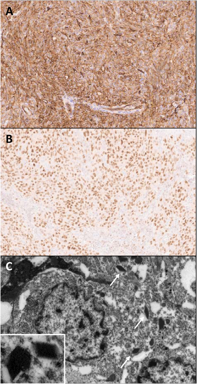
Immunohistochemical stains show tumor cells are diffusely positive for CD34 (A) and GATA3 (B). Electron microscopy shows tumor cells with cytoplasmic electron-dense rhomboid renin protogranule crystals (white arrows, inset) (C).
References
-
- Martin S.A., Mynderse L.A., Lager D.J., Cheville J.C. Juxtaglomerular cell tumor: a clinicopathologic study of four cases and review of the literature. Am J Clin Pathol. 2001;116(6):854–863. - PubMed
-
- Dong D., Li H., Yan W., Xu W. Juxtaglomerular cell tumor of the kidney--a new classification scheme. Urol Oncol. 2010;28(1):34–38. - PubMed
-
- Hagiya A., Zhou M., Hung A., Aron M. Juxtaglomerular cell tumor with atypical pathological features: report of a case and review of literature. Int J Surg Pathol. 2020;28(1):87–91. - PubMed
-
- Kuroda N., Maris S., Monzon F.A., et al. Juxtaglomerular cell tumor: a morphological, immunohistochemical and genetic study of six cases. Hum Pathol. 2013;44(1):47–54. - PubMed
Publication types
LinkOut - more resources
Full Text Sources
