. 2022 Aug 15;132(16):e156250.
doi: 10.1172/JCI156250.

Upregulated PD-1 signaling antagonizes glomerular health in aged kidneys and disease

Affiliations

Upregulated PD-1 signaling antagonizes glomerular health in aged kidneys and disease

Jeffrey W Pippin et al. J Clin Invest. .

Abstract

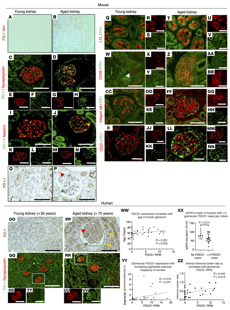
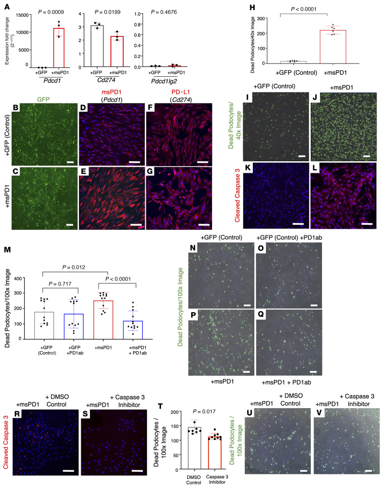
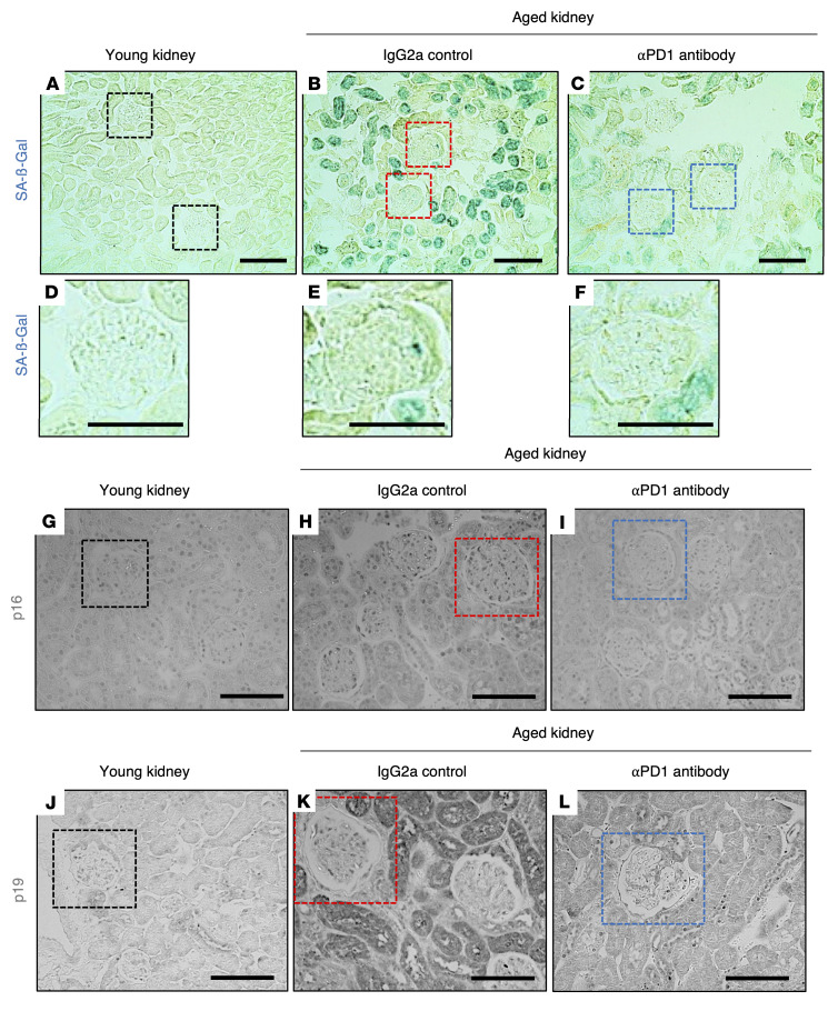
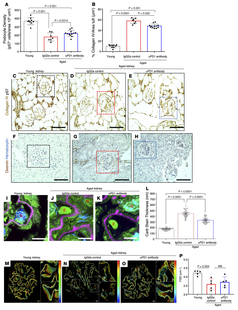
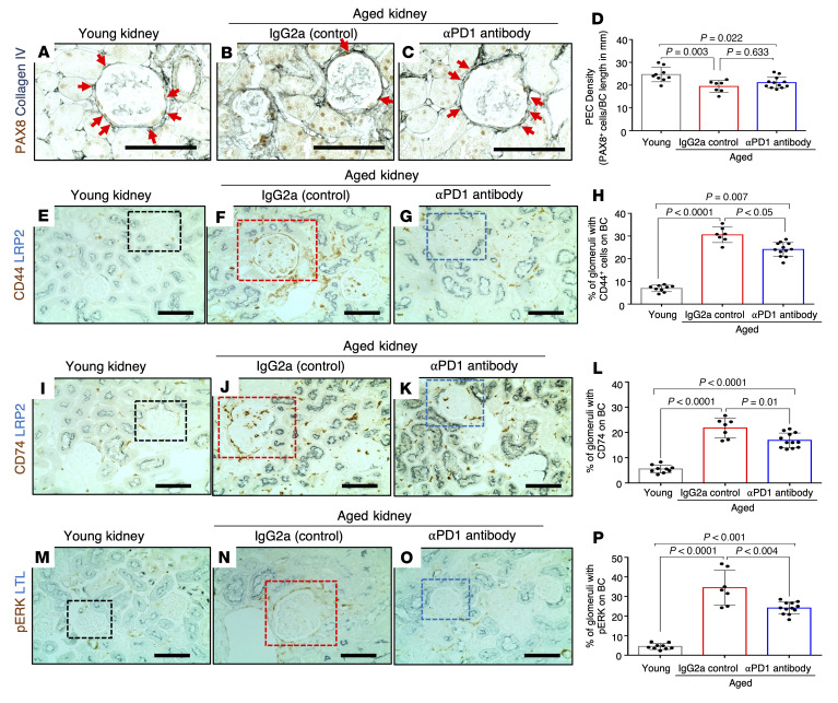
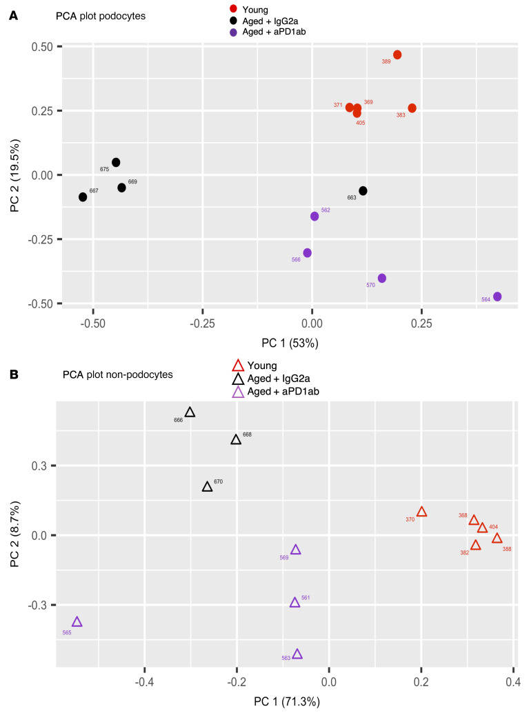
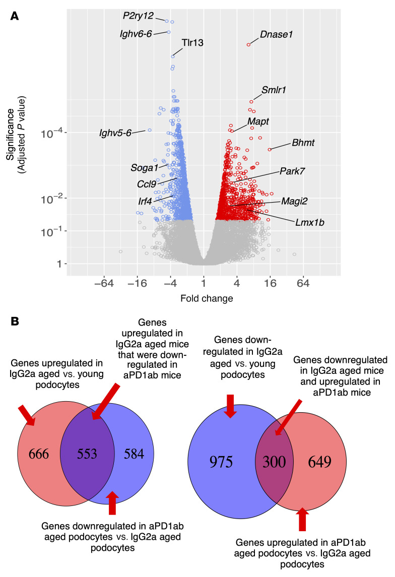
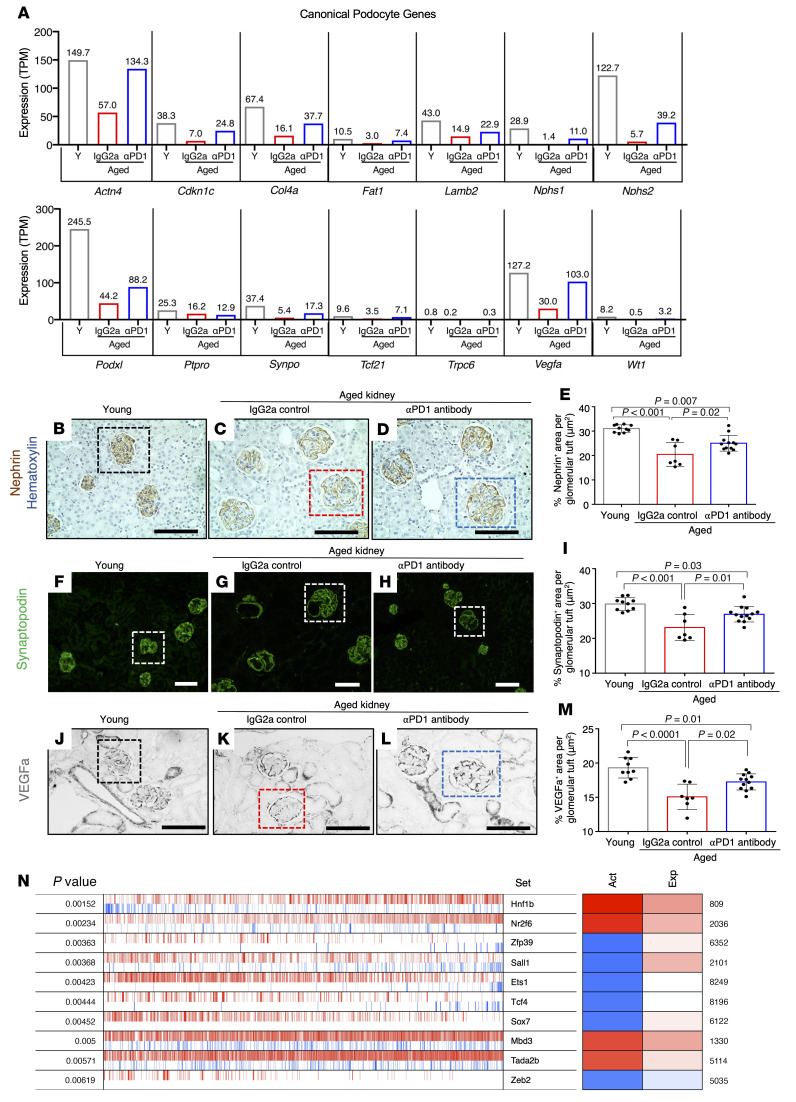
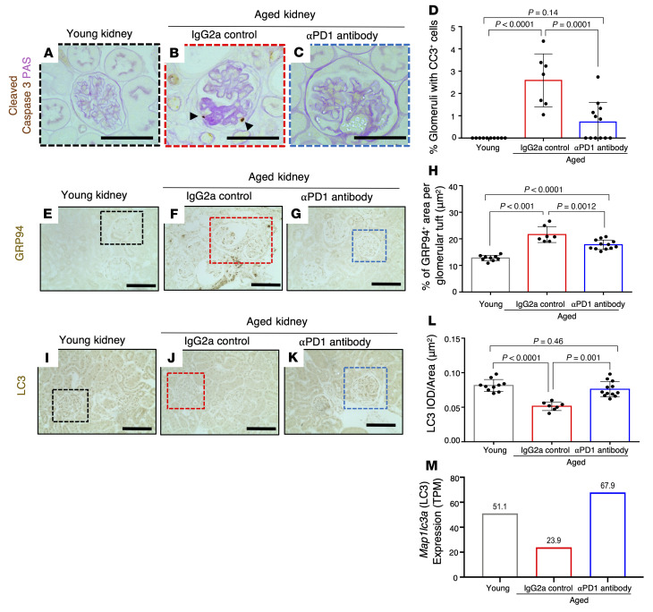
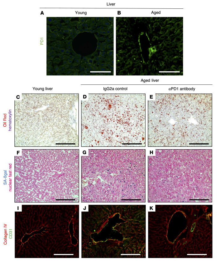
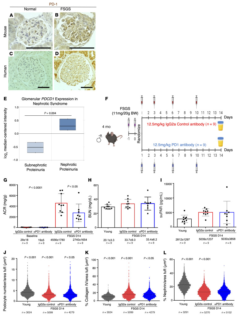
With an aging population, kidney health becomes an important medical and socioeconomic factor. Kidney aging mechanisms are not well understood. We previously showed that podocytes isolated from aged mice exhibit increased expression of programmed cell death protein 1 (PD-1) surface receptor and its 2 ligands (PD-L1 and PD-L2). PDCD1 transcript increased with age in microdissected human glomeruli, which correlated with lower estimated glomerular filtration rate and higher segmental glomerulosclerosis and vascular arterial intima-to-lumen ratio. In vitro studies in podocytes demonstrated a critical role for PD-1 signaling in cell survival and in the induction of a senescence-associated secretory phenotype. To prove PD-1 signaling was critical to podocyte aging, aged mice were injected with anti-PD-1 antibody. Treatment significantly improved the aging phenotype in both kidney and liver. In the glomerulus, it increased the life span of podocytes, but not that of parietal epithelial, mesangial, or endothelial cells. Transcriptomic and immunohistochemistry studies demonstrated that anti-PD-1 antibody treatment improved the health span of podocytes. Administering the same anti-PD-1 antibody to young mice with experimental focal segmental glomerulosclerosis (FSGS) lowered proteinuria and improved podocyte number. These results suggest a critical contribution of increased PD-1 signaling toward both kidney and liver aging and in FSGS.

Keywords: Aging; Chronic kidney disease; Expression profiling.


Figures

Figure 1
Figure 1. Podocyte PD-1 immunostaining and transcripts.
(ANN) Mouse kidney. (A and B) In situ hybridization shows that compared with young kidney (A), PD-1 transcript (brown) increases in aged kidney (B). (CH) PD-1 (green) and synaptopodin (red) staining shows that PD-1 in mouse glomerulus (C) merges with synaptopodin-positive podocytes in aged kidney (D, yellow). PD-1 stains PECs along Bowman’s capsule (D, green). (EH) Fluorescence channels from C and D. (IN) PD-1 (green) and nephrin (red) staining in young glomeruli (I) merges with nephrin in aged kidney (J, yellow). PD-1 increased in PECs and proximal tubular epithelial cells (J, green). (KN) Single channels from I and J. (O and P) PD-L1 (brown) in young mouse kidney (O) is detected in a podocyte (green arrowhead), PECs (red arrowhead), and proximal tubules in the aged mouse (P). (QV) Lotus tetragonolobus lectin (LTL) (red) stains proximal epithelial cells and merges with PD-1 (green) in aged kidney (T). (WBB) CD45+ interstitial lymphocytes (red) merge with PD-1 (green) in aged kidney. (CCHH) PD-1 (green) does not merge with the mesangial cell marker α8 integrin (red). (IINN) PD-1 (green) does not merge with the endothelial cell marker CD31 (red). (OOVV) Human kidney. (OO and PP) PD-1 (brown) in young human kidney (OO) increases in podocytes (green arrowhead), PECs (red arrowhead), and tubular epithelial cells (orange arrowheads) in aged human kidney (PP). (QQVV) PD-1 (green) and synaptopodin (red) in young human glomerulus merges with synaptopodin (yellow) in aged human glomerulus. (WWZZ) PDCD1 transcripts from microdissected human glomeruli. Expression of PDCD1 (corresponding to human PD-1) increased with age (WW), accompanied by lower eGFR (XX), higher glomerulosclerosis (YY), and vascular injury (ZZ). Scale bars: 25 μm (CNN), 100 μm (OO, PP, and SSVV), and 200 μm (QQ and RR). Statistical analysis was performed by t test, χ2 test, and quasi-Poisson regression modeling.
Figure 2
Figure 2. Overexpression of PD-1 induces podocyte death in cell culture.
(AG) Following overexpression of PD-1 using a lentiviral expression vector (msPD1, red bars) in immortalized mouse podocytes, mRNA levels increased significantly for Pdcd1 (PD-1) in comparison with GFP control–infected podocytes, without changes to Cd274 or Pdcd1lg2. GFP expression (green) of the GFP control (B) and PD-1–overexpressing (C) lentiviral vectors confirms efficient transfection. Immunocytochemistry for PD-1 protein (red) in GFP control–infected podocytes (D) was increased in msPD1-overexpressing podocytes (E). DAPI stains nuclei blue. Staining for PD-L1 (red) was not different between GFP control–infected (F) and PD-1–overexpressing (G) podocytes. (HL) Overexpression of PD-1 (red bar) increased podocyte death in comparison with GFP control–infected podocytes (gray bar). Representative images of dead podocytes (I and J). Cleaved caspase-3 staining (red) was barely detected in GFP control–infected podocytes (K) but was markedly increased in PD-1–overexpressing podocytes (L). Nuclei were counterstained with DAPI (blue). (MQ) Application of aPD1ab (blue bar, second column) did not impact cell death in GFP control–infected podocytes (gray bar). The increased podocyte death induced by overexpression of PD-1 (red bar) was reduced when anti–PD-1 antibody was applied (blue bar, fourth column). (NQ) Representative images of dead podocytes encircled with green annotations used for M. (R and S) Immunocytochemistry for cleaved caspase-3 (red) was increased in PD-1–overexpressing podocytes treated with DMSO (R) but was markedly decreased following treatment with the caspase-3–specific inhibitor Z-DEVD-FMK (S). (TV) Treatment of PD-1–overexpressing podocytes with the caspase-3 inhibitor significantly decreased podocyte death (red bar) compared with the DMSO control (gray bar). (U and V) Representative images of dead podocytes encircled with green annotations in DMSO-treated (U) and caspase-3 inhibitor–treated (V) PD-1–overexpressing podocytes. Statistical analysis was performed by t test. Scale bars represent 100 μm.
Figure 3
Figure 3. Podocyte senescence.
(AF) Representative images of SA-β-gal staining (blue). SA-β-gal was barely detected in young kidneys (A and D) but increased in glomeruli (red boxes) and tubular epithelial cells in IgG2a-injected aged mice (B and E) and was lowered by aPD1ab in glomeruli (blue boxes) and tubules (C and F). (GI) p16 staining (black) was occasionally detected in glomeruli and tubular epithelial cells of young kidneys (G). It was increased in glomerular (red box) and tubular epithelial cells in IgG2a-injected mice (H) but was lower in aPD1ab-injected mice (I). (JL) p19 staining (black) was barely detected in young kidneys (J) but was increased in glomerular (red box) and tubular epithelial cells in IgG2a-injected mice (K) and was lower in aPD1ab-injected mice (L). Scale bars: 50 μm.
Figure 4
Figure 4. Podocyte density, scarring, stress, and ultrastructure.
(AE) Podocyte density measured by p57 staining (dark blue, CE) and summarized in A. Each circle represents an individual mouse. Density was lower in aged IgG2a-injected mice compared with young mice and was increased in aged aPD1ab-injected mice. Glomerular scarring was measured by glomerular collagen IV staining (brown, CE) and is summarized in B. It was higher in IgG2a-injected aged mice compared with young mice and was lowered by aPD1ab. (FH) The podocyte stress marker desmin (brown) was increased in aged IgG2a-injected mice compared with young and was lower in aged aPD1ab mice. (IL) The filtration barrier ultrastructure was assessed by expansion microscopy of FLARE-labeled glomeruli, which demonstrated that glomerular basement membrane (GBM) thickness (pink) was significantly increased in aged IgG2a-injected mice (J) compared with young mice (I) and reduced in aged aPD1ab mice (K). Representative images are shown in IK, and GBM thickness is quantified in L. N, nuclei; E, erythrocytes. (MP) Podocyte ultrastructure was characterized by the podocyte exact morphology measurement procedure (PEMP). Representative images are shown in MO, and filtration slit density (FSD) is quantified in P. This analysis shows a significant decrease in FSD in aged IgG2a-injected mice compared with young mice (M, N, and P). Elevation of FSD was observed in aged aPD1ab mice but did not reach significance (O and P). Scale bars: 5 μm (MO), 10 μm (IK), and 50 μm (CH). Statistical analysis was performed by 1-way ANOVA.
Figure 5
Figure 5. Parietal epithelial cell changes.
(AD) Representative images of immunoperoxidase staining for the PEC marker PAX8 (brown) and collagen IV (blue, outlines Bowman’s capsule [BC]) (AC) and quantification thereof (D). PEC density was lower in aged IgG2a-injected mice (red bar) compared with young mice (gray bar) but did not change with aPD1ab treatment (blue bar). (EP) Representative images of immunoperoxidase double staining with antibodies against the PEC activation markers CD44 (EG, brown), CD74 (IK, brown), and p-ERK (MO, brown) counterstained with the proximal tubular cell markers LRP2 (EG and IK, blue) and LTL (MO, blue). Quantification (H, L, and P) shows that all 3 PEC activation markers were elevated in aged IgG2a-injected mice (red bars) compared with young mice (gray bars) and lowered by aPD1ab injections (blue bars). Scale bars represent 50 μm. Statistical analysis was performed by t test.
Figure 6
Figure 6. Principal component analysis.
Principal component analysis (PCA) of the mRNA-Seq data showed excellent clustering of the individual treatment groups for both podocytes (A) and non-podocytes (B).
Figure 7
Figure 7. Podocyte transcripts in aged mouse podocytes altered by anti–PD-1 antibody.
(A) Volcano plot shows the transcripts in aged podocytes that were decreased (blue circles), increased (red circles), or not changed (gray circles) by treatment of mice with aPD1ab for 8 weeks. (B) Summary of the number of genes upregulated and downregulated in podocytes from aged control IgG2a mice compared with young mice, and from aged aPD1ab-injected mice compared with age-matched IgG2a-injected mice.
Figure 8
Figure 8. Changes to podocyte canonical genes, proteins, and transcription factors.
(A) Expression of individual canonical podocyte genes from mRNA-Seq data of young mice (Y, gray bars), aged mice injected with IgG2a (red bars), and aged mice injected with anti–PD-1 antibody (αPD1, blue bars). The levels of all genes were lower in aged IgG2a-injected mice compared with young mice, and all except Ptpro were increased in aPD1ab-injected mice. TPM, transcripts per million. (BM) Protein validation of select genes by immunostaining. Representative immunoperoxidase staining for nephrin (BD, brown), immunofluorescent staining for synaptopodin (FH, green), and immunoperoxidase staining for VEGFA (JL, black). The box in each panel shows an example of a glomerulus. Bar graphs show the quantification of nephrin (E), synaptopodin (I), and VEGFA (M) staining with each circle representing an individual mouse. Compared with young mice (gray bars), immunostaining was lower for each in aged IgG2a-injected mice (red bars) but was higher in aged aPD1ab-injected mice (blue bars). (N) VIPER (virtual inference of protein activity by enriched regulon) analysis of transcription factor activity. The top 10 transcription factors impacted by aPD1ab treatment are shown in the third column, along with their significance (first column), representative activity (second column), conferred activity (fourth column), and expression (fifth column), with red showing increased and blue decreased levels/activities. The conferred activity of Hnf1b, Nr2f6, Mbd3, and Tada2b increased, with increased expression. Zfp39, Sall1, and Sox7 activity was decreased despite higher expression levels in aPD1ab-injected mice. While the lower activity of Ets1 and the lower activity of Tcf4 were independent of their expression levels, the lower activity of Zeb2 correlated with its lower expression. Scale bars represent 50 μm (BD and JL) and 100 μm (FH). Statistical analysis was performed by t test.
Figure 9
Figure 9. Apoptosis, ER stress, and autophagy.
(AD) Representative images of immunoperoxidase staining for the apoptosis marker cleaved caspase-3 (brown) and periodic acid counterstain (pink). Glomerular apoptotic cells in aged IgG2a-injected mice are indicated by black arrowheads (B). Quantification of the percentage of glomeruli with at least a single cleaved caspase-3–positive (CC3+) cell (D) shows an increase in aged IgG2a-injected mice (red bar) compared with young mice (gray bar) and a decrease with aPD1ab treatment (blue bar). (EH) Representative images of immunoperoxidase staining for the ER stress marker GRP94 (brown) and quantification thereof. GRP94 shows higher staining in aged IgG2a-injected mice (red bar), which was lowered upon aPD1ab treatment (blue bar). Individual mice are represented by circles. (IM) Measurement of autophagic activity measured by LC3. Representative images of immunoperoxidase staining for microtubule-associated protein 1 light chain 3 (LC3) (IK, brown) and quantification of the staining (L). Compared with young mice (gray bar), LC3 staining was lower in IgG2a-injected mice (red bar), indicating reduced autophagy, but was higher with aPD1ab treatment (blue bar). IOD, integrated optical density. (M) mRNA-Seq data from isolated podocytes showed a decrease in the Map1lc3a (LC3) transcript in IgG2a-injected mice (red bar) compared with young mice (gray bar) but an increase with aPD1ab treatment (blue bar). Scale bars: 50 μm. Statistical analysis was performed by t test.
Figure 10
Figure 10. Anti–PD-1 antibody decreases liver aging in mice.
(A and B) Representative images of PD-1 immunofluorescence staining, which is higher in aged mouse liver (B) compared with young liver (A). (CE) Oil Red staining (red) as a marker of fat deposition was barely detected in young livers but was increased in the livers of aged IgG2a-injected mice and decreased in aPD1ab-injected mice. (FH) SA-β-gal staining (blue) used as a marker of senescence was increased in aged IgG2a-injected livers and decreased by aPD1ab treatment. (IK) Double immunostaining for collagen IV (red) and the endothelial cell marker CD31 (green) shows increased collagen IV deposition in the blood vessels of the liver from aged IgG2a-injected mice compared with young mice, which was decreased by aPD1ab injection. Scale bars represent 50 μm (A and B) and 100 μm (CK).
Figure 11
Figure 11. Anti–PD-1 antibody improves experimental FSGS.
(AD) PD-1 immunoperoxidase staining (brown) of glomeruli from normal young mice (A) is increased in young mice with experimental FSGS (B). Similarly, in humans, PD-1 staining of glomeruli of a young human kidney (C) is increased in podocytes, PECs, and tubular epithelial cells of a kidney from an FSGS patient (D). Scale bars represent 50 μm. (E) Analysis of the Nephroseq data from NEPTUNE shows that PDCD1 mRNA levels in human microdissected glomeruli are significantly higher in patients with nephrotic-range versus sub-nephrotic-range proteinuria (n = 38). (F) To study PD-1 signaling in FSGS, 4-month-old mice were injected twice i.p. with a sheep anti-glomerular antibody. Mice were randomized into 2 groups, which received either an anti–PD-1 antibody (n = 8) or the isotype control IgG2a (n = 7) on days 1, 3, 6, and 10 after FSGS induction. (GI) At the conclusion of the experiment, kidney function analyses showed that albumin/creatinine ratio (ACR) values significantly increased following FSGS induction in IgG2a control–injected but were reduced in aPD1ab-injected mice (G). Blood urea nitrogen (BUN) levels were not significantly different between the groups (H), while plasma soluble urokinase plasminogen activator receptor (suPAR) levels were significantly increased in FSGS mice injected with control IgG2a compared with young mice but were not reduced in mice injected with aPD1ab (I). (J) Quantification of podocyte density was lower in FSGS mice injected with IgG2a compared with young mice and was increased upon aPD1ab injection. (K) Glomerular scarring measured by glomerular collagen IV staining was higher in FSGS mice injected with IgG2a compared with young mice and was lowered by aPD1ab injection. (L) Nephrin immunostaining was lower in FSGS mice injected with IgG2a compared with young mice but was not significantly changed by aPD1ab injection. Each circle in JL represents an individual glomerulus, and the number of glomeruli quantified is indicated. Statistical analysis was performed by t test.

Comment in

References

    1. Lang J, et al. Association of serum albumin levels with kidney function decline and incident chronic kidney disease in elders. Nephrol Dial Transplant. 2018;33(6):986–992. doi: 10.1093/ndt/gfx229. - DOI - PMC - PubMed
    1. Pottel H, et al. An estimated glomerular filtration rate equation for the full age spectrum. Nephrol Dial Transplant. 2016;31(5):798–806. doi: 10.1093/ndt/gfv454. - DOI - PMC - PubMed
    1. Denic A, et al. The substantial loss of nephrons in healthy human kidneys with aging. J Am Soc Nephrol. 2017;28(1):313–320. doi: 10.1681/ASN.2016020154. - DOI - PMC - PubMed
    1. Hoy WE, et al. A stereological study of glomerular number and volume: preliminary findings in a multiracial study of kidneys at autopsy. Kidney Int Suppl. 2003;(83):31–37. - PubMed
    1. Hughson MD, et al. Progressive nephron loss in aging kidneys: clinical-structural associations investigated by two anatomical methods. Anat Rec (Hoboken) 2020;303(10):2526–2536. doi: 10.1002/ar.24249. - DOI - PMC - PubMed

Publication types