. 2022 Aug 30;13(4):e0210722.
doi: 10.1128/mbio.02107-22. Epub 2022 Aug 15.

T Cell-Specific STAT1 Expression Promotes Lytic Replication and Supports the Establishment of Gammaherpesvirus Latent Reservoir in Splenic B Cells

Affiliations

T Cell-Specific STAT1 Expression Promotes Lytic Replication and Supports the Establishment of Gammaherpesvirus Latent Reservoir in Splenic B Cells

P A Sylvester et al. mBio. .

Abstract

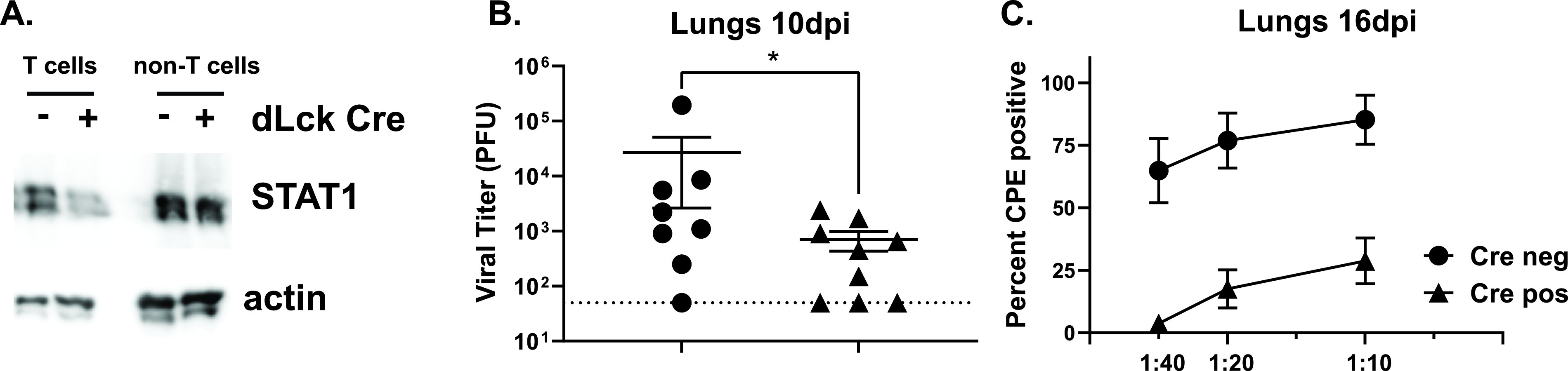
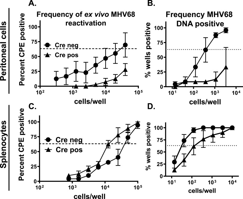
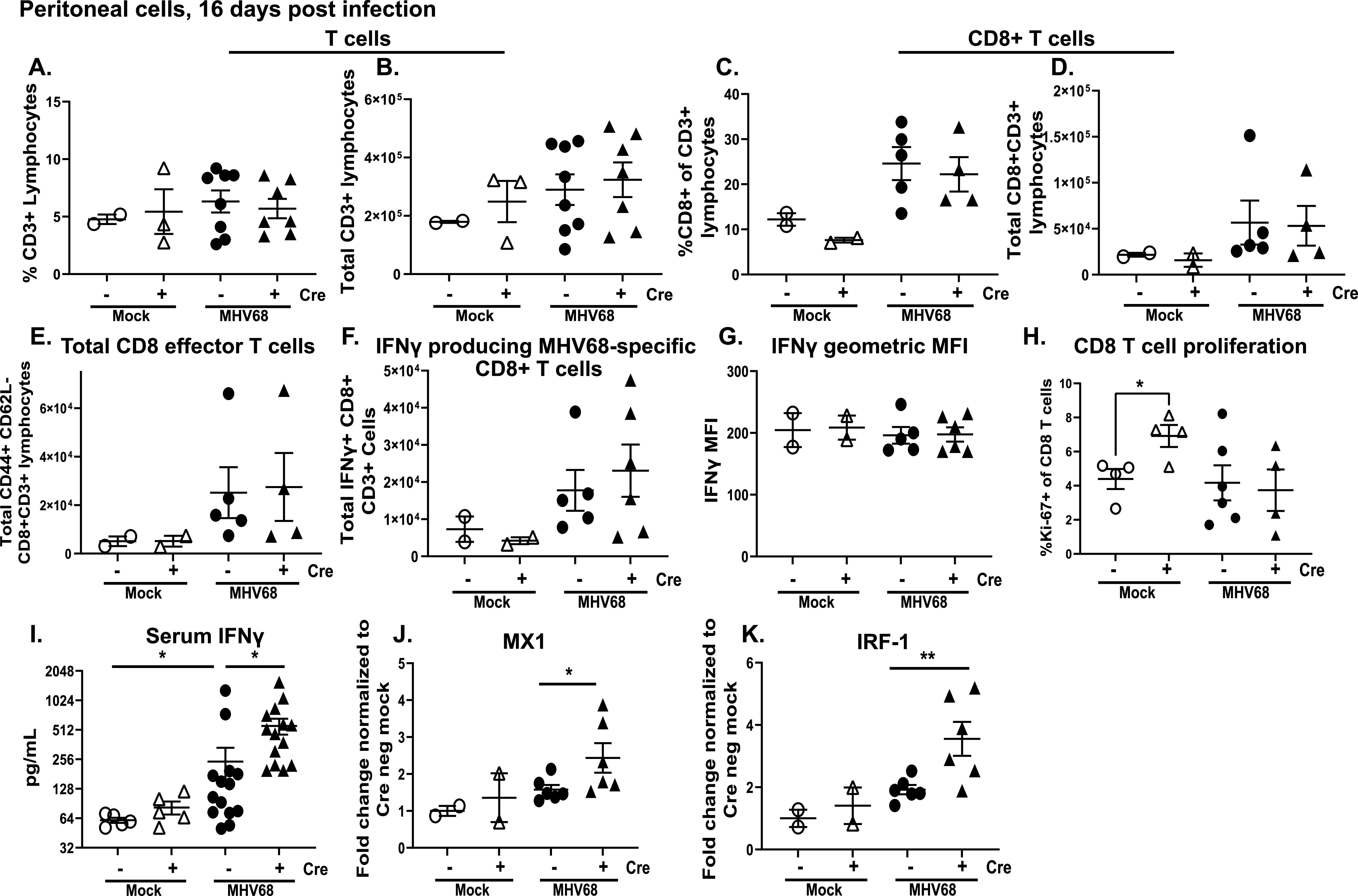
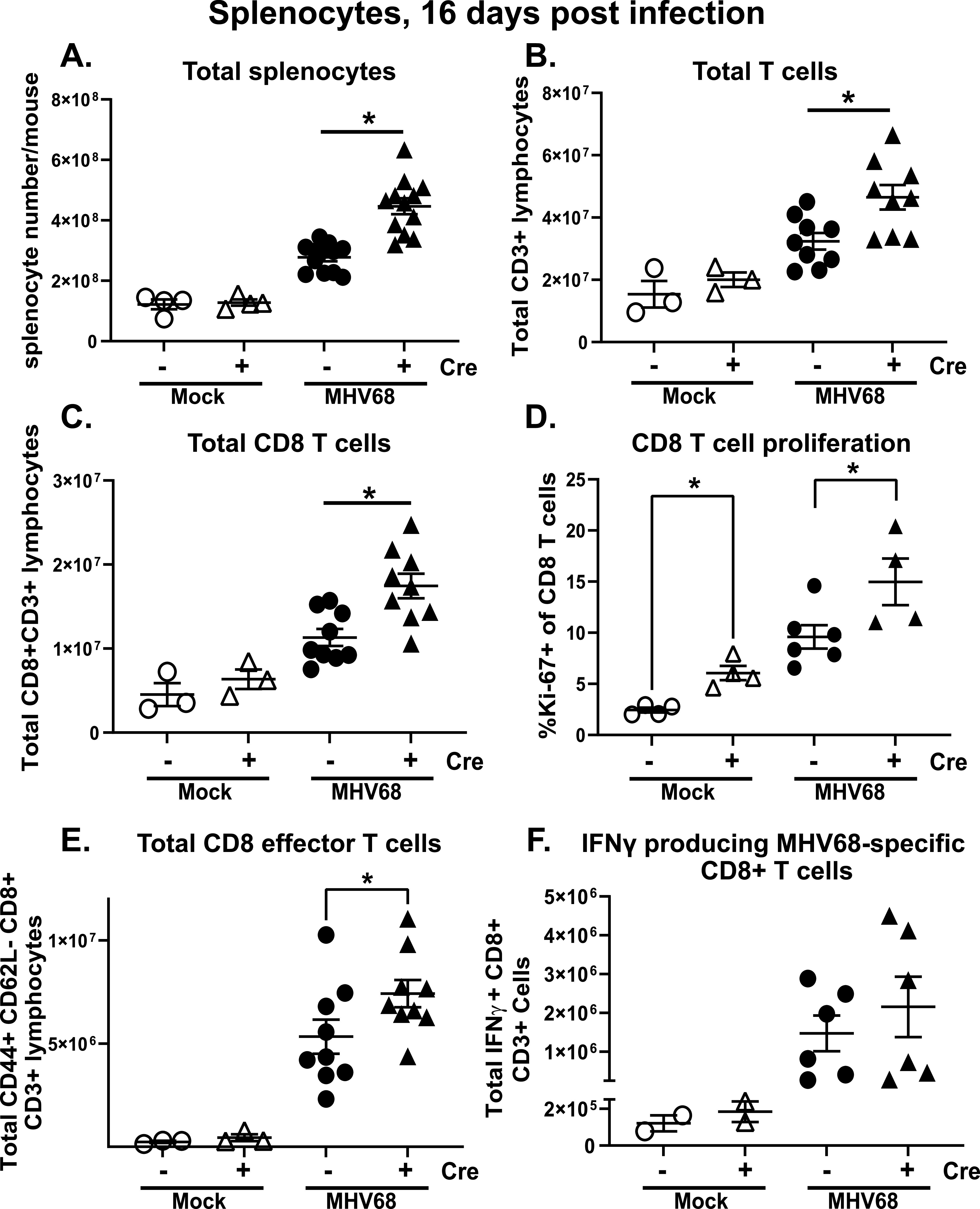
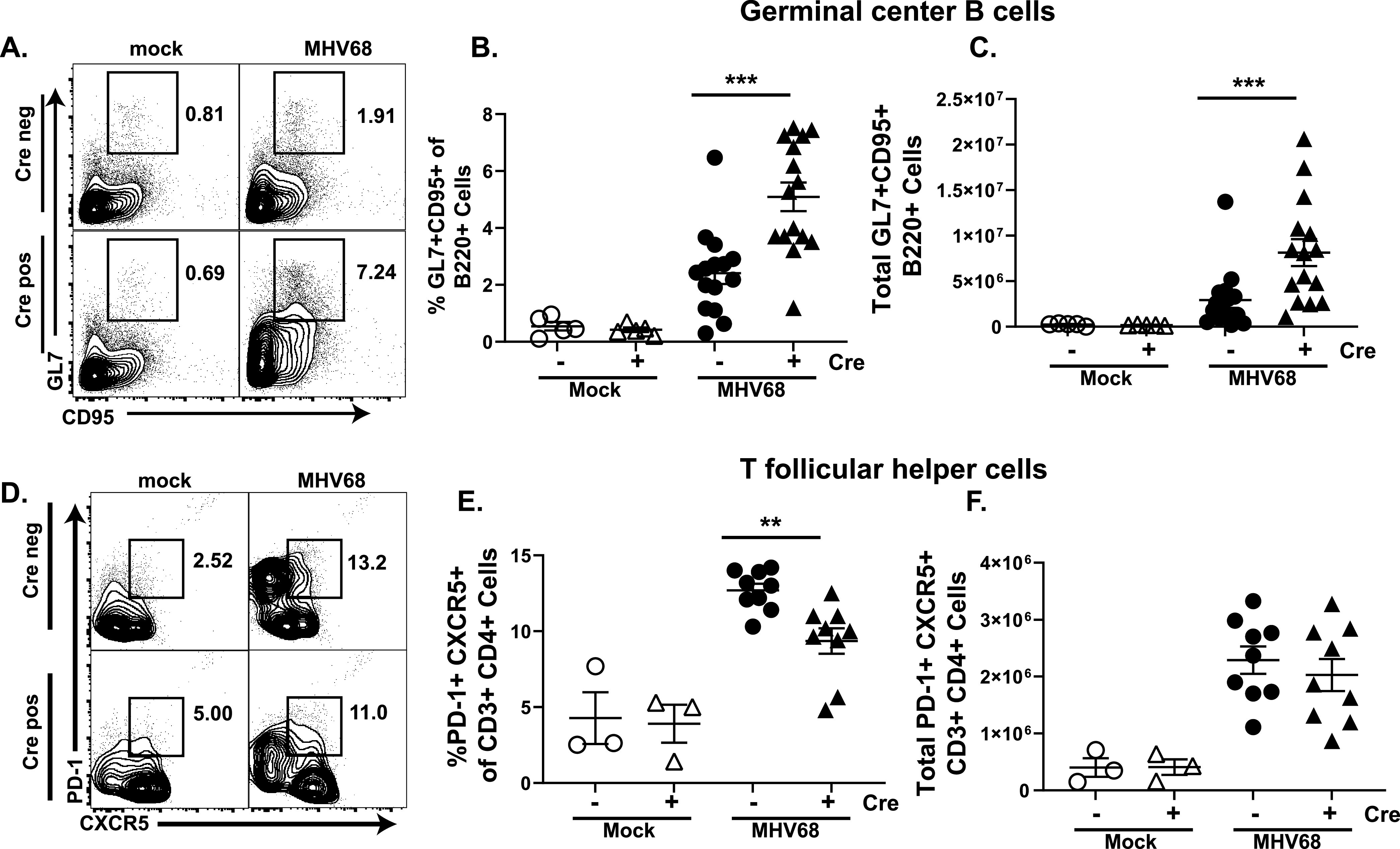
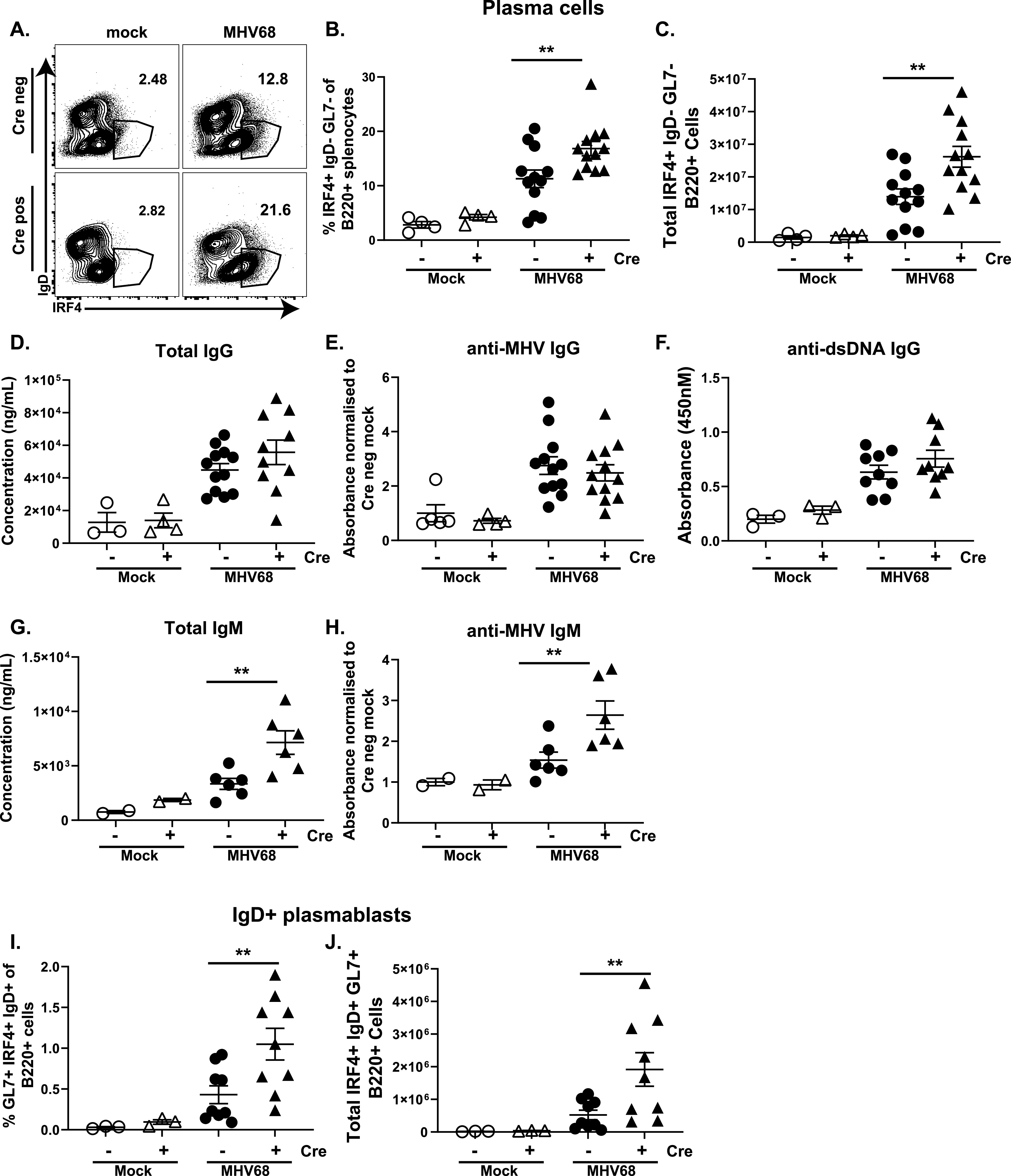
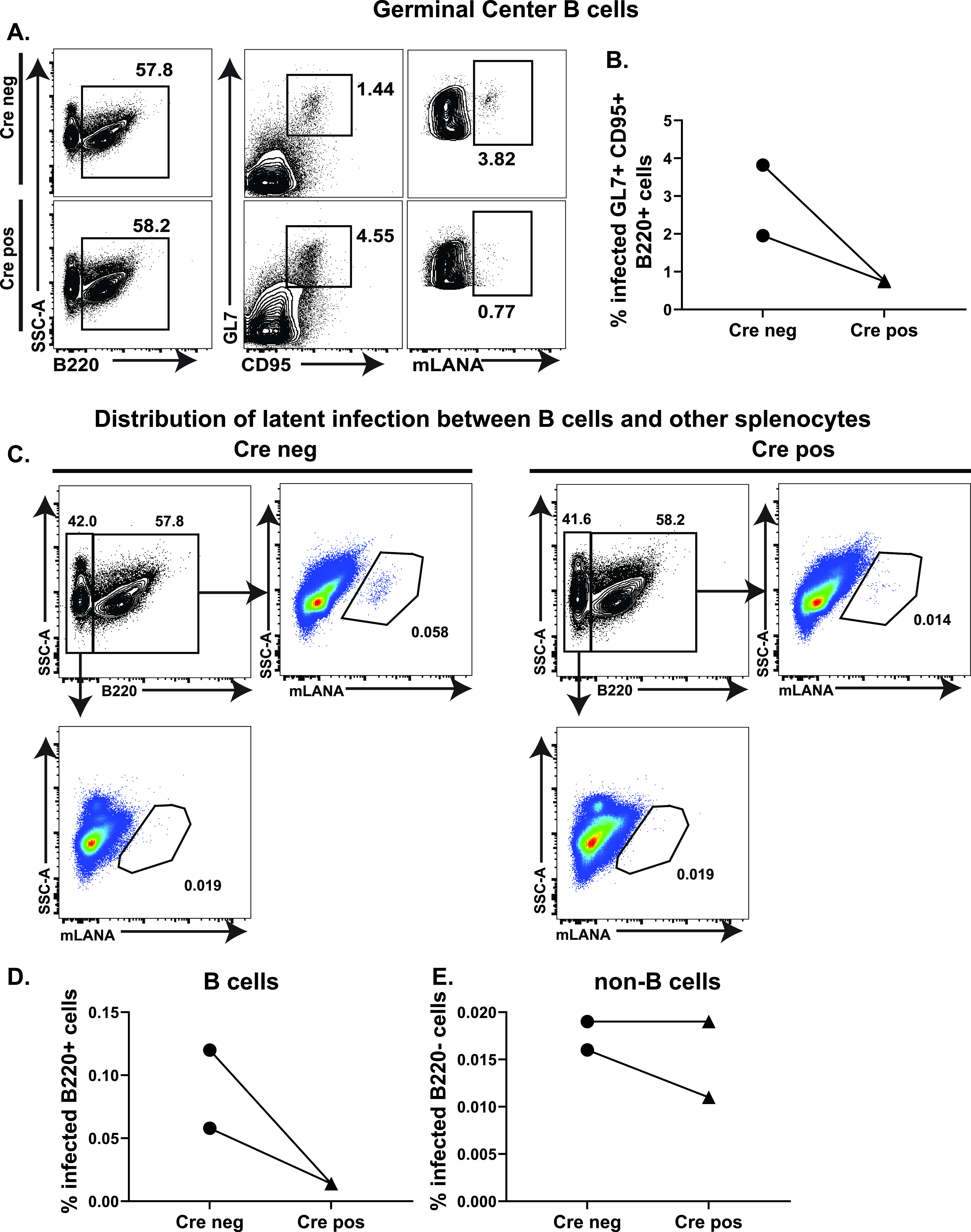
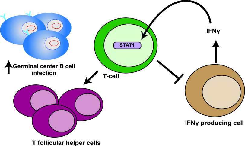
Gammaherpesviruses establish lifelong infections in most vertebrate species, including humans and rodents, and are associated with cancers, including B cell lymphomas. While type I and II interferon (IFN) systems of the host are critical for the control of acute and chronic gammaherpesvirus infection, the cell type-specific role(s) of IFN signaling during infection is poorly understood and is often masked by the profoundly altered viral pathogenesis in the hosts with global IFN deficiencies. STAT1 is a critical effector of all classical IFN responses along with its involvement in other cytokine signaling pathways. In this study, we defined the effect of T cell-specific STAT1 deficiency on the viral and host parameters of infection with murine gammaherpesvirus 68 (MHV68). MHV68 is a natural rodent pathogen that, similar to human gammaherpesviruses, manipulates and usurps B cell differentiation to establish a lifelong latent reservoir in B cells. Specifically, germinal center B cells host the majority of latent MHV68 reservoir in the lymphoid organs, particularly at the peak of viral latency. Unexpectedly, T cell-specific STAT1 expression, while limiting the overall expansion of the germinal center B cell population during chronic infection, rendered these B cells more effective at hosting the latent virus reservoir. Further, T cell-specific STAT1 expression in a wild type host limited circulating levels of IFNγ, with corresponding increases in lytic MHV68 replication and viral reactivation. Thus, our study unveils an unexpected proviral role of T cell-specific STAT1 expression during gammaherpesvirus infection of a natural intact host. IMPORTANCE Interferons (IFNs) represent a major antiviral host network vital to the control of multiple infections, including acute and chronic gammaherpesvirus infections. Ubiquitously expressed STAT1 plays a critical effector role in all classical IFN responses. This study utilized a mouse model of T cell-specific STAT1 deficiency to define cell type-intrinsic role of STAT1 during natural gammaherpesvirus infection. Unexpectedly, T cell-specific loss of STAT1 led to better control of acute and persistent gammaherpesvirus replication and decreased establishment of latent viral reservoir in B cells, revealing a surprisingly diverse proviral role of T cell-intrinsic STAT1.

Keywords: STAT1; T cell; acute infection; chronic infection; gammaherpesvirus; germinal center B cell.


Conflict of interest statement

The authors declare no conflict of interest.

Figures

FIG 1
FIG 1
T cell-specific STAT1 deficiency leads to decreased acute and persistent MHV68 replication in the lungs. (A) Splenocytes were harvested from Cre negative and Cre positive mice. Total STAT1 levels in sorted T and non-T cells in the spleen were measured via Western blot. (B, C) Cre negative and positive littermates were infected intranasally with 1000 PFU of WT MHV68. (B) Viral lytic titers in lungs at 10 days postinfection, dotted line represents limit of detection, each symbol represents an individual animal. *, P < 0.05. (C) Persistent MHV68 replication in the lungs at 16 days postinfection was determined by semi-quantitative dilution assay. Data were pooled from 9 mice/group.
FIG 2
FIG 2
T cell-specific STAT1 expression affects the latent reservoir and MHV68 reactivation in an anatomic site-specific manner. Cre negative and positive littermates were infected as in Fig. 1. Peritoneal cells (A, B) or splenocytes (C, D) were harvested at 16 days postinfection and pooled from 3–5 mice within each group. (A, C) Intact pooled cellular populations were subjected to limiting dilution assay to determine the frequencies of ex vivo reactivation. (B, D) Frequencies of MHV68 DNA positive cells in each pooled population were determined by limiting dilution PCR. Data were pooled from 4–5 independent experiments. In the limiting dilution assays of this the dotted line is drawn at 63.2% and the x-coordinate of intersection of this line with the sigmoid graph represents an inverse of frequency of positive events.
FIG 3
FIG 3
T cell-specific STAT1 deficiency leads to increased systemic gamma production. Cre negative and positive littermates were infected as in Fig. 1. Peritoneal cells were harvested at 16 days postinfection or mock treatment and subjected to flow cytometry to determine the frequencies (A, C, H) and absolute numbers (B, D, E, F) of total CD3+ T cells (A, B), CD8+ T cells (C, D), effector CD8 T cells (E), IFN gamma producing T cells (F), and proliferating CD8 T cells (H). (G) Flow cytometry was used to determine the mean fluorescence intensity of IFN gamma expression. (I) IFN gamma serum concentrations were determined by ELISA. (J, K) Gene expression of MX1 (J) and IRF-1 (K) from total peritoneal cells was determined by qRT-PCR. Each symbol represents an individual animal. *, P < 0.05.
FIG 4
FIG 4
T cell-specific STAT1 deficiency leads to polyclonal CD8 T cell activation in the spleen. Cre negative and positive littermates were infected as in Fig. 1. Splenocytes were harvested at 16 days postinfection or mock treatment and subjected to flow cytometry to determine the frequencies (D) and absolute numbers (A-C, E, F) of total splenocytes (A), T cells (B), CD8+ T cells (C), proliferating CD8 T cells (D), effector CD8 T cells (E), and MHV68-specific IFN gamma producing CD8 T cells (F). Each symbol represents an individual animal. *, P < 0.05.
FIG 5
FIG 5
T cell-specific STAT1 deficiency leads to an increased germinal center B cell but not T follicular helper cell population during chronic MHV68 infection. Cre negative and positive littermates were infected as in Fig. 1. Splenocytes were harvested at 16 days postinfection or mock treatment and flow cytometry used to determine the frequencies (A, B, D, E) and absolute numbers (C, F) of germinal center B cells defined as B220+ GL7+ CD95+ (A–C) and T follicular helper cells defined as CD3+ CD4+ PD1+ CXCR5+ (D–F).
FIG 6
FIG 6
T cell-specific STAT1 deficiency leads to an increased abundance of terminally differentiated B cells. Cre negative and positive littermates were infected as in Fig. 1. Splenocytes were harvested at 16 days postinfection or mock treatment and flow cytometry used to determine the frequencies (A, B, I) and absolute numbers (D, J) of plasma cells defined as B220+ IRF4+ IgD- GL7- (A, B, C) and IgD+ plasmablasts defined as B220+ IgD+ IRF4+ GL7+ (I, J). Serum levels of total (D, G), virus-specific (E, H), IgG (D, E), and IgM (G, H) and anti-double stranded DNA IgG (F) were determined by ELISA. Each symbol represents an individual animal. **, P < 0.01.
FIG 7
FIG 7
T cell-specific STAT1 deficiency alters MHV68 cell tropism in the spleen. Cre negative and positive littermates were infected intranasally with 1000 PFU of MHV68.orf73βla. At 16 days postinfection, splenocytes were harvested and pooled within each experimental group. The cells were then stained with cell surface markers and loaded with the fluorescent β-lactamase substrate CCF2-AM. Flow cytometry was used to determine mLANA expression as a result of the fluorescent signal from cleaved CCF2 in (A, B) germinal center B cells defined as B220+ GL7+ CD95+, (C, D) B220+ B cells, or (C, F) B220-non-B cells. Data in A and C are representative of two independent experiments.
FIG 8
FIG 8
Working model of the role of T-cell intrinsic STAT1 expression during chronic MHV68 infection. T-cell intrinsic STAT1 expression serves two distinct functions during chronic MHV68 infection. First, T cell-intrinsic STAT1 is necessary for quantitative and qualitative features of CD4 T follicular helper population that support latently infected germinal center B cells. Second, STAT1 dependent IFNγ signaling is a negative-feedback loop whereby, in response to high systemic IFNγ levels, T cells suppress the production of IFNγ by other immune cells.

Similar articles

Cited by

References

    1. Cesarman E. 2014. Gammaherpesviruses and lymphoproliferative disorders. Annu Rev Pathol 9:349–372. doi:10.1146/annurev-pathol-012513-104656. - DOI - PubMed
    1. Efstathiou S, Ho YM, Minson AC. 1990. Cloning and molecular characterization of the murine herpesvirus 68 genome. J Gen Virol 71:1355–1364. doi:10.1099/0022-1317-71-6-1355. - DOI - PubMed
    1. Efstathiou S, Ho YM, Hall S, Styles CJ, Scott SD, Gompels UA. 1990. Murine herpesvirus 68 is genetically related to the gammaherpesviruses Epstein-Barr virus and herpesvirus saimiri. J Gen Virol 71:1365–1372. doi:10.1099/0022-1317-71-6-1365. - DOI - PubMed
    1. Virgin HW, Latreille P, Wamsley P, Hallsworth K, Weck KE, Dal Canto AJ, Speck SH. 1997. Complete sequence and genomic analysis of murine gammaherpesvirus 68. J Virol 71:5894–5904. doi:10.1128/JVI.71.8.5894-5904.1997. - DOI - PMC - PubMed
    1. Weck KE, Kim SS, Virgin HW, Speck SH. 1999. Macrophages are the major reservoir of latent murine gammaherpesvirus 68 in peritoneal cells. J Virol 73:3273–3283. doi:10.1128/JVI.73.4.3273-3283.1999. - DOI - PMC - PubMed

Publication types

LinkOut - more resources