. 2022 Sep 20;66(9):e0017122.
doi: 10.1128/aac.00171-22. Epub 2022 Aug 15.

Lysocin E Targeting Menaquinone in the Membrane of Mycobacterium tuberculosis Is a Promising Lead Compound for Antituberculosis Drugs

Affiliations

Lysocin E Targeting Menaquinone in the Membrane of Mycobacterium tuberculosis Is a Promising Lead Compound for Antituberculosis Drugs

Geberemichal Geberetsadik et al. Antimicrob Agents Chemother. .

Abstract

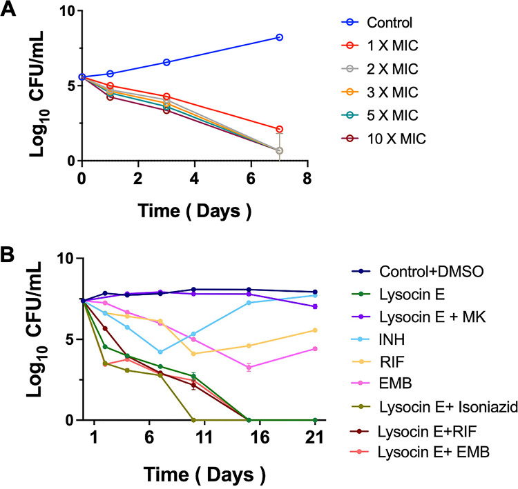
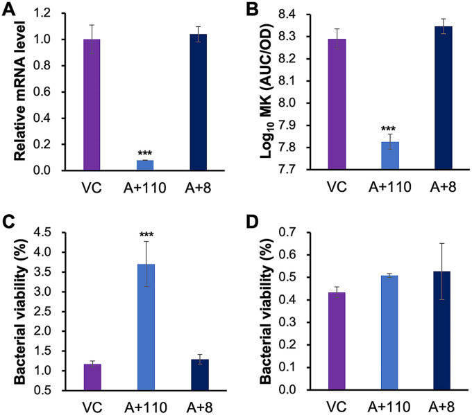
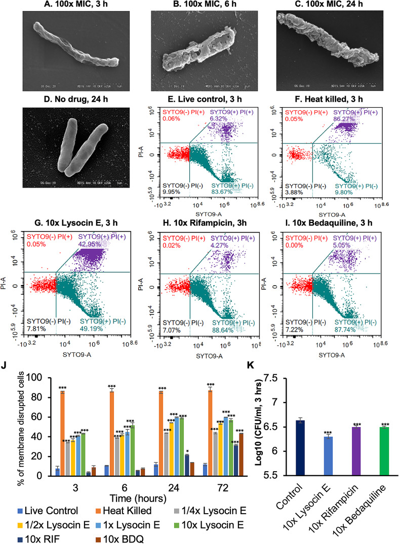
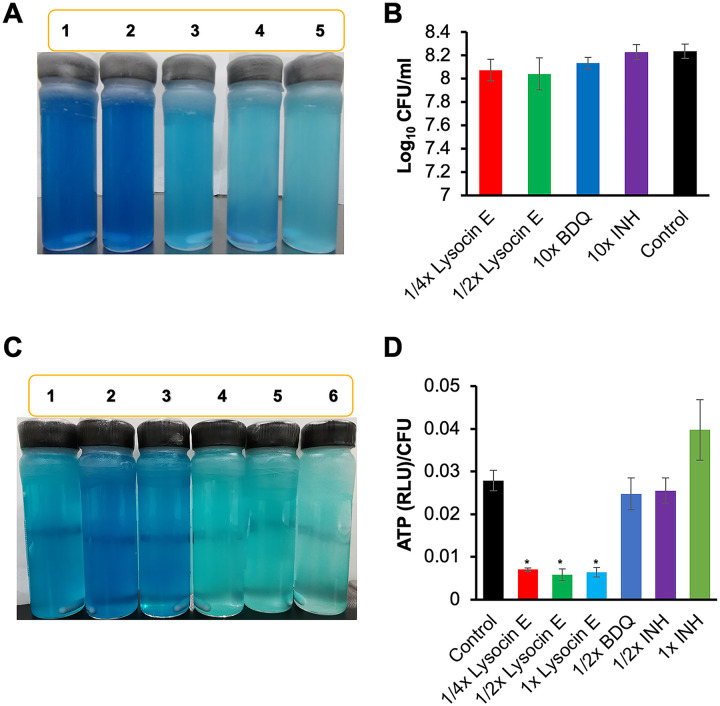
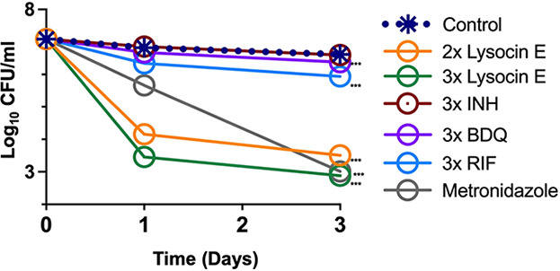
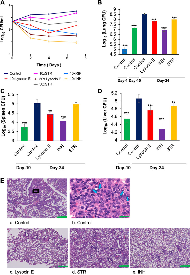
Tuberculosis remains a public health crisis and a health security threat. There is an urgent need to develop new antituberculosis drugs with novel modes of action to cure drug-resistant tuberculosis and shorten the chemotherapy period by sterilizing tissues infected with dormant bacteria. Lysocin E is an antibiotic that showed antibacterial activity against Staphylococcus aureus by binding to its menaquinone (commonly known as vitamin K2). Unlike S. aureus, menaquinone is essential in both growing and dormant Mycobacterium tuberculosis. This study aims to evaluate the antituberculosis activities of lysocin E and decipher its mode of action. We show that lysocin E has high in vitro activity against both drug-susceptible and drug-resistant Mycobacterium tuberculosis var. tuberculosis and dormant mycobacteria. Lysocin E is likely bound to menaquinone, causing M. tuberculosis membrane disruption, inhibition of oxygen consumption, and ATP synthesis. Thus, we have concluded that the high antituberculosis activity of lysocin E is attributable to its synergistic effects of membrane disruption and respiratory inhibition. The efficacy of lysocin E against intracellular M. tuberculosis in macrophages was lower than its potent activity against M. tuberculosis in culture medium, probably due to its low ability to penetrate cells, but its efficacy in mice was still superior to that of streptomycin. Our findings indicate that lysocin E is a promising lead compound for the development of a new tuberculosis drug that cures drug-resistant and latent tuberculosis in a shorter period.

Keywords: Mycobacterium tuberculosis; antituberculosis; lysocin E; tuberculosis.


Conflict of interest statement

The authors declare no conflict of interest.

Figures

FIG 1
FIG 1
Efficacy of lysocin E against mycobacteria in vitro. (A) MBC of lysocin E. M. tuberculosis harvested at exponential phase was adjusted to an OD600 of 0.001 and then exposed to different concentrations of lysocin E for 7 days at 37°C. The CFU was counted at the indicated time points. (B) Twenty-one-day kill kinetics of lysocin E. M. tuberculosis harvested at mid-log phase and adjusted to an OD600 of 0.1 was incubated with 10× MIC of lysocin E, INH, RIF, and EMB. At the indicated time points, CFU with a detection limit of 2 log10/mL were counted. Data represent the mean ± SD of triplicates. These results are representative of three independent experiments.
FIG 2
FIG 2
Effect of MK biosynthesis CKD on the susceptibility of M. tuberculosis to lysocin E. The menA-CKD strains, constructed using a TetR-regulated CRISPR interference system optimized for mycobacteria, were cultured in the presence of aTC, which was supplied at a final concentration of 200 ng/mL every 2 days. (A) Four weeks later, the menA mRNA level was quantified by real-time PCR. VC, vector control (wild-type strain with empty vector); A+8 and A+110 strains, M. tuberculosis H37Rv menA CKD strains constructed using a TetR-regulated CRISPR interference system targeting two different nontemplate strands of menA (G8GAGACCCACTGTGCGAAAC27 and T110GTGGTGGAAAGCGCTGTTG129, respectively). (B) Four weeks after achieving menA CKD, the MK amount (AUC) was quantified and normalized to bacterial OD600. (C and D) Bacterial susceptibility to lysocin E and INH was also examined 4 weeks after menA CKD. To examine susceptibility, the bacteria (OD600 of 0.1) were incubated at 37°C in the presence or absence of 50× MIC of lysocin E or 50× MIC of INH. After incubating the bacteria with the drugs for 24 h, bacterial CFU was determined in lysocin E-treated bacteria (C) or INH-treated bacteria (D). The bacterial viability was calculated by comparison of the CFU counts of treated samples with those of their corresponding nontreated samples (same recombinant bacteria). Statistical differences were analyzed by ANOVA with Dunnett’s multiple-comparison test. ***, P < 0.0001. Data represent the mean ± SD of triplicates. This experiment was repeated two times, and reproducible data were observed.
FIG 3
FIG 3
Lysocin E causes mycobacterial membrane disruption. (A to D) SEM observation of M. tuberculosis exposed to 100× MIC of lysocin E for 3 h (A), 6 h (B), or 24 h (C) and untreated M. tuberculosis at 24 h (D). All micrographs are at ×35,000 magnification. (E to I) After treating BCG (OD600 of 0.05) with 1/4×, 1/2×, 1×, and 10× MIC of lysocin E and 1/2×, 1×, and 10× MIC of RIF and BDQ, the effect of drugs on membrane permeability was evaluated by FACS analysis using Live/Dead (Syto9/PI) staining at 3, 6, 24, and 72 h. Representative flow cytometric dot plots of the live control (E), the heat-killed control (F), and samples treated for 3 h with 10× MIC lysocin E (G), 10× MIC RIF (H), and 10× MIC BDQ (I) are shown. (J) The percentages of membrane-disrupted (PI-positive) cells were calculated and summarized. RFP, RIF. (K) In parallel, the activity of drugs was evaluated by CFU count after treating BCG with 10× MIC of lysocin E, RIF, and BDQ for 3 h. Statistical differences were analyzed by one-way ANOVA with Dunnett’s multiple-comparison test. *, P < 0.05; ***, P < 0.0001. Data represent the mean ± SD of triplicates, and each picture is representative of two reproducible biological experiments.
FIG 4
FIG 4
Effect of lysocin E on mycobacterial energy production. (A and B) Oxygen consumption in M. tuberculosis H37Rv (OD600 of 0.5) was evaluated using 0.001% methylene blue as an oxygen sensor. BDQ, a drug that targets oxidative phosphorylation, and INH, a drug that targets cell wall synthesis but not cell membranes, were used as positive controls. (A) Tube 1, M. tuberculosis treated with 1/4× MIC (0.125 μg/mL) of lysocin E; tube 2, M. tuberculosis treated with 1/2× MIC (0.25 μg/mL) of lysocin E; tube 3, M. tuberculosis treated with 10× MIC (2.5 μg/mL) of BDQ; tube 4, M. tuberculosis treated with 10× MIC (0.31 μg/mL) of INH; tube 5, control without drug. Cultures were incubated at 37°C for 24 h. (B) CFU of the cultures was also counted. (C) A similar assay was performed using M. smegmatis MC2155 cultures. Tube 1, M. smegmatis (OD600 of 0.5) treated with 1/8× MIC (1.25 μg/mL) of lysocin E; tube 2, M. smegmatis treated with 1/4× MIC (2.5 μg/mL) of lysocin E; tube 3, M. smegmatis treated with 1/2× MIC (5 μg/mL) of lysocin E; tube 4, M. smegmatis treated with 10× MIC (160 μg/mL) of INH; tube 5, M. smegmatis treated with 10× MIC (0.625 μg/mL) of BDQ; tube 6, control without drug. Cultures were incubated at 37°C for 2 h. (D) Effect of the drug treatment on ATP synthesis. M. tuberculosis was treated with the indicated concentrations of lysocin E, INH and BDQ. After 24 h of incubation, the intracellular ATP was quantified using the BacTiter-Glo microbial cell viability assay kit (Promega). The RLU were normalized by CFU. The statistical differences between the control and each treatment were analyzed by one-way ANOVA with Dunnett’s multiple-comparison test. *, P < 0.01. All experiments were repeated two times, and reproducible data were observed.
FIG 5
FIG 5
Bactericidal activity of lysocin E against NRP mycobacteria. The bactericidal activity against NPR bacteria was determined by exposing M. smegmatis subjected to growth in the Wayne hypoxia model to the indicated antibiotics. Statistical differences between the control and each treatment were analyzed by one-way ANOVA with Dunnett’s multiple-comparison test. ***, P < 0.0001. Data represent the mean ± SD of triplicates. This experiment was repeated two times, and reproducible data were observed.
FIG 6
FIG 6
Efficacy of lysocin E against M. tuberculosis in macrophages and mice. (A) Efficacy of lysocin E against intracellular M. tuberculosis. The THP-1 cells were infected with M. tuberculosis (multiplicity of infection [MOI], 1:1) for 4 h. Subsequently, the cultures were washed twice with warm serum-free DMEM. The infected macrophages were treated by adding 100 μL DMEM containing 10× or/and 50× MIC of lysocin E, INH, STR, and RIF (RFP). The cells were incubated at 37°C in a humidified 5% CO2 incubator. CFU on day 0, day 2, day 4, and day 7 were determined. (B to D) Female C57BL/6J mouse (17 to 19 g) infection was initiated by intratracheal instillation of M. tuberculosis H37Rv. For this purpose, M. tuberculosis was grown to an OD600 of 0.5. The inoculum (5.5 × 105 CFU per mouse, suspended in 50 μL of PBS) was delivered to the lung through forced inhalation with a syringe at day 0. Six of the infected mice were then randomly assigned to one of the indicated groups to evaluate the bactericidal activities of antibiotics. Five mice from each group were sacrificed on the day after infection, on the day of treatment initiation, and after 14 days of treatment to determine the number of CFU implanted in the lungs, the pretreatment baseline CFU counts, and the CFU counts after treatment, respectively. A mouse was also sacrificed from each group for histopathological study after treatment. Lung CFU (B), spleen CFU (C), and liver CFU (D) were counted. Data represent the mean ± SD of triplicates. Statistical differences were analyzed by one-way ANOVA with Dunnett’s multiple-comparison test. **, P < 0.005; ***, P < 0.001. (E) Lung tissue images after treatment. Lung tissue sections of untreated control (a and b), lysocin E-treated (c), STR-treated (d), and INH-treated (e) mice were stained with hematoxylin and eosin (a, c, and d, scale bars, 500 μm; b, scale bar, 50 μm). Image b is an example of the magnified image (the area indicated as a black box in image a) used for mononuclear cell counting. Arrows in image b indicate the examples of mononuclear cells counted. Counted data of untreated and treated groups are shown and compared in Fig. S4F in the supplemental material.

Similar articles

Cited by

References

    1. World Health Organization. 2021. Global tuberculosis report 2021. World Health Organization, Geneva, Switzerland. https://www.who.int/publications/i/item/9789240037021.
    1. Sukheja P, Kumar P, Mittal N, Li S, Singleton E, Russo R, Perryman AL, Shrestha R, Awasthi D, Husain S, Soteropoulos P, Brukh R, Connell N, Freundlich JS, Alland DA. 2017. A novel small-molecule inhibitor of the Mycobacterium tuberculosis demethylmenaquinone methyltransferase MenG is bactericidal to both growing and nutritionally deprived persister cells. mBio 8:e02022-16. 10.1128/mBio.02022-16. - DOI - PMC - PubMed
    1. Machado D, Girardini M, Viveiros M, Pieroni M. 2018. Challenging the drug-likeness dogma for new drug discovery in tuberculosis. Front Microbiol 9:1367. 10.3389/fmicb.2018.01367. - DOI - PMC - PubMed
    1. World Health Organization. 2020. Global tuberculosis report 2020. World Health Organization, Geneva, Switzerland. https://www.who.int/publications/i/item/9789240013131.
    1. Berube BJ, Russell D, Castro L, Choi S, Narayanasamy P, Parish T. 2019. Novel MenA inhibitors are bactericidal against Mycobacterium tuberculosis and synergize with electron transport chain inhibitors. Antimicrob Agents Chemother 63:e02661-18. 10.1128/AAC.02661-18. - DOI - PMC - PubMed

Publication types

MeSH terms

LinkOut - more resources