. 2022 Sep 14;96(17):e0102822.
doi: 10.1128/jvi.01028-22. Epub 2022 Aug 15.

IFI16 Partners with KAP1 to Maintain Epstein-Barr Virus Latency

Affiliations

IFI16 Partners with KAP1 to Maintain Epstein-Barr Virus Latency

Huanzhou Xu et al. J Virol. .

Abstract

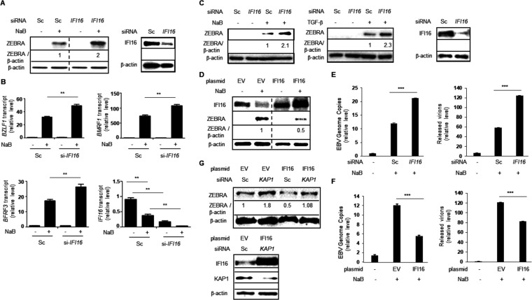
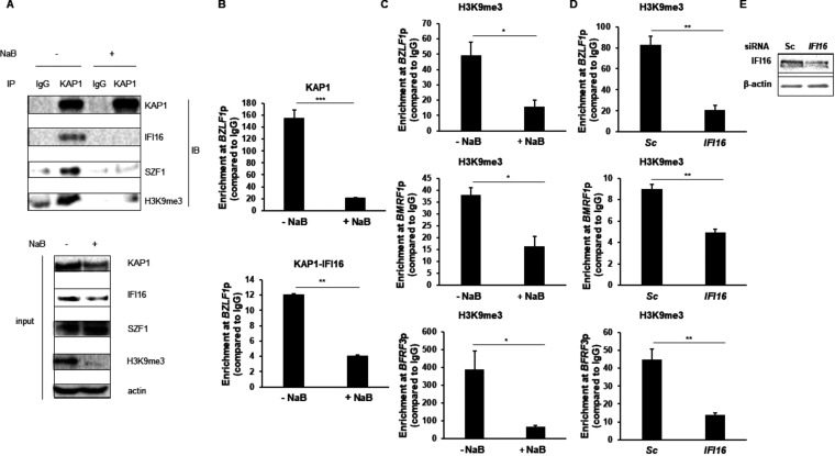
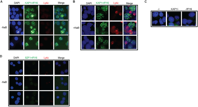
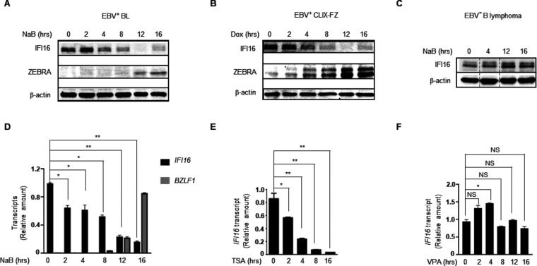
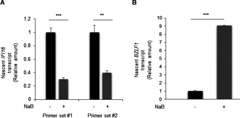
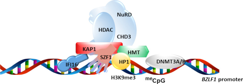
Herpesviruses establish latency to ensure permanent residence in their hosts. Upon entry into a cell, these viruses are rapidly silenced by the host, thereby limiting the destructive viral lytic phase while allowing the virus to hide from the immune system. Notably, although the establishment of latency by the oncogenic herpesvirus Epstein-Barr virus (EBV) requires the expression of viral latency genes, latency can be maintained with a negligible expression of viral genes. Indeed, in several herpesviruses, the host DNA sensor IFI16 facilitated latency via H3K9me3 heterochromatinization. This silencing mark is typically imposed by the constitutive heterochromatin machinery (HCM). The HCM, in an antiviral role, also silences the lytic phase of EBV and other herpes viruses. We investigated if IFI16 restricted EBV lytic activation by partnering with the HCM and found that IFI16 interacted with core components of the HCM, including the KRAB-associated protein 1 (KAP1) and the site-specific DNA binding KRAB-ZFP SZF1. This partnership silenced the EBV lytic switch protein ZEBRA, encoded by the BZLF1 gene, thereby favoring viral latency. Indeed, IFI16 contributed to H3K9 trimethylation at lytic genes of all kinetic classes. In defining topology, we found that IFI16 coenriched with KAP1 at the BZLF1 promoter, and while IFI16 and SZF1 were each adjacent to KAP1 in latent cells, IFI16 and SZF1 were not. Importantly, we also found that disruption of latency involved rapid downregulation of IFI16 transcription. These findings revealed a previously unknown partnership between IFI16 and the core HCM that supports EBV latency via antiviral heterochromatic silencing. IMPORTANCE The interferon-gamma inducible protein 16 (IFI16) is a nuclear DNA sensor that mediates antiviral responses by activating the inflammasome, triggering an interferon response, and silencing lytic genes of herpesviruses. The last, which helps maintain latency of the oncoherpesvirus Epstein-Barr virus (EBV), is accomplished via H3K9me3 heterochromatinization through unknown mechanisms. Here, we report that IFI16 physically partners with the core constitutive heterochromatin machinery to silence the key EBV lytic switch protein, thereby ensuring continued viral latency in B lymphocytes. We also find that disruption of latency involves rapid transcriptional downregulation of IFI16. These findings point to hitherto unknown physical and functional partnerships between a well-known antiviral mechanism and the core components of the constitutive heterochromatin machinery.

Keywords: Epstein-Barr virus; H3K9me3; IFI16; KAP1; TRIM28; ZEBRA; ZTA; herpesvirus; heterochromatin machinery; latency.


Conflict of interest statement

The authors declare no conflict of interest.

Figures

FIG 1
FIG 1
IFI16 partners with KAP1 to silence the EBV lytic state. (A and B) HH514-16 BL cells were transfected with control scrambled siRNA (Sc) or siRNA targeting IFI16, exposed to NaB (sodium butyrate; lytic trigger) after 18 h, harvested after another 24 h, and immunoblotted with indicated antibodies (A) or subjected to RT-qPCR analysis of EBV lytic genes of all kinetic classes (BZLF1, immediate early; BMRF1, early; and BFRF3, late) using the ΔΔCT method after normalization to 18S rRNA (B). Error bars, SEM of three technical replicates and two biological replicates; **, P < 0.01. (C) Mutu I BL cells were transfected with control scrambled siRNA (Sc) or siRNA targeting IFI16, exposed to NaB (left) or TGF-β1 (middle) after 18 h, harvested after another 24 h, and immunoblotted with indicated antibodies. (D) HH514-16 BL cells were transfected with empty vector (EV) or IFI16 plasmid, exposed to NaB after 18 h, and harvested after another 24 h before performing immunoblotting with indicated antibodies. (E and F) HH514-16 BL cells were transfected with siRNA targeting IFI16 (versus scrambled RNA as control) (E) or IFI16 plasmid (versus empty vector [EV] as control) (F), exposed to NaB after 20 h, and harvested after another 36 h (F, left) or 48 h (E and F, right) for qPCR analysis of cell-associated viral genomes (left) or released viral genomes following DNase treatment (right). (G) HH514-16 BL cells were transfected with empty vector (EV), IFI16 plasmid, control scrambled siRNA, or siRNA targeting KAP1, exposed to NaB after 18 h, and harvested after another 24 h before immunoblotting with indicated antibodies. Numbers indicate the relative amounts of ZEBRA protein after normalization to β-actin levels. These experiments were performed at least twice.
FIG 2
FIG 2
KAP1 and IFI16 coenriched with H3K9me3 and at the BZLF1 promoter, preferentially in latent cells. (A) HH514-16 BL cells were left untreated or exposed to NaB. After 24 h, lysates were immunoprecipitated (IP) with anti-KAP1 antibody versus control IgG and immunoblotted with indicated antibodies. Input represents 5% of the sample. (B) HH514-16 BL cells were treated with or without NaB for 24 h followed by ChIP-re-ChIP. Chromatin was precipitated using rabbit anti-KAP1 or a control rabbit IgG antibody (top) and subjected to the second round of ChIP using mouse anti-IFI16 or a control mouse IgG antibody (bottom). Extracted DNA was analyzed by qPCR using primers spanning the known KAP1 enrichment site on the BZLF1 promoter and normalized to input. (C) HH514-16 BL cells were treated with or without NaB for 6 h followed by ChIP using an anti-H3K9me3 antibody or control IgG. (D) HH514-16 BL cells were transfected with control scrambled siRNA (Sc) or siRNA targeting IFI16 for 18 h (and harvested for immunoblotting in E) or exposed to NaB, harvested after 12 and 24 h, and chromatin was immunoprecipitated with an anti-H3K9me3 antibody or control IgG. Primer sets targeting BZLF1, BMRF1, and BFRF3 promoters were used for qPCR in C and D. Error bars, SEM of the means of three independent experiments; *, P < 0.05; **, P < 0.01; ***, P < 0.001.
FIG 3
FIG 3
IFI16 was adjacent to KAP1 but not SZF1 in latent cells. HH514-16 BL cells were not induced or induced with NaB for 24 h before performing a proximity ligation assay (PLA) using antibodies targeting KAP1, IFI16, and SZF1. EBV seropositive human reference serum followed by APC-conjugated goat anti-human IgG was used to stain lytic cells (A, B, and D). Negative-control staining in the absence of anti-KAP1, anti-IFI16, or both antibodies is shown in (C). Green foci, in situ interactions between KAP1 and IFI16. The experiment was performed at least 3 times. Three representative fields from two independent experiments are shown in A, B, and D.
FIG 4
FIG 4
IFI16 message and protein abundance rapidly decrease with lytic cycle activation in EBV+ BL cells. (A and B) HH514-16 (EBV+ BL; A), CLIX-FZ (doxycycline-inducible EBV+ BL; B), and EBV B lymphoma (BJAB; C) cells were exposed to lytic triggers (NaB in A and C; doxycycline in B) for different durations followed by immunoblotting with indicated antibodies. (D to F) HH514-16 cells were induced with NaB (D), TSA (E), or VPA as mock induction control (F) for different durations followed by RT-qPCR analysis to determine the relative amounts of IFI16 message after normalization to 18S rRNA using the ΔΔCT method. Error bars, SEM of three technical replicates and two biological replicates; *, P < 0.05; **, P < 0.01; NS, not significant.
FIG 5
FIG 5
Reduced transcription contributes to a drop in abundance of IFI16 messages after lytic cycle activation. HH514-16 BL cells were left untreated or induced with NaB for 10 h, before performing BrU-PCR. Nascent RNA was precipitated with an anti-BrdU antibody followed by RT-qPCR analysis with two primer sets (number 1 and number 2) to determine the relative amounts of newly synthesized IFI16 message (A) and BZLF1 message (B) after normalization to 18S rRNA. Error bars, SEM of three technical replicates and two biological replicates; **, P < 0.01; ***, P < 0.001; NS, not significant.
FIG 6
FIG 6
Model of IFI16’s partnership with the constitutive heterochromatin machinery (HCM) that blocked EBV latent-to-lytic transition. N-terminal KRAB domain of the KRAB-ZFP SZF1 interacted with the corepressor KAP1 while C-terminal zinc fingers bind viral DNA. KAP1 recruited histone-lysine methyltransferases (HMT), the deacetylase complex NuRD-HDAC, and the heterochromatin amplification factor HP1 to silence target genes via H3K9me3 marks. Shown also is the proposed involvement of the DNA sensor IFI16 which directly contacts KAP1 but not SZF1.

Similar articles

Cited by

References

    1. Gariano GR, Dell'Oste V, Bronzini M, Gatti D, Luganini A, De Andrea M, Gribaudo G, Gariglio M, Landolfo S. 2012. The intracellular DNA sensor IFI16 gene acts as restriction factor for human cytomegalovirus replication. PLoS Pathog 8:e1002498. 10.1371/journal.ppat.1002498. - DOI - PMC - PubMed
    1. Lo Cigno I, De Andrea M, Borgogna C, Albertini S, Landini MM, Peretti A, Johnson KE, Chandran B, Landolfo S, Gariglio M. 2015. The nuclear DNA sensor IFI16 acts as a restriction factor for human papillomavirus replication through epigenetic modifications of the viral promoters. J Virol 89:7506–7520. 10.1128/JVI.00013-15. - DOI - PMC - PubMed
    1. Johnson KE, Bottero V, Flaherty S, Dutta S, Singh VV, Chandran B. 2014. IFI16 restricts HSV-1 replication by accumulating on the hsv-1 genome, repressing HSV-1 gene expression, and directly or indirectly modulating histone modifications. PLoS Pathog 10:e1004503. 10.1371/journal.ppat.1004503. - DOI - PMC - PubMed
    1. Abe T, Marutani Y, Shoji I. 2019. Cytosolic DNA-sensing immune response and viral infection. Microbiol Immunol 63:51–64. 10.1111/1348-0421.12669. - DOI - PMC - PubMed
    1. Diner BA, Lum KK, Cristea IM. 2015. The emerging role of nuclear viral DNA sensors. J Biol Chem 290:26412–26421. 10.1074/jbc.R115.652289. - DOI - PMC - PubMed

Publication types

MeSH terms