

Intestinal Epithelial Regeneration in Response to Ionizing Irradiation
- PMID: 35969101
- PMCID: PMC9631267
- DOI: 10.3791/64028
Intestinal Epithelial Regeneration in Response to Ionizing Irradiation
Abstract
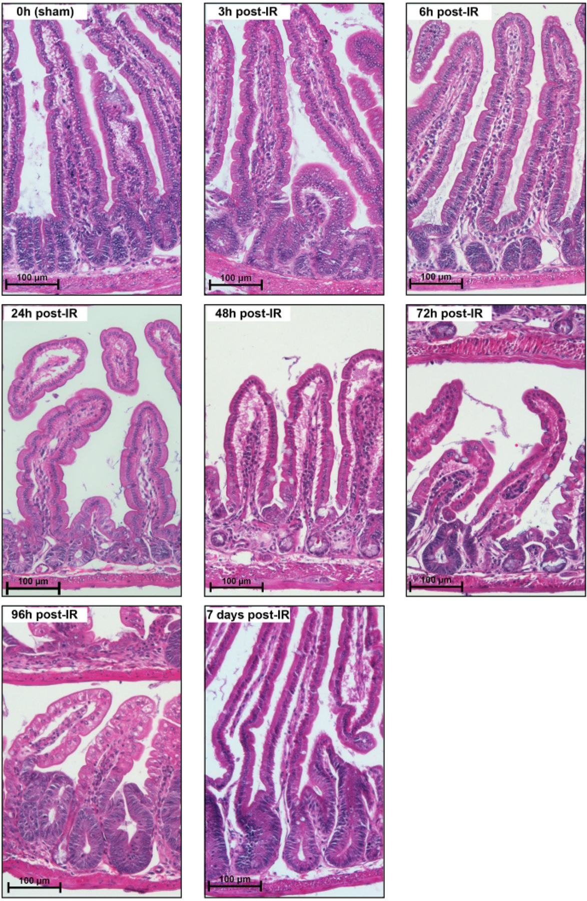
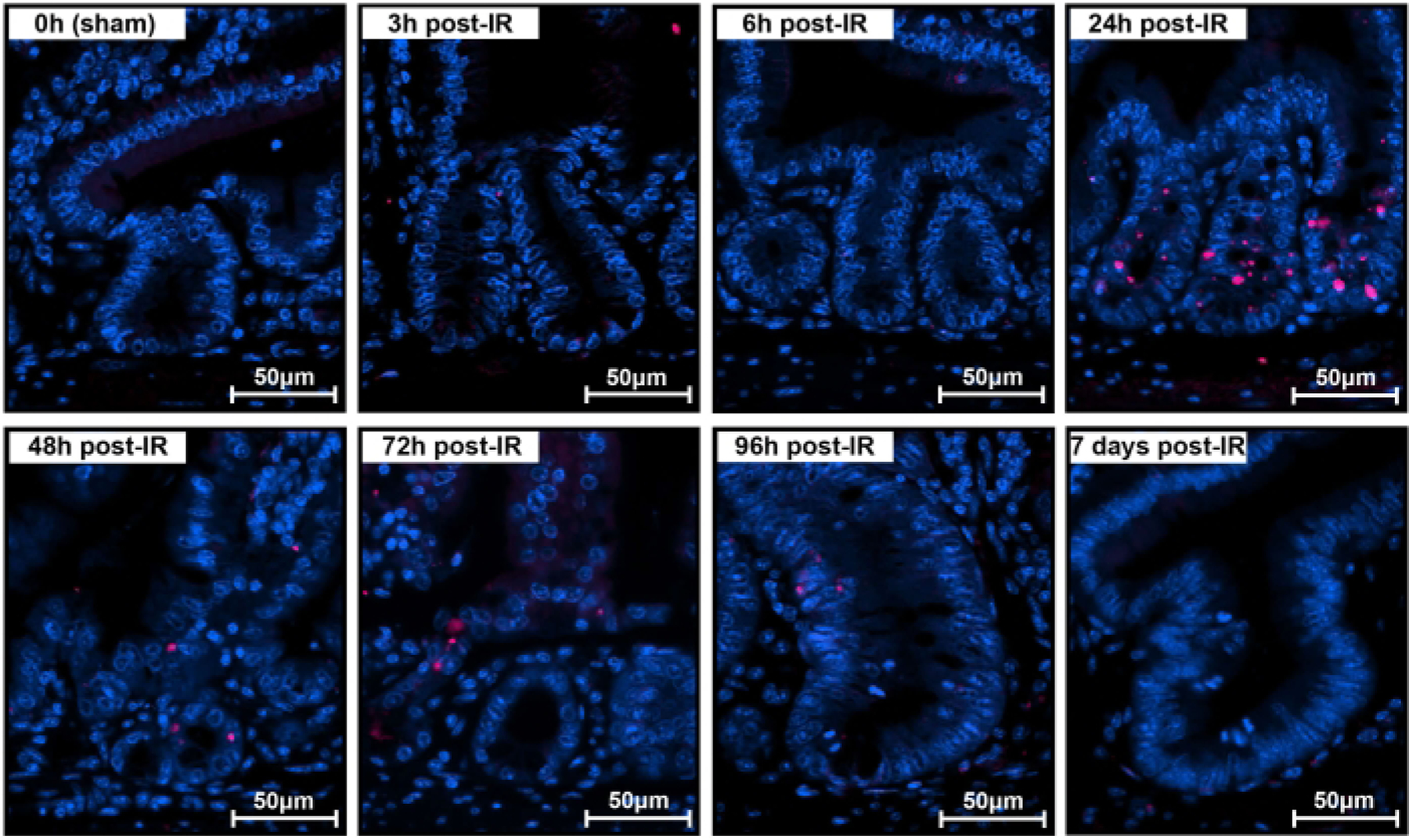
The intestinal epithelium consists of a single layer of cells yet contains multiple types of terminally differentiated cells, which are generated by the active proliferation of intestinal stem cells located at the bottom of intestinal crypts. However, during events of acute intestinal injury, these active intestinal stem cells undergo cell death. Gamma irradiation is a widely used colorectal cancer treatment, which, while therapeutically efficacious, has the side effect of depleting the active stem cell pool. Indeed, patients frequently experience gastrointestinal radiation syndrome while undergoing radiotherapy, in part due to active stem cell depletion. The loss of active intestinal stem cells in intestinal crypts activates a pool of typically quiescent reserve intestinal stem cells and induces dedifferentiation of secretory and enterocyte precursor cells. If not for these cells, the intestinal epithelium would lack the ability to recover from radiotherapy and other such major tissue insults. New advances in lineage-tracing technologies allow tracking of the activation, differentiation, and migration of cells during regeneration and have been successfully employed for studying this in the gut. This study aims to depict a method for the analysis of cells within the mouse intestinal epithelium following radiation injury.
Conflict of interest statement
Disclosures
The authors have no conflicts of interest.
Figures

Similar articles
-
Nutrient sensing by absorptive and secretory progenies of small intestinal stem cells.Am J Physiol Gastrointest Liver Physiol. 2017 Jun 1;312(6):G592-G605. doi: 10.1152/ajpgi.00416.2016. Epub 2017 Mar 23. Am J Physiol Gastrointest Liver Physiol. 2017. PMID: 28336548 Free PMC article.
-
BHLHA15-Positive Secretory Precursor Cells Can Give Rise to Tumors in Intestine and Colon in Mice.Gastroenterology. 2019 Mar;156(4):1066-1081.e16. doi: 10.1053/j.gastro.2018.11.024. Epub 2018 Nov 15. Gastroenterology. 2019. PMID: 30448068 Free PMC article.
-
SOX9 maintains reserve stem cells and preserves radioresistance in mouse small intestine.Gastroenterology. 2015 Nov;149(6):1553-1563.e10. doi: 10.1053/j.gastro.2015.07.004. Epub 2015 Jul 11. Gastroenterology. 2015. PMID: 26170137 Free PMC article.
-
Injury-associated reacquiring of intestinal stem cell function.World J Gastroenterol. 2015 Feb 21;21(7):2005-10. doi: 10.3748/wjg.v21.i7.2005. World J Gastroenterol. 2015. PMID: 25717233 Free PMC article. Review.
-
Plasticity of Intestinal Epithelium: Stem Cell Niches and Regulatory Signals.Int J Mol Sci. 2020 Dec 31;22(1):357. doi: 10.3390/ijms22010357. Int J Mol Sci. 2020. PMID: 33396437 Free PMC article. Review.
Cited by
-
MIIST305 mitigates gastrointestinal acute radiation syndrome injury and ameliorates radiation-induced gut microbiome dysbiosis.bioRxiv [Preprint]. 2024 Oct 22:2024.10.22.619652. doi: 10.1101/2024.10.22.619652. bioRxiv. 2024. Update in: Gut Microbes. 2025 Dec;17(1):2458189. doi: 10.1080/19490976.2025.2458189. PMID: 39484519 Free PMC article. Updated. Preprint.
-
MIIST305 mitigates gastrointestinal acute radiation syndrome injury and ameliorates radiation-induced gut microbiome dysbiosis.Gut Microbes. 2025 Dec;17(1):2458189. doi: 10.1080/19490976.2025.2458189. Epub 2025 Feb 10. Gut Microbes. 2025. PMID: 39930324 Free PMC article.
References
-
- Helander HF, Fandriks L Surface area of the digestive tract - Revisited. Scandinavian Journal of Gastroenterology. 49 (6), 681–689 (2014). - PubMed
-
- van der Flier LG, Clevers H Stem cells, self-renewal, and differentiation in the intestinal epithelium. Annual Review of Physiology. 71, 241–260 (2009). - PubMed
-
- Clevers H The intestinal crypt, a prototype stem cell compartment. Cell. 154 (2), 274–284 (2013). - PubMed
-
- Barker N et al. Identification of stem cells in small intestine and colon by marker gene Lgr5. Nature. 449 (7165), 1003–1007 (2007). - PubMed
Publication types
MeSH terms
Grants and funding
LinkOut - more resources
Full Text Sources
Medical
