. 2023 May;21(3):257-267.
doi: 10.2450/2022.006-22. Epub 2022 Aug 9.

The effect of exosomes released from apheresis platelet concentrates under the impact of gamma irradiation and storage time upon platelet aggregation and hemostasis

Affiliations

The effect of exosomes released from apheresis platelet concentrates under the impact of gamma irradiation and storage time upon platelet aggregation and hemostasis

Salih Haldun Bal et al. Blood Transfus. 2023 May.

Abstract

Background: Blood components should be gamma-irradiated (γ-IR) in order to prevent transfusion-associated graft-versus-host disease. The aim of this study is to determine the effect of γ-IR and storage time on the exosomes released from apheresis platelet concentrates (aPC) and to investigate their impact on the maximum platelet aggregation (MPA) and hemostasis.

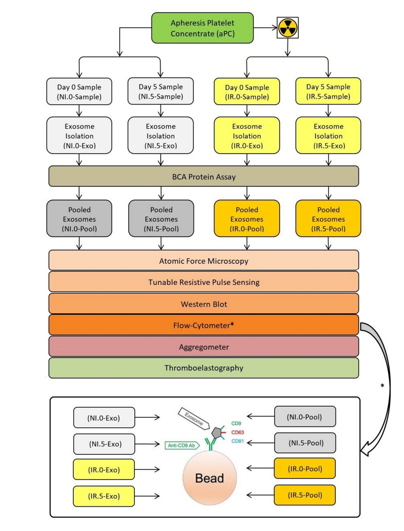
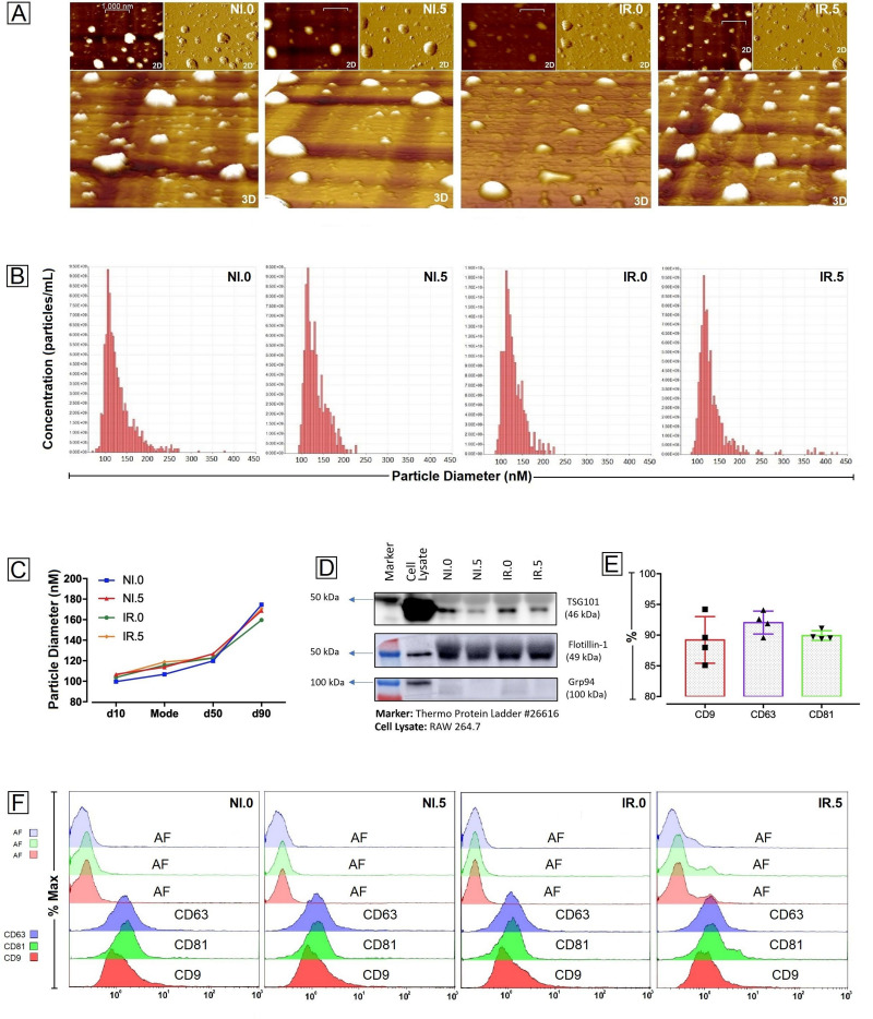
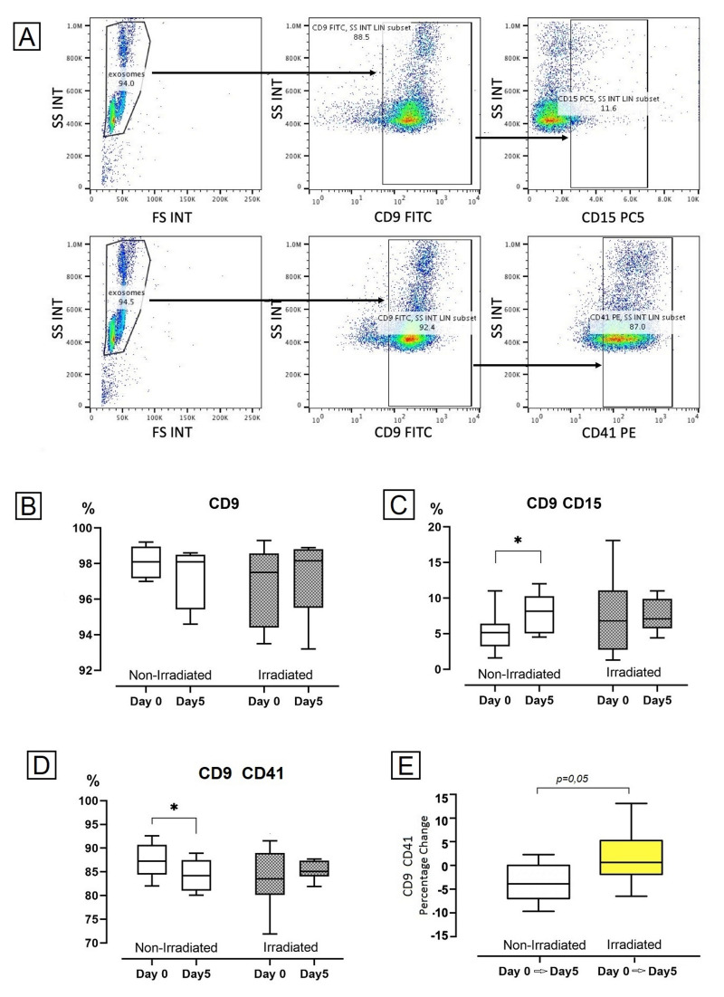
Materials and methods: Eight units of aPC were included in this study. These were divided into four equal portions. Two portions were irradiated before storage while the other two were not. Thus, irradiated and non-irradiated aPC samples for storage Days 0 (D0) and 5 (D5) were obtained. Exosomes were isolated from these samples using a commercial kit and were evaluated to ascertain their parent cells by flow cytometry. For the following steps, exosomes were pooled according to their features. Pooled exosomes were then used for aggregometry and thromboelastography.

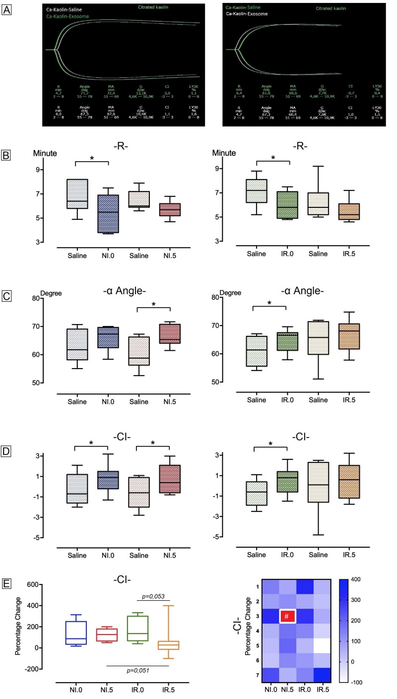
Results: Platelet-derived exosome (PD-EX) levels decreased in D5 compared to D0 in NI-aPC, whereas granulocyte-derived exosome (GD-EX) levels increased. Exosome pools had no effect on MPA compared to saline groups. Exosome pools decreased the time to initial fibrin formation (R), whereas they increased the rate of clot formation (α-angle) and coagulation index (CI) compared to saline groups.

Discussion: Storage time and γ-IR each have almost the opposite effects on PD-EX and GD-EX. Exosomes have no impact on MPA, but enhance the clot strength. The impact of exosomes on aPC quality and effectiveness can be ignored or considered as a positive effect.


Conflict of interest statement

The Authors declare no conflicts of interest.

Figures

Figure 1
Figure 1
Study algorithm
Figure 2
Figure 2
Flow-cytometry gating strategy and parental analysis results for exosome samples (A) Exosomes were gated by forward (FS) and side scatter (SS) properties, and also, by SS and surface expression of CD9. Their subgroups were gated and assessed surface expressions (CD41, CD15, etc.) in CD9 positive population. Representative flow-cytometry dot plots showing the granulocyte (CD9+CD15+) and platelet (CD9+CD41+) derived exosomes’ gating strategies. (B, C and D) Exosome levels in the non-irradiated and irradiated apheresis platelet concentrates samples. Graphs showing the percentage of the total exosomes (B) and the exosomes derived from CD15 and CD41 positive cells (C and D, respectively) on day 0 and day 5. n=8 for each column. *p=0.049. (E) Percentage changes of CD9+ CD41+ exosomes during the storage time in the non-irradiated and irradiated apheresis platelet concentrates. n=8 for each column.
Figure 3
Figure 3
Characterization of exosomes (A) Atomic Force Microscopy (AFM) image of pooled exosome samples. Scale bar represents 1,000 nm. (B and C) Size distribution of pooled exosome samples. Tunable Resistive Pulse Sensing (TRPS) were used for this purpose and graphs for each analysis are shown in (B). No difference in size was detected among groups (C). (D) Western blot analysis of exosomal markers TSG101, Flottilin-1, and GRP94 expression in pooled exosome samples. The cell lysate (RAW264.7 cell line) was used as a positive control, whereas GRP94 was used as a negative control. (E and F) Surface marker expressions of pooled exosome samples. CD9, CD63, CD81 were analysed using flow cytometry. While percentage values of surface markers were shown in bar graphs (E), flow cytometry graphs for each analysis are given at the bottom of figure (F). NI.0: non-irradiated Day 0 pooled exosome sample; NI.5: non-irradiated Day 5 pooled exosome sample; IR.0: irradiated Day 0 pooled exosome sample; IR.5: irradiated Day 5 pooled exosome sample; AF: auto-flourescense.
Figure 4
Figure 4
Impact of pooled exosomes on maximum platelet aggregation (MPA) (A) Representative aggregometer images from two different experiments. (B) Impact of saline and exosomes on MPA. There were no significant changes in the exosome groups compared to saline groups (n=8). (C) Percentage change between saline and exosome groups was calculated. The percentage changes between the exosome groups did not show any significance. The percentage change was demonstrated with both a graph and a heat map. The graph presents cumulative effects; the heat map indicates individual results (n=8). (D) Dose-related impact of exosomes on MPA. No dose-dependent increase or decrease was found. A similar impact was detected via both (1×) and (4×) exosome volumes (n=5). (E) Evaluation of individual or pooled exosomes effects on MPA. Their effect was similar on MPA, a significant difference was not determined (n=9). (F) Percentage change between saline and exosome groups for the impacts of ADP and Collagen on the stimulation of aggregation. They both had a similar impact on MPA (n=4). NI.0: non-irradiated Day 0 pooled exosome sample; NI.5: non-irradiated Day 5 pooled exosome sample; IR.0: irradiated Day 0 pooled exosome sample; IR.5: irradiated Day 5 pooled exosome sample.
Figure 5
Figure 5
Impact of pooled exosomes on hemostasis (A) Representative thromboelastography images from two different experiments. (B, C and D) The impact of saline and exosomes on hemostasis (n=7). There was a significant decrease in the NI.0 and IR.0 groups compared to saline groups on R-value (B). The significant increases were detected in the NI.5 and IR.0 groups compared to saline groups on α-angle value (C), and in the NI.0, NI.5, IR.0 groups compared to saline groups on CI value (D) *p=0.018. (E) The percentage change between saline and exosome groups was calculated. There were no significant changes in in-group and intergroup analysis via percentage changes. Only CI value in the IR.5-Pool group was close to significance compared to the NI.5-Pool and IR.0-Pool groups (p=0.051 and p=0.053, respectively). This result was demonstrated with both a graph and a heat map. The graph presents cumulative effects; the heat map indicates individual results (n=7). #Percentage change was not calculated, since the related saline group result was “0”. NI.0: non-irradiated Day 0 pooled exosome sample; NI.5: non-irradiated Day 5 pooled exosome sample; IR.0: irradiated Day 0 pooled exosome sample; IR.5: irradiated Day 5 pooled exosome sample.

Similar articles

References

    1. Dwyre DM, Holland PV. Transfusion-associated graft-versus-host disease. Vox Sang. 2008;95:85–93. doi: 10.1111/j.1423-0410.2008.01073.x. - DOI - PubMed
    1. Manduzio P. Transfusion-associated graft-versus-host disease: A concise review. Hematol Rep. 2018;10:98–102. doi: 10.4081/hr.2018.7724. - DOI - PMC - PubMed
    1. Bahar B, Tormey CA. Prevention of transfusion-associated graft-versus-host disease with blood product irradiation the past, present, and future. Arch Pathol Lab Med. 2018;142:662–667. doi: 10.5858/arpa.2016-0620-RS. - DOI - PubMed
    1. Pelszynski MM, Moroff G, Luban NL, Taylor BJ, Quinones RR. Effect of gamma irradiation of red blood cell units on T-cell Inactivation as assessed by limiting dilution analysis: implications for preventing transfusion-associated graft-versus-host disease. Blood. 1994;83:1683–1689. - PubMed
    1. Góes EG, Borges JC, Covas DT, Orellana MD, Palma PV, Morais FR, et al. Quality control of blood irradiation: determination T cells radiosensitivity to cobalt-60 gamma rays. Transfusion. 2006;46:34–40. doi: 10.1111/j.1537-2995.2005.00669.x. - DOI - PubMed

LinkOut - more resources