Review
. 2023 Feb;96(1142):20211302.
doi: 10.1259/bjr.20211302. Epub 2022 Aug 22.

Benign incidental cardiac findings in chest and cardiac CT imaging

Affiliations
Review

Benign incidental cardiac findings in chest and cardiac CT imaging

Keke Zhao et al. Br J Radiol. 2023 Feb.

Abstract

With the continuous expansion of the disease scope of chest CT and cardiac CT, the number of these CT examinations has increased rapidly. In addition to their common indications, many incidental cardiac findings can be observed when carefully evaluating the coronary arteries, valves, pericardium, ventricles, and large vessels. These findings may have clinical significance or risk of complications, but they are sometimes overlooked or may not be described in the final reports. Although most of the incidental findings are benign, timely detection and treatment can improve the management of chronic diseases or reduce the possibility of severe complications. In this review, we summarized the imaging findings, incidence rate, and clinical relevance of some benign cardiac findings such as coronary artery calcification, aortic and mitral valve calcification, aortic calcification, cardiac thrombus, myocardial bridge, aortic dilation, cardiac myxoma, pericardial cyst, and coronary artery fistula. Reporting incidental cardiac findings will help reduce the risk of severe complications or disease deterioration and contribute to the recovery of patients.


Figures

Figure 1.
Figure 1.
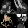
Incidental finding of coronary artery calcification in noncontrast chest CT and coronary CTA. A 58-year-old male had cough and expectoration. He needed to rule out pulmonary diseases such as tuberculosis and pneumonia, so he received a chest CT examination. (a, b) The axial image of noncontrast chest CT and multiplanar planar reformation shows sheet-like calcified plaques in the left anterior descending branch.
Figure 2.
Figure 2.
Incidental finding of aortic valve calcification in contrast-enhanced chest CT. A 59-year-old male had a history of diabetes and high blood pressure, chest tightness and pain for 8 months and aggravated for 10 days. The patient had chest distress, chest pain, breathlessness and lower limb edema. The purpose of enhanced CT was to seek the cause of breathlessness (a, b) The axial and coronal plane images show the heavily calcified aortic valves.
Figure 3.
Figure 3.
Incidental finding of calcified mitral valves in contrast-enhanced chest CT. A 62-year-old male had breath shortness for 6 months and aggravated for 15 days. Non-contrast chest CT showed a nodule in the basal segment of the left lower lobe. Then, the purpose of enhanced CT is to stage possible malignancy. (a, b, c) Long axial view, short axial view, and transverse view show the calcified mitral valves.
Figure 4.
Figure 4.
(a, b) Incidental finding of aortic wall calcification in noncontrast and contrast-enhanced chest CT. An 80-year-old female had elevated serum creatinine and chest tightness for 1 year, breathlessness and aggravated edema for 7 days. (a, b) Axial and coronal images of noncontrast chest CT show the aortic wall calcification. (c, d) An 85-year-old female, after resection of right upper lung cancer. Contrast-enhanced CT performed was to see if there were any metastases. Axial and coronal images of contrast-enhanced chest CT demonstrates the aortic wall calcification.
Figure 5.
Figure 5.
Myocardial bridging in CT angiography. A 49-year-old male had progressive dysuria and high blood pressure for over 5 years. CTA incidentally showed the myocardial bridge. (a) The axial image of CTA shows the deep myocardial bridge in the middle segment of the left anterior descending artery. (b, c) Curved planar reformation image and 3D reconstruction image demonstrates the deep myocardial bridge.
Figure 6.
Figure 6.
Left atrial appendage thrombus incidentally found by contrast-enhanced chest CT. A 63-year-old male, 1 year after mitral valve replacement, with symptoms of chest tightness and breath shortness. (a, b, c) The axial, coronal, and sagittal images show a low-density region in the left atrial appendage, without contrast-enhancement. The diagnosis of left atrial appendage thrombosis has been confirmed by esophageal ultrasound.
Figure 7.
Figure 7.
Incidental finding of aortic dilatation in noncontrast and contrast-enhanced chest CT. An 86-year-old male presented hematuria for more than 1 month. (a, b) The axial and coronal views of the noncontrast chest CT show aortic dilation, with a diameter >5 cm. A 50-year-old male was found to have a right maxillary sinus cyst for more than 2 months. (c, d) The axial and coronal views of the contrast-enhanced chest CT show aortic dilation, with a diameter >5 cm.
Figure 8.
Figure 8.
Incidental finding of aortic dissection in noncontrast chest CT and the reference CTA images. A 57-year-old male had coronary artery calcification and stenosis. Because he needed to be hospitalized, he received a noncontrast chest CT scan to rule out chest diseases, such as lung cancer and tuberculosis. (a) Noncontrast CT reveals the inward moving calcified plaque. (b, c) The subsequent axial and sagittal images of aortic CTA confirms the aortic dissection, which originates from the ascending aorta with a thin layer separating the true and false lumen. The true lumen is small and the false lumen is large.
Figure 9.
Figure 9.
Incidental finding of cardiac myxoma in coronary CTA. A 64-year-old male had chest tightness and palpitation for 2 months after physical activities. (a, b) The axial and four-chamber images show a hypo-attenuation lesion in the left atrium, with a maximum cross-section of 23 × 59 mm and a CT attenuation value of 51HU. The lesion is locally close to the atrial septum and extends into the left ventricle.
Figure 10.
Figure 10.
Incidental finding of pericardial cyst. A 75-year-old female with pulmonary nodules was admitted to the hospital. A noncontrast chest CT scan was performed to follow-up the nodules. (a, b) The axial and coronal images showed a sac of 49 × 30 mm at the pericardium edge of the left anterior mediastinum.
Figure 11.
Figure 11.
Incidental finding of coronary artery fistula in CTA. A 73-year-old male had repeated chest tightness and palpitation for more than half a year. The patient has previously been diagnosed with coronary artery calcification and stenosis and CTA was used to assess progression of stenosis. (a, b) The axial and coronal views of coronary CTA demonstrates the left anterior descending coronary artery-pulmonary artery fistula. (c) 3D reconstruction shows the whole varicose vessels and (d) the curved planar reformation displays the origin of coronary artery fistula.

Similar articles

Cited by

References

    1. Chen Y, Hu Z, Li M, Jia Y, He T, Liu Z, et al. . Comparison of nongated chest CT and dedicated calcium scoring CT for coronary calcium quantification using a 256-dector row CT scanner. Academic Radiology 2019; 26: e267–74. doi: 10.1016/j.acra.2018.12.005 - DOI - PubMed
    1. Jacobs PC, Prokop M, van der Graaf Y, Gondrie MJ, Janssen KJ, de Koning HJ, et al. . Comparing coronary artery calcium and thoracic aorta calcium for prediction of all-cause mortality and cardiovascular events on low-dose non-gated computed tomography in a high-risk population of heavy smokers. Atherosclerosis 2019; 209: 455–62. doi: 10.1016/j.atherosclerosis.2009.09.031 - DOI - PubMed
    1. Haller C, Vandehei A, Fisher R, Boster J, Shipley B, Kaatz C, et al. . Incidence and implication of coronary artery calcium on non-gated chest computed tomography scans: A large observational cohort. Cureus 2019. doi: 10.7759/cureus.6218 - DOI - PMC - PubMed
    1. Shim SS, Oh YW, Kong KA, Ryu YJ, Kim Y, Jang DH. Pulmonary nodule size evaluation with chest tomosynthesis and CT: A phantom study. BJR 2019; 88: 20140040. doi: 10.1259/bjr.20140040 - DOI - PMC - PubMed
    1. Liang CH, Liu YC, Wu MT, Garcia-Castro F, Alberich-Bayarri A, Wu FZ. Identifying pulmonary nodules or masses on chest radiography using deep learning: external validation and strategies to improve clinical practice. Clinical Radiology 2019; 75: 38–45. doi: 10.1016/j.crad.2019.08.005 - DOI - PubMed

MeSH terms