
Syndrome of Combined Pulmonary Fibrosis and Emphysema: An Official ATS/ERS/JRS/ALAT Research Statement

Vincent Cottin et al. Am J Respir Crit Care Med. .

Abstract

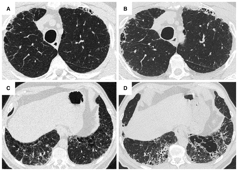
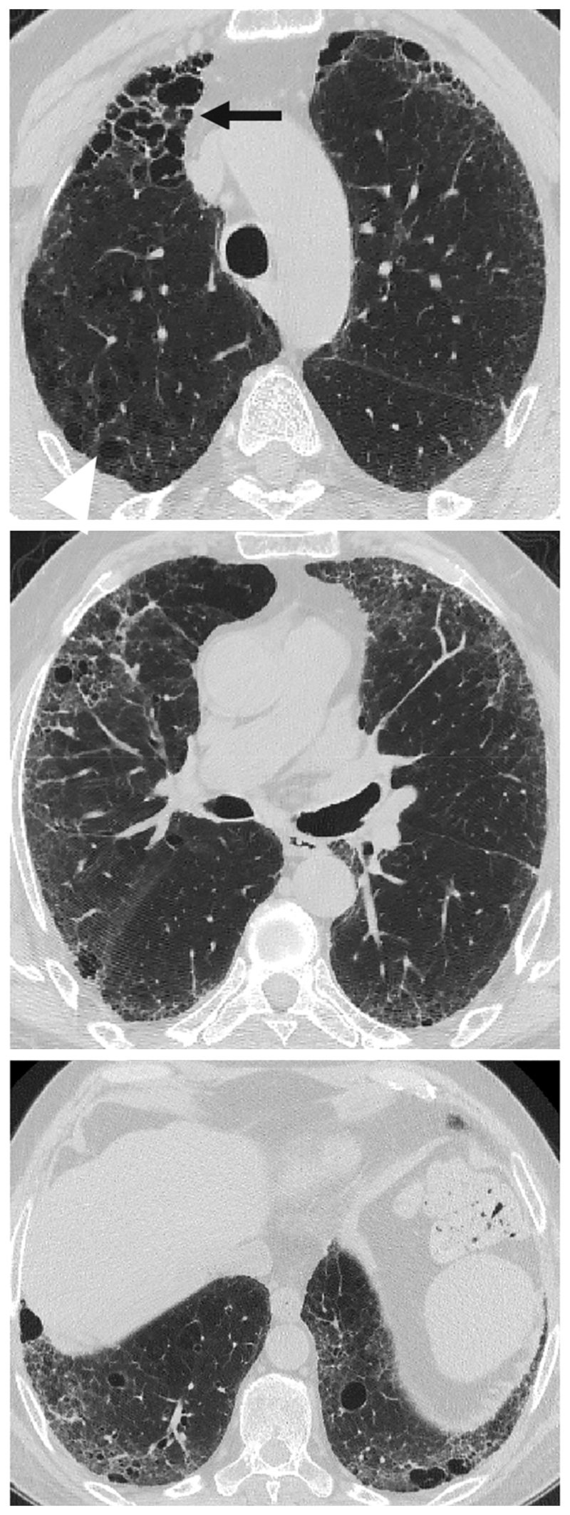
Background: The presence of emphysema is relatively common in patients with fibrotic interstitial lung disease. This has been designated combined pulmonary fibrosis and emphysema (CPFE). The lack of consensus over definitions and diagnostic criteria has limited CPFE research. Goals: The objectives of this task force were to review the terminology, definition, characteristics, pathophysiology, and research priorities of CPFE and to explore whether CPFE is a syndrome. Methods: This research statement was developed by a committee including 19 pulmonologists, 5 radiologists, 3 pathologists, 2 methodologists, and 2 patient representatives. The final document was supported by a focused systematic review that identified and summarized all recent publications related to CPFE. Results: This task force identified that patients with CPFE are predominantly male, with a history of smoking, severe dyspnea, relatively preserved airflow rates and lung volumes on spirometry, severely impaired DlCO, exertional hypoxemia, frequent pulmonary hypertension, and a dismal prognosis. The committee proposes to identify CPFE as a syndrome, given the clustering of pulmonary fibrosis and emphysema, shared pathogenetic pathways, unique considerations related to disease progression, increased risk of complications (pulmonary hypertension, lung cancer, and/or mortality), and implications for clinical trial design. There are varying features of interstitial lung disease and emphysema in CPFE. The committee offers a research definition and classification criteria and proposes that studies on CPFE include a comprehensive description of radiologic and, when available, pathological patterns, including some recently described patterns such as smoking-related interstitial fibrosis. Conclusions: This statement delineates the syndrome of CPFE and highlights research priorities.

Keywords: diagnosis; emphysema; fibrosis; interstitial lung disease; management.


Figures

Figure 1
Figure 1
HRCT showing a typical distribution of disease seen in combined pulmonary fibrosis and emphysema (separate emphysema and fibrosis pattern). Paraseptal and centrilobular emphysema is localized to the upper lobes, whilst fibrosis characterized by traction bronchiectasis is localized to the lower lobes.
Figure 2
Figure 2
HRCT showing a typical distribution of disease seen in combined pulmonary fibrosis and emphysema (progressive transition pattern). A predominant pattern of centrilobular emphysema is seen in the upper lobes, extending to the midzones of the lungs in a 72-year-old patient with idiopathic pulmonary fibrosis. No emphysema is seen in the lower zones. The appearances are in keeping with a progressive transition pattern of combined pulmonary fibrosis and emphysema
Figure 3
Figure 3
HRCT showing a typical distribution of disease seen in combined pulmonary fibrosis and emphysema (paraseptal emphysema pattern). In this 67 year-old male diagnosed with idiopathic pulmonary fibrosis, extensive isolated paraseptal and centrilobular emphysema is present in the upper zones. Whilst the centrilobular emphysema is mostly isolated in the midzones, paraseptal emphysema is increasingly admixed resembling honeycomb cysts in the left lung. Within the left lower lobe, paraseptal emphysema mimicking honeycomb cysts lies adjacent to more centrally placed irregularly shaped centrilobular emphysema (arrow).
Figure 4
Figure 4
Chest HRCT showing worsening of admixed destructive emphysema (combined pulmonary fibrosis and emphysema, admixed pattern). In the pair of axial images of the upper (A, B) and lower zones (C, D), taken 2 years apart (A, C: baseline; B, D: follow-up) in a 66-year old male patient with idiopathic pulmonary fibrosis, isolated emphysema in the right upper lobe becomes admixed with fibrosis over time. In the left lower lobe, centrally placed emphysema becomes pulled apart (‘traction emphysema’) and expands as the surrounding fibrosis evolves.
Figure 5
Figure 5
Chest HRCT showing emphysema admixed with desquamative interstitial pneumonia confirmed by lung biopsy (combined pulmonary fibrosis and emphysema, admixed pattern at HRCT). Areas of low attenuation are admixed with ground glass opacities (high attenuation) and thickening of peri-emphysematous areas.
Figure 6
Figure 6
Chest HRCT showing admixed emphysema and fibrosis with thick-walled large cysts (combined pulmonary fibrosis and emphysema, thick-walled large cysts pattern). Histopathology demonstrated predominantly smoking-related interstitial fibrosis (SRIF).
Figure 7
Figure 7
Sagittal CT images of the lungs performed over the course of 4 years in a 66-year-old male ex-smoker diagnosed with idiopathic pulmonary fibrosis and emphysema (combined pulmonary fibrosis and emphysema, thick-walled large cysts pattern). In the top left image low-attenuation lesions without clearly visible walls in keeping with emphysema (arrow) are visible adjacent to the diaphragm and lie within fibrotic regions of lung. Over the next 4 years, as the fibrosis matures, the low-attenuation lesions coalesce (arrow) and enlarge in size forming a thick-walled cystic lesion.
Figure 8
Figure 8
HRCT in a 69-year-old male with idiopathic pulmonary fibrosis, showing diffuse emphysema through the lung zones and lower zone predominant fibrosis (combined pulmonary fibrosis and emphysema, unclassifiable pattern). Emphysema in the right upper lobe is a combination of admixed (black arrow) and isolated emphysema (white arrowhead). Admixed emphysema is visible in the midzones, whilst in the lower zones a mixture of admixed and isolated paraseptal and centrilobular emphysema is apparent.
Figure 9
Figure 9
Visual scoring of emphysema. Axial section through the upper lobes in a patient with idiopathic pulmonary fibrosis and emphysema (top). Emphysema is distributed irregularly through the lobe making visual quantification difficult. Visually combining the emphysematous foci together (bottom) and estimating the fraction of the lobe that it comprises (i.e. 50%, 33%, 25%, 20%, 15%, 10%, 5%) can simplify quantitation in challenging cases (online supplement).
Figure 10
Figure 10
Centrilobular emphysema in wedge excision in a heavy smoker with a peripheral small cell carcinoma. An intermediate magnification photomicrograph shows enlarged airspaces with destruction of bronchiolar walls evidenced by detached free-floating connective tissue fragments. Hematoxylin and eosin staining.
Figure 11
Figure 11. Respiratory bronchiolitis (RB) in a patient with RB-ILD. RB is a common finding in patients with CPFE who have concomitant pulmonary fibrosis.
A. Low magnification photomicrograph showing RB characterized by clusters of lightly pigmented macrophages in the lumens of distal bronchioles and peribronchiolar air spaces. B. Higher magnification view showing pigmented intraluminal macrophages in respiratory bronchiole and surrounding air spaces with no significant inflammation or fibrosis. Hematoxylin and eosin staining.
Figure 12
Figure 12
Smoking-related interstitial fibrosis (SRIF) in upper lobe biopsy from 120-pack-year smoker with CPFE characterized by a combination of emphysema and usual interstitial pneumonia in middle and lower lobe biopsies. A. Low magnification photomicrograph showing mild expansion of subpleural parenchyma by paucicellular, densely eosinophilic (“amyloid-like”) collagen with preservation of lung architecture. B. Higher magnification photomicrograph showing subpleural fibrosis without honeycomb change or fibroblast foci. There is mild associated emphysema.
Figure 13
Figure 13. Subpleural cysts of smoking-related interstitial fibrosis (SRIF) contrasted with honeycomb change in usual interstitial pneumonia (UIP).
A. Low magnification photomicrograph of SRIF with associated distal acinar (“paraseptal”) emphysema forming thick-walled cysts. Overall lung architecture is preserved and the paucicellular fibrosis lacks the qualitative variability more characteristic of UIP. The cystic spaces are mainly lined by attenuated pneumocytes. B. Low magnification photomicrograph of honeycomb change in UIP. The fibrosis has a patchy distribution with collapse and distortion of normal lung architecture. The fibrosis includes fibroblast foci (arrow), and a mild, patchy infiltrate of lymphocytes resulting in a variegated appearance that contrasts with the uniform, paucicelluar, densely eosinophilic fibrosis in SRIF. Cystic honeycomb spaces (*) are mainly lined by columnar bronchiolar type epithelium rather than pneumocytes. Hematoxylin and eosin stain.
Figure 14
Figure 14. Desquamative interstitial pneumonia (DIP).
A. Low magnification photomicrograph showing a relatively uniform interstitial pneumonia compounded by prominent clusters of pigments alveolar macrophages. B. Higher magnification photomicrograph shows the interstitial inflammation that distinguishes DIP from SRIF (compare to Figure 14B). Hematoxylin and eosin stain.

References

    1. Cottin V, Nunes H, Brillet PY, et al. Combined pulmonary fibrosis and emphysema: a distinct underrecognised entity. Eur Respir J. 2005;26:586–93. - PubMed
    1. Cottin V, Cordier JF. The syndrome of combined pulmonary fibrosis and emphysema. Chest. 2009;136:1–2. - PubMed
    1. Wong AW, Liang J, Cottin V, Ryerson CJ. Diagnostic Features in Combined Pulmonary Fibrosis and Emphysema: A Systematic Review. Ann Am Thorac Soc. 2020;17:1333–6. - PubMed
    1. Kawabata Y, Hoshi E, Murai K, et al. Smoking-related changes in the background lung of specimens resected for lung cancer: a semiquantitative study with correlation to postoperative course. Histopathology. 2008;53:707–14. - PubMed
    1. Katzenstein AL. Smoking-related interstitial fibrosis (SRIF): pathologic findings and distinction from other chronic fibrosing lung diseases. J Clin Pathol. 2013;66:882–7. - PubMed

Publication types