. 2022 Oct;40(5):1479-1498.
doi: 10.1007/s12640-022-00555-x. Epub 2022 Aug 15.

Neuroprotective Potential of Intranasally Delivered Sulforaphane-Loaded Iron Oxide Nanoparticles Against Cisplatin-Induced Neurotoxicity

Affiliations

Neuroprotective Potential of Intranasally Delivered Sulforaphane-Loaded Iron Oxide Nanoparticles Against Cisplatin-Induced Neurotoxicity

Ghadha Ibrahim Fouad et al. Neurotox Res. 2022 Oct.

Abstract

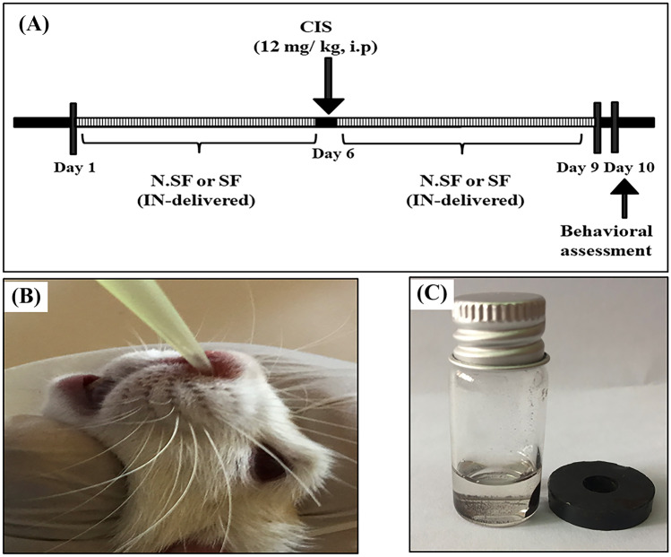
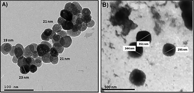
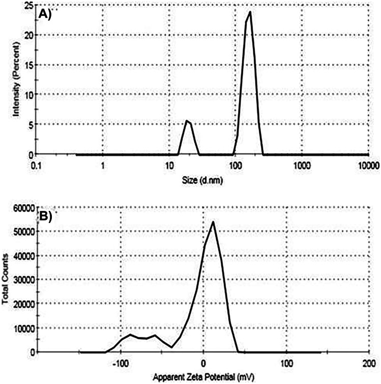
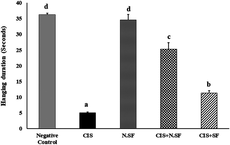
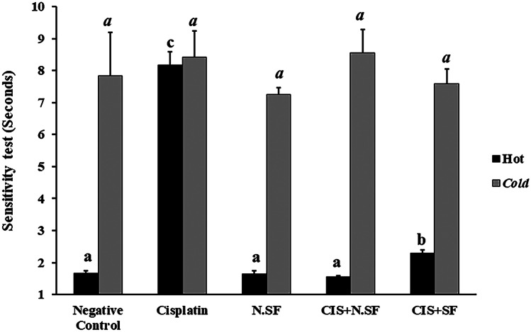
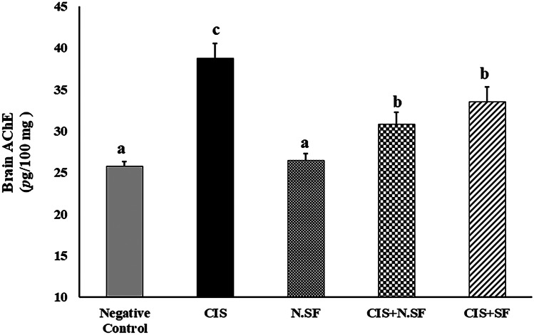
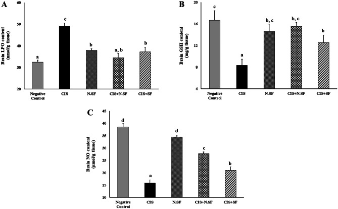
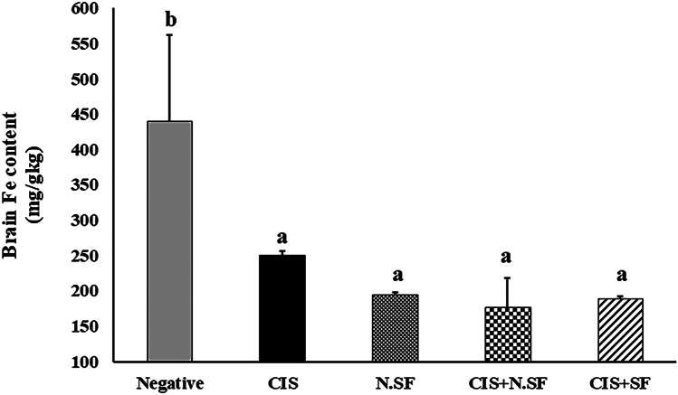
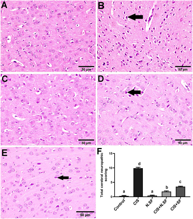
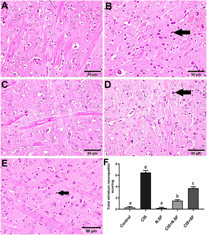
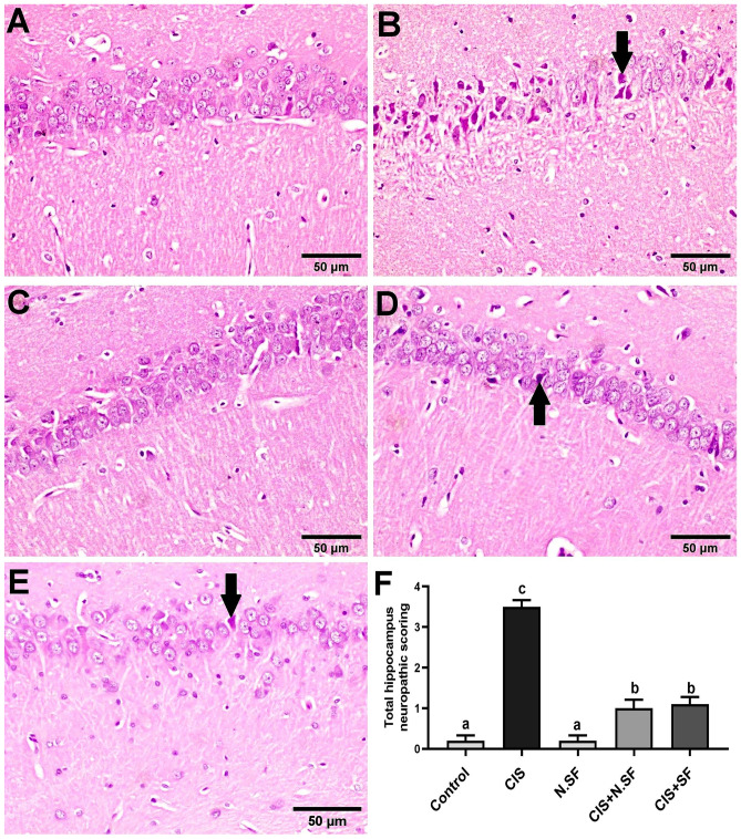
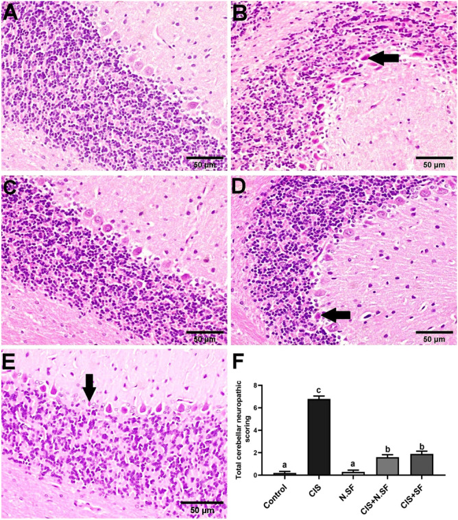
Cisplatin (CIS) is a platinum-based chemotherapeutic drug that is widely used to treat cancer. However, its therapeutic efficiency is limited due to its potential to provoke neurotoxicity. Sulforaphane (SF) is a natural phytochemical that demonstrated several protective activities. Iron oxide nanoparticles (Fe3O4-NPs) could be used as drug carriers. This study aimed to explore the nanotoxic influence of SF-loaded within Fe3O4-NPs (N.SF), and to compare the neuroprotective potential of both N.SF and SF against CIS-induced neurotoxicity. N.SF or SF was administrated intranasally for 5 days before and 3 days after a single dose of CIS (12 mg/kg/week, i.p.) on the 6th day. Neuromuscular coordination was assessed using hanging wire and tail-flick tests. Acetylcholinesterase (AChE) activities and markers of oxidative stress were measured in the brain. In addition, the brain iron (Fe) content was estimated. CIS significantly induced a significant increase in AChE activities and lipid peroxides, and a significant decrement in glutathione (GSH) and nitric oxide (NO) contents. CIS elicited impaired neuromuscular function and thermal hyperalgesia. CIS-induced brains displayed a significant reduction in Fe content. Histopathological examination of different brain regions supported the biochemical and behavioral results. Contradict, treatment of CIS-rats with either N.SF or SF significantly decreased AChE activity, mitigated oxidative stress, and ameliorated the behavioral outcome. The histopathological features supported our results. Collectively, N.SF demonstrated superior neuroprotective activities on the behavioral, biochemical, and histopathological (striatum and cerebral cortex) aspects. N.SF could be regarded as a promising "pre-clinical" neuroprotective agent. Furthermore, this study confirmed the safe toxicological profile of Fe3O4-NPs.

Keywords: Acetylcholinesterase; Cisplatin; Intranasal; Iron oxide nanoparticles; Neurotoxicity; Sulforaphane.


Conflict of interest statement

The authors declare no competing interests.

Figures

Fig. 1
Fig. 1
A Schematic representation of timeline and experimental design, B intranasal dosage (IN) administration in rats, and C demonstration of magnetic nature of iron oxide nanoparticles (Fe3O4-NPs) used for SF loading
Fig. 2
Fig. 2
TEM images of (A) magnetic iron oxide nanoparticles (Fe3O4-NPs) and (B) sulforaphane (SF)-loaded iron oxide nanoparticles (Fe3O4-NPs)
Fig. 3
Fig. 3
A Particle size distribution curve of the prepared sulforaphane (SF) loaded within iron oxide nanoparticles (Fe3O4-NPs) and B zeta potential profile of the prepared sulforaphane (SF) loaded within iron oxide nanoparticles (Fe3O4-NPs) recorded by the Zetasizer device
Fig. 4
Fig. 4
Effect of intranasal administration of either sulforaphane (SF)-loaded within iron oxide nanoparticles (N.SF) or free sulforaphane (SF) on neuromuscular coordination “hanging wire” test in CIS-induced rats. Groups: negative control rats, cisplatin (CIS)-exposed rats, N.SF-alone exposed rats, CIS + N.SF-treated rats, and CIS + SF-treated rats. Results are presented as mean ± standard error of the mean (SEM). Mean with different letters (a–d) is significant at p ≤ 0.05
Fig. 5
Fig. 5
Effect of intranasal administration of sulforaphane (SF)-loaded within iron oxide nanoparticles (N.SF) or free sulforaphane (SF) on thermal sensitivity: tail-flick test in CIS-induced rats. Groups: negative control rats, cisplatin (CIS)-exposed rats, N.SF-alone exposed rats, CIS + N.SF-treated rats, and CIS + SF-treated rats. Results are presented as mean ± standard error of the mean (SEM). Mean with different letters (a–c) is significant at p ≤ 0.05
Fig. 6
Fig. 6
Effect of intranasal administration of sulforaphane (SF)-loaded within iron oxide nanoparticles (N.SF) or free sulforaphane (SF) on brain acetylcholinesterase (AChE) activities in CIS-induced rats. Groups: negative control rats, cisplatin (CIS)-exposed rats, N.SF-alone exposed rats, CIS + N.SF treated rats, and CIS + SF-treated rats. Results are presented as mean ± standard error of the mean (SEM). Mean with different letters (a–c) is significant at p ≤ 0.05
Fig. 7
Fig. 7
Effect of intranasal administration of sulforaphane (SF) loaded within iron oxide nanoparticles (N.SF) or free sulforaphane (SF) on brain contents of lipid peroxides (LPO), glutathione reduced (GSH), and nitric oxide (NO) in CIS-induced rats. Groups: negative control rats, cisplatin (CIS)-exposed rats, N.SF-alone exposed rats, CIS + N.SF-treated rats, and CIS + SF-treated rats. Results are presented as mean ± standard error of the mean (SEM). Mean with different letters (a–d) is significant at p ≤ 0.05
Fig. 8
Fig. 8
Iron (Fe) distribution in the brain of different experimental groups: negative control rats, cisplatin (CIS)-exposed rats, N.SF-alone exposed rats, CIS + N.SF-treated rats, and CIS + SF-treated rats. Results are presented as mean ± standard error of the mean (SEM). Mean with different letters (a–b) is significant at p ≤ 0.05
Fig. 9
Fig. 9
Representative photomicrographs of H&E-stained cerebral cortex of different experimental groups. A Negative control brains showing the normal histological architecture with intact neurons. B CIS-neurotoxicated brains showing prominent degeneration and necrosis of neurons (arrow). C N.SF alone-control brains showing no histopathological alterations. D CIS + N.SF-treated brains, showing necrosis of sporadic neurons (arrow). E CIS + SF-treated brains, showing necrosis of some neurons (arrow) (scale bar: 50 μm). F Total histological scoring of cerebral cortex damage. Results are presented as mean ± standard error of the mean (SEM). Mean with different letters (a–d) is significant at p ≤ 0.05
Fig. 10
Fig. 10
Representative photomicrographs of H&E-stained striata of different experimental groups. A Negative control brains showing the normal histological architecture with intact neurons. B CIS-induced brains showing necrosis of neurons (arrow). C N.SF-alone treated brains, showing no histopathological alterations. D CIS + N.SF-treated brains showing shrunken and necrosis of sporadic neurons (arrow). E CIS + SF-treated brains showing necrosis of some neurons (arrow) (scale bar: 50 μm). F Total histological scoring of striatum damage. Results are presented as mean ± standard error of the mean (SEM). Mean with different letters (a–d) is significant at p ≤ 0.05
Fig. 11
Fig. 11
Representative photomicrographs of H&E-stained hippocampus of different experimental groups. A Negative control showing the normal histological architecture. B CIS-neurotoxicated brains, showing marked shrunken and pyknosis of pyramidal neurons (arrow). C N.SF-alone treated brains showing no histopathological changes. D and E CIS + N.SF- and CIS + SF-treated brains respectively showing sparse necrosis of pyramidal neurons (arrow) (scale bar: 50 μm). F Total histological scoring of hippocampus damage. Results are presented as mean ± standard error of the mean (SEM). Mean with different letters (a–c) is significant at p ≤ 0.05
Fig. 12
Fig. 12
Representative photomicrographs of H&E-stained cerebellum of different experimental groups. A Negative control brains showing the normal histological architecture. B CIS-neurotoxicated brains showing pyknotic and shrunken Purkinje cells (arrow). C N.SF alone treated brains showing normal histological structure. D and E CIS + N.SF- and CIS + SF-treated brains, respectively, showing degeneration of sporadic Purkinje cells (arrow) (scale bar: 50 μm). F Total histological scoring of cerebellum damage. Results are presented as mean ± standard error of the mean (SEM). Mean with different letters (a–c) is significant at p ≤ 0.05

Similar articles

Cited by

References

    1. Adesso S, Magnus T, Cuzzocrea S, Campolo M, et al. Indoxyl sulfate affects glial function increasing oxidative stress and neuroinflammation in chronic kidney disease: interaction between astrocytes and microglia. Front Pharmacol. 2017;8:370. doi: 10.3389/fphar.2017.00370. - DOI - PMC - PubMed
    1. Ahmad N, Subhan F, Islam NU, Shahid M, Rahman FU, Sewell RD. Gabapentin and its salicylaldehyde derivative alleviate allodynia and hypoalgesia in a cisplatin-induced neuropathic pain model. Eur J Pharmacol. 2017;814:302–312. doi: 10.1016/j.ejphar.2017.08.040. - DOI - PubMed
    1. Akbar S, Subhan F, Shahid M, Wadood A, et al. 6-Methoxyflavanone abates cisplatin-induced neuropathic pain apropos anti-inflammatory mechanisms: a behavioral and molecular simulation study. Eur J Pharmacol. 2020;872:172972. doi: 10.1016/j.ejphar.2020.172972. - DOI - PubMed
    1. Al Moundhri MS, Al-Salam S, Al Mahrouqee A, Beegam S, Ali BH. The effect of curcumin on oxaliplatin and cisplatin neurotoxicity in rats: some behavioral, biochemical, and histopathological studies. J Med Toxicol. 2013;9(1):25–33. doi: 10.1007/s13181-012-0239-x. - DOI - PMC - PubMed
    1. Ali BH, Ramkumar A, Madanagopal TT, Waly MI, Tageldin M et al (2014) Motor and behavioral changes in mice with cisplatin-induced acute renal failure. Physiological Res 63(1) - PubMed

LinkOut - more resources