. 2022 Aug 15;18(8):e1010386.
doi: 10.1371/journal.ppat.1010386. eCollection 2022 Aug.

Early and mid-gestation Zika virus (ZIKV) infection in the olive baboon (Papio anubis) leads to fetal CNS pathology by term gestation

Affiliations

Early and mid-gestation Zika virus (ZIKV) infection in the olive baboon (Papio anubis) leads to fetal CNS pathology by term gestation

Sunam Gurung et al. PLoS Pathog. .

Abstract

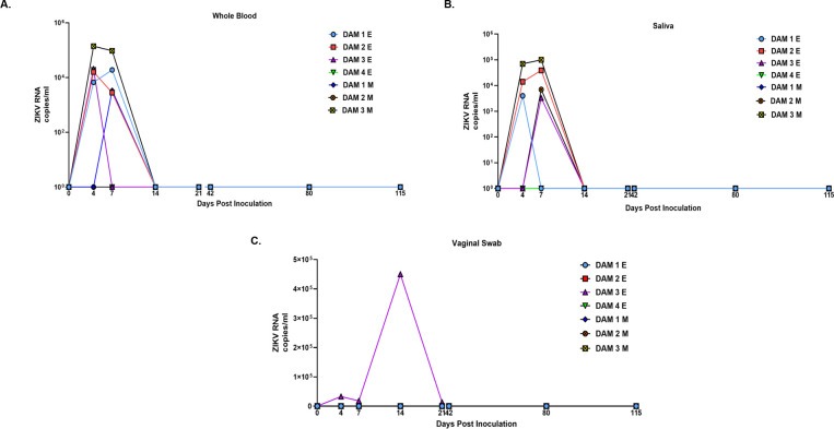
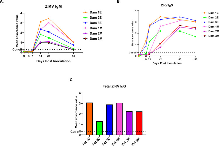
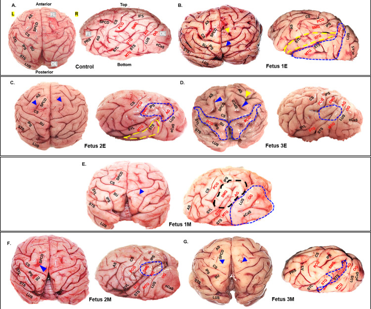
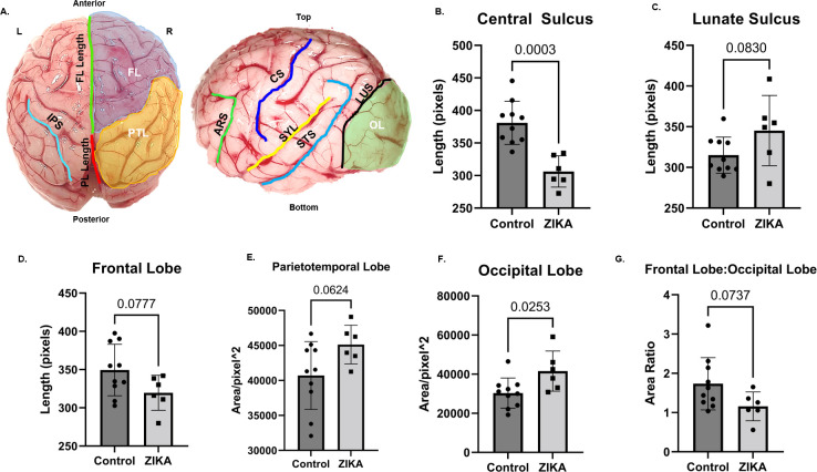
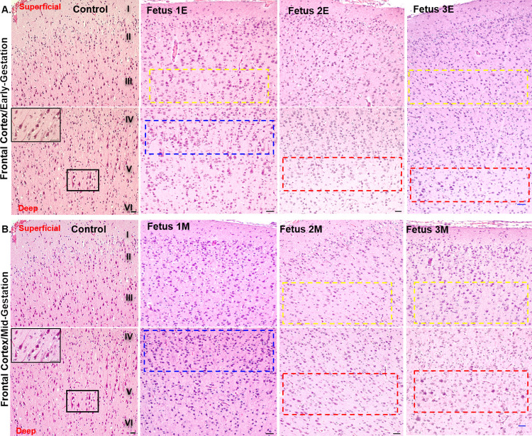
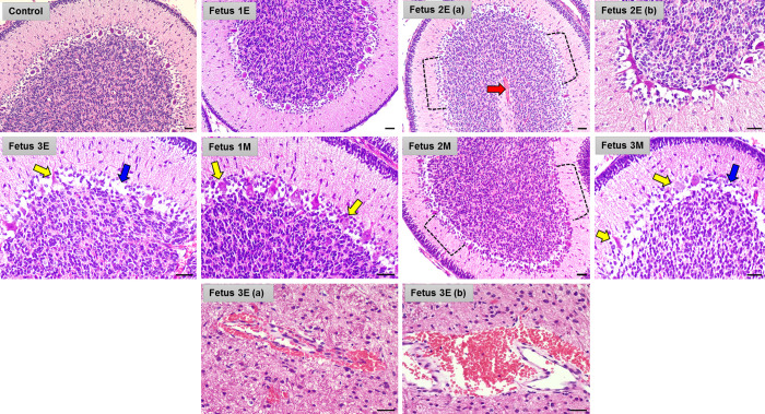
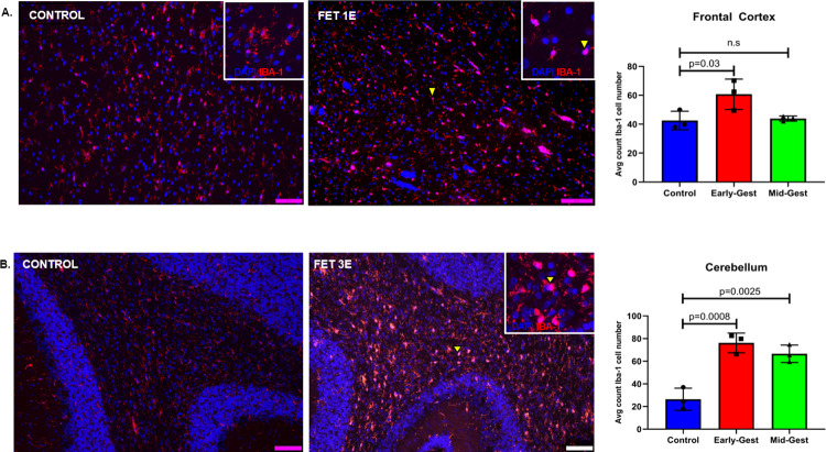
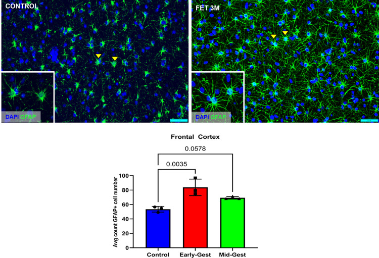
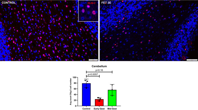
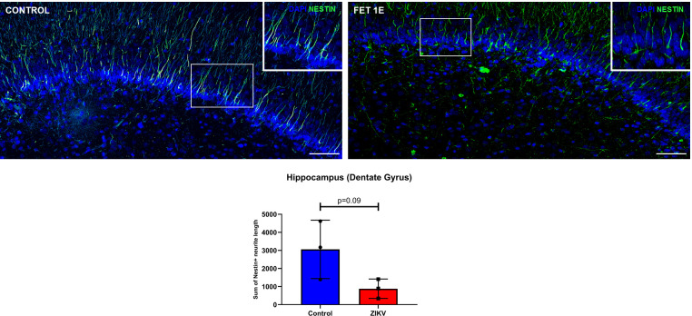
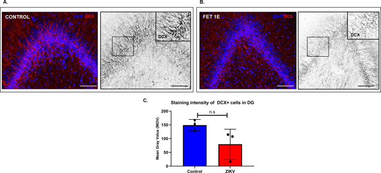
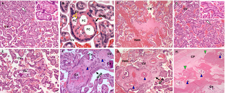
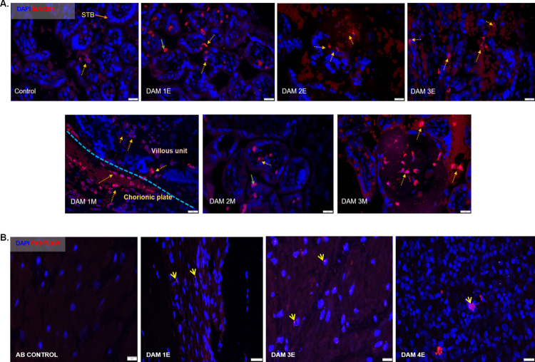
Zika virus (ZIKV) infection in pregnancy can produce catastrophic teratogenic damage to the developing fetus including microcephaly and congenital Zika syndrome (CZS). We previously described fetal CNS pathology occurring by three weeks post-ZIKV inoculation in Olive baboons at mid-gestation, including neuroinflammation, loss of radial glia (RG), RG fibers, neuroprogenitor cells (NPCs) resulting in disrupted NPC migration. In the present study, we explored fetal brain pathologies at term gestation resulting from ZIKV exposure during either first or second trimester in the Olive baboon. In all dams, vRNA in whole blood resolved after 7 days post inoculation (dpi). One first trimester infected dam aborted at 5 dpi. All dams developed IgM and IgG response to ZIKV with ZIKV IgG detected in fetal serum. Placental pathology and inflammation were observed including disruption of syncytiotrophoblast layers, delayed villous maturation, partially or fully thrombosed vessels, calcium mineralization and fibrin deposits. In the uterus, ZIKV was detected in ¾ first trimester but not in second trimester infected dams. While ZIKV was not detected in any fetal tissue at term, all fetuses exhibited varying degrees of neuropathology. Fetal brains from ZIKV inoculated dams exhibited a range of gross brain pathologies including irregularities of the major gyri and sulci of the cerebral cortex and cerebellar pathology. Frontal cortices of ZIKV fetuses showed a general disorganization of the six-layered cortex with degree of disorganization varying among the fetuses from the two groups. Frontal cortices from ZIKV inoculation in the first but not second trimester exhibited increased microglia, and in both trimester ZIKV inoculation, increased astrocyte numbers (white matter). In the cerebellum, increased microglia were observed in fetuses from both first and second trimester inoculation. In first trimester ZIKV inoculation, decreased oligodendrocyte precursor cell populations were observed in fetal cerebellar white matter. In general, our observations are in accordance with those described in human ZIKV infected fetuses.


Conflict of interest statement

The authors have declared that no competing interests exist.

Figures

Fig 1
Fig 1
ZIKV RNA (copies per milliliter) in whole blood (A), saliva (B) and vaginal swab (C) samples in baboons inoculated subcutaneously with PR ZIKV isolate during early and mid-gestation. ZIKV RNA was prepared from samples collected from each animal at the indicated days post inoculation and quantitated by one-step qRT-PCR.
Fig 2
Fig 2
Detection of anti-ZIKV antibody responses in pregnant baboon serum. Antibodies against ZIKV was determined by ELISA for maternal IgM (A) and IgG (B) and fetal IgG (C). anti-ZIKV IgM were detected at 14 days post-inoculation in all six baboons sampled at this time point. In the early-gestation group, dams 1E and 3E had ZIKV IgG at 14 dpi and dam 2E had at 21 dpi. In the mid-gestation inoculation group, all three dams had IgG by 21dpi. All six fetuses had anti-ZIKV IgG at levels similar to the mothers indicating efficient FcγR transfer of IgG across the placenta. Mean absorbance value was calculated from duplicates for each sample. Absorbance for all serum samples was read at 450nm for IgM and 405nm for ZIKV IgG assays. Cut off values for both assays were set per manufacturer’s criteria for each assay.
Fig 3
Fig 3
Gyri/sulci malformations in brains from fetuses form early and mid-gestation ZIKV inoculated dams compared to controls (A). Top and side view of fetal brains showing major gyri and sulci formations. Abnormal sulci (abnormal sulcal length and depth, asymmetry across cerebral hemispheres) or missing sulci are marked by arrows. Gyri malformations (enlargement, asymmetric across left and right cerebral hemispheres) are highlighted by dotted areas compared to the control fetal brain (A). Fetuses from early gestation inoculated dams (B, C, D); fetuses from mid-gestation inoculated dams (E, F, G). Frontal Lobe (FL), Occipital Lobe (OL), Parietal Lobe (PL), Left Cerebral Hemisphere (L), Right Cerebral Hemisphere (R), Arcuate Sulcus (ARS), Central Sulcus (CS), External calcarine Sulcus (eCaS), Inferior Parietal Sulcus (IPS), Lunate Sulcus (LUS), Superior Pre-central dimple (SPCD), Superior Post-central dimple (SU), Principal Sulcus (PRS), Superior Temporal Sulcus (STS), Sylvian Fissure/Lateral Sulcus (SYL), Postcentral Gyrus (PCG), Supramarginal Gyrus (SMG), Superior Temporal Gyrus (STG), Angular Gyrus (AG), Middle Temporal Gyrus (MTG), Occipital Gyrus (OG).
Fig 4
Fig 4. Differences in sulcal length and cerebral lobe surface area in brains from fetuses from early and mid-gestation ZIKV inoculated dams compared to controls.
Image showing lengths of different sulcis and surface areas of different cerebral lobes measured using Image J (A). Measurements of CS, ARS, SYL, STS, LUS length and OL surface area were done on left hemisphere image (side view) of each brain. Measurements of right and left IPS length, FL and PTL surface areas were done on top view image of the brain. FL length was measured by drawing a line from tip of the frontal lobe to CS (A, top view, green line) and PL length was measured by drawing a line from CS to LUS (A, top view, red line). Significant decrease in length of central sulcus (B) and increase in surface area of Occipital lobe (F) was seen in fetal brains from ZIKV inoculated dams compared to control brains (n = 10). In fetal brains from ZIKV inoculated dams, a trend towards reduction in frontal lobe length (D), in frontal lobe to occipital lobe area ratio (G) and increase in length of lunate sulcus (C), and in surface area of parieto-temporal lobe surface area (E) was seen compared to control brains. (Error bars depict SD, unpaired t-test, p<0.05 considered significant). Frontal Lobe (FL), Occipital Lobe (OL), Parietal Lobe (PL), Parieto Temporal area (PTL), Left Cerebral Hemisphere (L), Right Cerebral Hemisphere (R), Arcuate Sulcus (ARS), Central Sulcus (CS), Inferior Parietal Sulcus (IPS), Lunate Sulcus (LUS), Superior Temporal Sulcus (STS), Sylvian Fissure/Lateral Sulcus (SYL).
Fig 5
Fig 5. ZIKV inoculation disrupts formation of six-layered cortex in a developing fetal brain.
Representative images from H&E staining of control and early-gestation (A) and mid-gestation (B) cortical plate of the fetal frontal cortex. The histological stain shows control fetal brain with orderly formation of six layers of cortex compared to the disturbance of this process in the fetal cortical plates from fetuses from early (A) and mid-gestation (B) inoculated dams. Yellow dotted areas highlight lack of small pyramidal neurons in Layer III. The blue dotted areas highlight presence of small pyramidal neurons normally present in Layer III seen instead shifted down to Layer IV which normally consists mostly of stellate cells and smaller portion of pyramidal cells. The red dotted areas highlight lack of large pyramidal neurons in Layer V of the cortical plate as seen in the control image (box and inset). In Fetus 3E and 3M, some of the cells highlighted in the red dotted areas could be cells other than the large pyramidal cells typical of Layer V since these cells appear lacking the characteristic apical dendrites emerging from the soma of the pyramidal neurons as seen in the control brain (inset). (Scale bar = 100μm).
Fig 6
Fig 6. ZIKV inoculation results in structural cerebellar damage in early and mid-gestation infected fetal brain compared to the control.
Representative images from H&E staining of control, early (Fetus 1E, 2E, 3E) and mid-gestation (Fetus 1M, 2M, 3M) cerebellum showing damaged and abnormal Purkinje cells in the ganglionic layer as marked by the yellow arrows whereas the blue arrows mark empty areas lacking Purkinje cells. The red arrow marks focal hemorrhage in the white matter of Fetus 2E cerebellum. In Fetus 2E (a) and Fetus 2M, the black dotted brackets highlight large areas in the ganglionic layer of the cerebellum lacking Purkinje cells. Fetus 3E (a and b) show large hemorrhagic areas in the cerebellar white matter. (Scale bar = 100μm).
Fig 7
Fig 7
Immunofluorescence (IF) for neuroinflammatory marker for microglia (Iba-1) in the frontal cortex (A), and cerebellum (B) of control and ZIKV fetuses. In the white matter of the frontal cortex (A), significant increase in Iba-1+ reactive microglial cell (arrow and inset) population was observed in the early but not in fetal brains from mid-gestation ZIKV inoculated dams compared to controls. Microglial cells undergo morphological changes from resting to reactive state. Microglia in brains from control fetuses mostly appear in resting state (arrow, inset) where Iba-1 staining is seen concentrated in the neuronal processes which are more ramified compared to reactive or activated microglia in the brains from fetuses from ZIKV inoculated dams (arrow, inset) which are unramified with a larger cell body in an amoeboid shape where Iba-1 staining is most concentrated. Fetal cerebellum from both early and mid-gestation inoculated dams (B) had significantly more Iba-1+ reactive microglia (arrow and inset) in the granular layer compared to control brains (Scale bar = 50μm; Error bars depict SD, Dunnett’s multiple comparison test was done for statistical analysis, p<0.05 considered significant, n.s, non-significant).
Fig 8
Fig 8. Immunofluorescence (IF) for GFAP representing astrogliosis in the frontal cortex of fetal baboons from early and mid-gestation ZIKV inoculated dams compared to the control fetal brains.
In fetal frontal cortices from both early and mid-gestation ZIKV inoculated dams, significant increase in GFAP+ cell population was seen. GFAP immunoreactive cells also visually appeared to have increased GFAP staining intensity and morphological changes such as longer and more ramified cellular processes compared to control brains (arrow and inset). (Scale bar = 50μm; Error bars depict SD, Dunnett’s multiple comparison test was done for statistical analysis, p<0.05 considered significant).
Fig 9
Fig 9. Immunofluorescence (IF) for Olig-2 (oligodendrocyte precursor) in the granular layer of the cerebellum.
In the control cerebellum, numerous Olig-2+ cells (arrow, inset) could be seen in the granular layer whereas in fetal cerebellum from early-gestation ZIKV inoculated dams, significant decrease in Olig-2+ cell population were noted. However, fetal cerebellum from mid-gestation ZIKV inoculation dams did not show difference in Olig-2+ cell population compared to the control. (Scale bar = 100μm (control), 20μm (Fet 3E); Error bars depict SD, Dunnett’s multiple comparison test was done for statistical analysis, p<0.05 considered significant).
Fig 10
Fig 10. Immunofluorescence (IF) for neural progenitor cell (NPC) marker Nestin in the sub granular zone (SGZ) of the hippocampal dentate gyrus (DG).
Control hippocampus show uniform and organized Nestin positive neurite processes of progenitor cells in the DG (box and inset). Hippocampal neurite lengths in the Nestin+ cells were reduced in fetuses from ZIKV inoculated dams with the appearance of non-uniform and disorganized distribution of Nestin+ neuronal processes of progenitor cells along the DG as compared to control hippocampi (inset) (Scale bar = 100μm; Error bars depict SD, unpaired t-test, p<0.05 considered significant).
Fig 11
Fig 11
Immunofluorescence (IF) for neuronal migration marker doublecortin (DCX) in the dentate gyrus (DG) (A) and hippocampus (B) in fetal brains from ZIKV inoculated and control dams. Staining intensity of DCX+ neurons localized in the DG was measured as mean grey value (MGV). Control DG showed uniform distribution of DCX+ neurons with strong DCX staining in neuronal processes that appeared uniform and abundant (inset) whereas in fetuses from ZIKV inoculated dams, the DG had DCX+ cells with decreased DCX staining suggesting reduction in dendritic arborization of immature neurons along the DG (inset). (C) Difference in MGV of DCX+ neurons between ZIKV and control hippocampus was not statistically significant. (Scale bar = 100μm, Error bars depict SD, unpaired t-test, p<0.05 considered significant).
Fig 12
Fig 12. Placental histopathology in control dams and dams inoculated with ZIKV.
(A, B) Control placenta with normal chorionic villi structure and mature vascular villi surrounded by intact MVM and containing numerous fetal capillaries (arrowhead, inset) and fetal capillaries lined with endothelial cells (B). (C, H) Placenta from ZIKV inoculated dams showed multiple changes in the villi and vascular structure such as immature and avascular villi (D; arrowhead, inset), hemorrhage (C, G), fetal capillary thickening, thrombosis and mineralization (F, G, H; blue arrowhead), disruption of MVM (F, G) and fibrin deposits (green arrowhead) surrounding fetal vessels undergoing thrombosis and mineralization (E, F, H). Fig C, H are from Dam 1E placenta and Fig D, E, F and G are from Dam 3E placenta. CV, chorionic villi; EC, endothelial cells; FC, fetal capillaries; Hem, hemorrhage; MVM, microvillous plasma membrane; Ca, calcification; CP, chorionic plate. (Scale bar = 50μm).
Fig 13
Fig 13. Immunofluorescence (IF) for placental macrophages (MAC387) and ZIKV (pan flavivirus) in the uterus.
(A) Macrophage staining in the placenta indicated with dotted yellow arrows was seen mostly in the villous and intervillous space in the placenta in Dams 1E, 2E, 3E, 2M and 3M and in chorionic plate on the fetal side in dam 1M (blue dotted line separating the fetal and villous sides). Only occasional macrophages were observed in Dam 1E placenta and abundant macrophages were observed in Dam 3M within villi compared to the control placenta (green arrows denote auto-fluorescing red blood cells). (B) Pan-flavivirus immunofluorescence staining in the uterus. ZIKV IF was observed in the endometrial stromal cells in Dams 1E, 3E and 4E (yellow arrows). Dam 4E had widespread ZIKV IF staining with clusters of ZIKV positive cells. None of the mid-gestation inoculated dams showed ZIKV IF staining in the uterus. (Scale bar = 20μm).

Similar articles

Cited by

References

    1. Dick GW. Zika virus. II. Pathogenicity and physical properties. Transactions of the Royal Society of Tropical Medicine and Hygiene. 1952;46(5):521–34. Epub 1952/09/01. doi: 10.1016/0035-9203(52)90043-6 . - DOI - PubMed
    1. Dick GW, Kitchen SF, Haddow AJ. Zika virus. I. Isolations and serological specificity. Transactions of the Royal Society of Tropical Medicine and Hygiene. 1952;46(5):509–20. Epub 1952/09/01. doi: 10.1016/0035-9203(52)90042-4 . - DOI - PubMed
    1. Brasil P, Pereira JP Jr., Moreira ME, Ribeiro Nogueira RM, Damasceno L, Wakimoto M, et al.. Zika Virus Infection in Pregnant Women in Rio de Janeiro. N Engl J Med. 2016;375(24):2321–34. doi: 10.1056/NEJMoa1602412 ; PubMed Central PMCID: PMC5323261. - DOI - PMC - PubMed
    1. Chan JF, Choi GK, Yip CC, Cheng VC, Yuen KY. Zika fever and congenital Zika syndrome: An unexpected emerging arboviral disease. J Infect. 2016;72(5):507–24. Epub 2016/03/05. doi: 10.1016/j.jinf.2016.02.011 . - DOI - PMC - PubMed
    1. Musso D, Ko AI, Baud D. Zika Virus Infection—After the Pandemic. New England Journal of Medicine. 2019;381(15):1444–57. doi: 10.1056/NEJMra1808246 . - DOI - PubMed

Publication types

Substances