. 2022 Aug 15;18(8):e1010543.
doi: 10.1371/journal.ppat.1010543. eCollection 2022 Aug.

Nonlytic cellular release of hepatitis A virus requires dual capsid recruitment of the ESCRT-associated Bro1 domain proteins HD-PTP and ALIX

Affiliations

Nonlytic cellular release of hepatitis A virus requires dual capsid recruitment of the ESCRT-associated Bro1 domain proteins HD-PTP and ALIX

Takayoshi Shirasaki et al. PLoS Pathog. .

Abstract

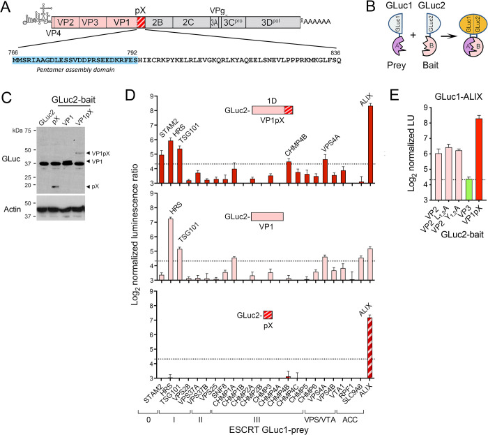
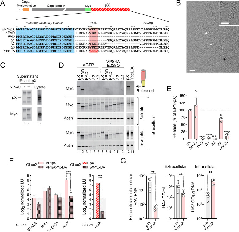
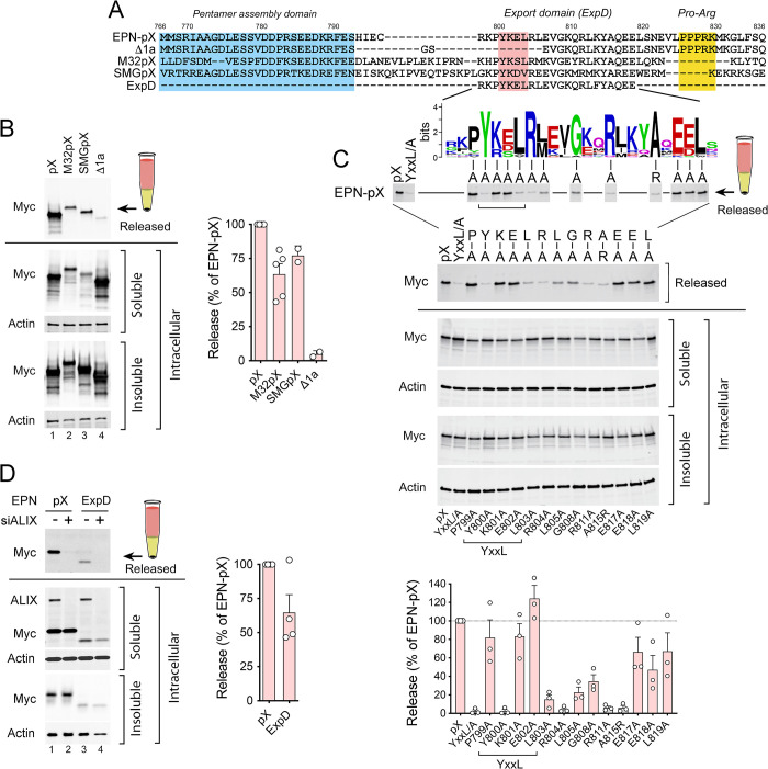
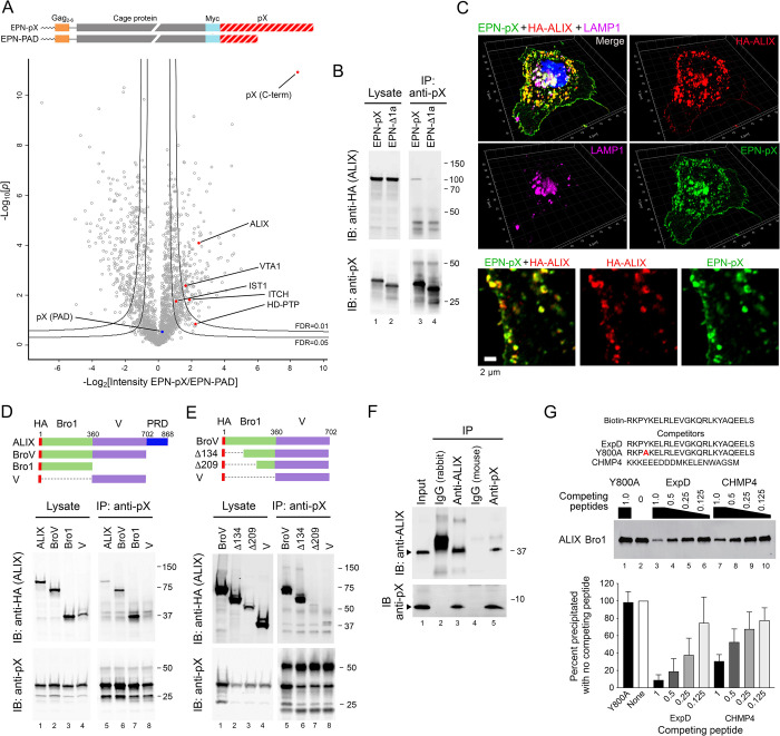
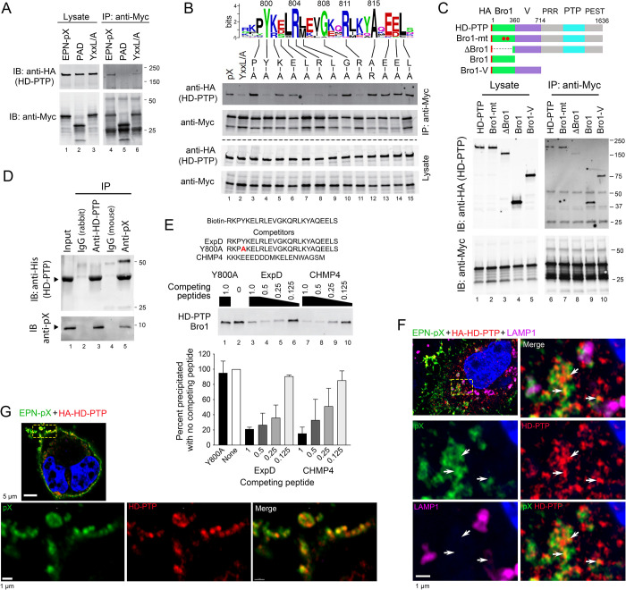
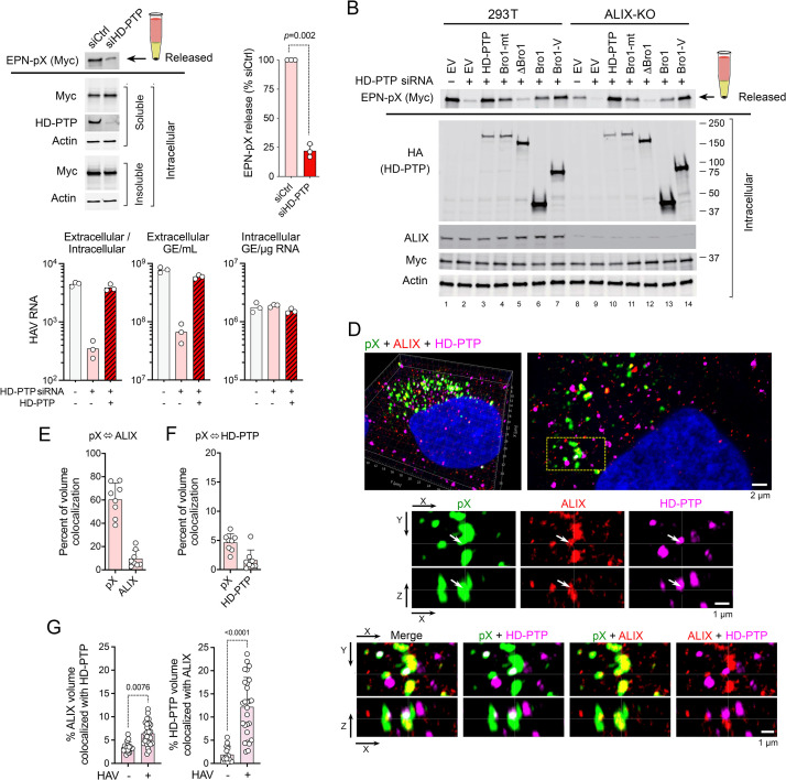
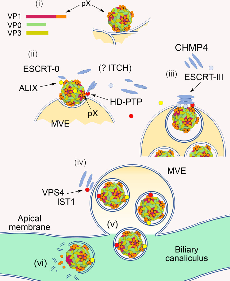
Although picornaviruses are conventionally considered 'nonenveloped', members of multiple picornaviral genera are released nonlytically from infected cells in extracellular vesicles. The mechanisms underlying this process are poorly understood. Here, we describe interactions of the hepatitis A virus (HAV) capsid with components of host endosomal sorting complexes required for transport (ESCRT) that play an essential role in release. We show release of quasi-enveloped virus (eHAV) in exosome-like vesicles requires a conserved export signal located within the 8 kDa C-terminal VP1 pX extension that functions in a manner analogous to late domains of canonical enveloped viruses. Fusing pX to a self-assembling engineered protein nanocage (EPN-pX) resulted in its ESCRT-dependent release in extracellular vesicles. Mutational analysis identified a 24 amino acid peptide sequence located within the center of pX that was both necessary and sufficient for nanocage release. Deleting a YxxL motif within this sequence ablated eHAV release, resulting in virus accumulating intracellularly. The pX export signal is conserved in non-human hepatoviruses from a wide range of mammalian species, and functional in pX sequences from bat hepatoviruses when fused to the nanocage protein, suggesting these viruses are released as quasi-enveloped virions. Quantitative proteomics identified multiple ESCRT-related proteins associating with EPN-pX, including ALG2-interacting protein X (ALIX), and its paralog, tyrosine-protein phosphatase non-receptor type 23 (HD-PTP), a second Bro1 domain protein linked to sorting of ubiquitylated cargo into multivesicular endosomes. RNAi-mediated depletion of either Bro1 domain protein impeded eHAV release. Super-resolution fluorescence microscopy demonstrated colocalization of viral capsids with endogenous ALIX and HD-PTP. Co-immunoprecipitation assays using biotin-tagged peptides and recombinant proteins revealed pX interacts directly through the export signal with N-terminal Bro1 domains of both HD-PTP and ALIX. Our study identifies an exceptionally potent viral export signal mediating extracellular release of virus-sized protein assemblies and shows release requires non-redundant activities of both HD-PTP and ALIX.


Conflict of interest statement

The authors declare no competing interests.

Figures

Fig 1
Fig 1. Protein fragment complementation screen for VP1pX interactions with ESCRT.
(A) HAV genome organization: capsid proteins are in color with pX hatched. Below is the pX sequence of p16 virus. (B) Protein-fragment complementation assay with ‘bait’ and ‘prey’ proteins fused to N-terminal GLuc1 and C-terminal GLuc2 Gaussia princeps luciferase fragments [26]. (C) Representative GLuc immunoblot showing expression levels of GLuc2 bait proteins fused to VP1pX (1D), VP1, or pX in 293T cells. Actin included as a loading control. (D) Screen for interactions of (top) VP1pX, (middle) VP1, and (bottom) pX with ESCRT components. Normalized light units (LU) >20 (dashed line) exceed values from a large panel of control prey proteins [27]. Acc: ESCRT accessory protein. (E) Results of protein-fragment complementation assay assessing interactions of GLuc1-ALIX prey with VP2, VP2 late domain mutants with Ala substitutions of Leu (L1-2A) and Tyr residues (Y1-2A) (14), VP3, or VP1pX fused to GLuc2 as bait. Data in C and E are means from 2 independent experiments, each with 3 technical replicates.
Fig 2
Fig 2. pX mediates ESCRT-dependent extracellular release of assembled EPN-pX nanocages and noncytopathic p16 virus.
(A) (top) EPN-pX, in which pX is fused to the C-terminus of the nanocage protein with an intervening Myc tag. (bottom) pX sequence in EPN-pX and related mutants. The pentamer assembly domain (PAD) [21] and YxxL motif are indicated. Δ1, Δ2 and Δ3 deletions were described previously [23]. (B) Representative electron micrograph showing multiple EPN-pX nanocages (white arrows) within a membrane-bound vesicle (black arrow) in extracellular fluids of transfected 293T cells. Scale bar = 20 nm. The inset shows nanocages in extracellular fluids treated with CHAPS. Scale bar = 30 nm. (C) pX and Myc immunoblots of anti-pX immunoprecipitates of extracellular fluids, with and without NP-40 treatment, versus lysate from EPN-pX transfected cells. (D) Myc immunoblot of released EPN protein recovered following centrifugation through a 20% sucrose cushion, with soluble and insoluble intracellular EPN proteins expressed by cells transfected with EPN-pX and related constructs shown below the solid horizontal line. Cells were co-transfected with vectors expressing eGFP (control, lanes 1–6) or the E228Q VPS4A mutant (lanes 7–12). (E) Quantitation of nanocage protein release normalized to intracellular protein expression. Results are means ±S.E.M. from 3 independent experiments. ****p<0.0001 by ANOVA with Sidak’s multiple comparison test. (F) Protein-fragment complementation assay with (left) VP1pX and (right) pX GLuc2 baits with and without YxxL/A substitutions and GLuc1 prey fused to ESCRT components. Results are means ± S.D. from two independent experiments, each with 3 technical replicates. ***p<0.001 by two-way ANOVA with Sidak’s multiple comparison test. (G) Extracellular HAV RNA determined by RT-PCR [genome equivalents (GE) per mL supernatant fluids] and intracellular HAV RNA [GE per μg total RNA], and calculated extracellular/intracellular RNA ratios, 12 days after transfection of cells with p16 or p16-YxxL/A RNA; GE = genome equivalents. Data are from n = 6 cultures from two transfections, and are representative of multiple independent experiments. **p<0.01 calculated by Mann-Whitney test.
Fig 3
Fig 3. A conserved centrally-located pX peptide sequence, ExpD, mediates ALIX-dependent nanocage release.
(A) Wild-type and mutant pX sequences of human p16 virus aligned with bat virus pX sequences of M32 (Hepatovirus H), recovered from Eidolon helvum, and SMG18520 (‘SMG’, Hepatovirus C), recovered from Miniopterus manavi [39]. The N-terminal pentamer assembly domain is highlighted in blue, central ExpD sequence in red, and C-terminal Pro-Arg motif in yellow. Below is shown a WebLogo for ExpD derived from an alignment of 22 pX sequences from different mammalian hepatoviruses (see S3 Fig). Note that the Δ1a deletion in the human pX sequence extends the Δ1 deletion (Fig 2) to Leu805 with a GlySer insertion resulting from the cloning strategy. (B) Immunoblots showing released EPN-pX, EPN-M32pX, and EPN-SMGpX nanocages recovered from beneath a sucrose cushion, and related soluble and insoluble intracellular EPN proteins. EPN-Δ1a was included as a negative control. Quantitation is shown on the right, with mean p16 and bat pX nanocage release normalized to intracellular protein expression, ± S.E.M., n = 2–5. (C) Alanine scanning of conserved amino acid residues within EPN-pX using the nanocage release assay. Shown below are immunoblots of released and intracellular EPN proteins, and at the bottom quantitation of immunoblots in 3 independent experiments. Brackets indicate residues within the YxxL motif. (D) Immunoblots showing released and soluble/insoluble intracellular EPN-pX and EPN-ExpD proteins following transfection with ALIX (PDCD6IP)-targeting (+) or non-targeting (-) scrambled siRNAs. Immunoblotting of soluble intracellular proteins was with anti-Myc and anti-ALIX antibodies. Quantitation of EPN-ExpD release is shown on the right.
Fig 4
Fig 4. pX interacts directly with the Bro1 domain of ALIX.
(A) EPN-pX constructs expressed in 293T cells for label-free quantitative (LFQ) proteomic analysis of the pX interactome. Below is a volcano plot showing differential abundance of proteins (LFQ intensities) identified in anti-Myc precipitates of EPN-pX versus EPN-PAD lysates. Differences in normalized LFQ peptide intensity are plotted versus significance (p value). Lines represent lower limits of FDR <0.01 and <0.05. Each protein is represented by a single data point with proteins of interest labelled. (B) Co-immunoprecipitation of HA-tagged ALIX with EPN-pX. Proteins in lysates of 293T cells transfected with EPN-pX or EPN-Δ1a were immunoprecipitated with anti-pX, followed by blotting with (top) anti-HA (ALIX) and (bottom) anti-pX. Whole cell lysates are on the left. IP: immunoprecipitation; IB, immunoblot. (C) Merged, single-channel Airyscan fluorescent images of a cell transfected with EPN-pX and HA-ALIX expression vectors, showing pX (green), ALIX (HA, red) and LAMP1 (magenta). At the bottom are shown enlarged dual- and single-channel recordings of the region delineated by the yellow rectangle in the merged image at the top. (D,E) Co-immunoprecipitation of HA-tagged ALIX or the indicated ALIX domain fragments (top) with EPN-pX expressed in 293T cells. (F) Co-immunoprecipitation of bacterially-expressed pX and 6XHis-tagged ALIX Bro1 protein (residues 1–392) mixed in buffer containing 0.7% BSA and 0.07% Tween-20. The input mixture (lane 1) was precipitated with the indicated antibodies, followed by immunoblotting as shown. IgG = isotype control. (G) Biotin-tagged ExpD peptide pulldown assay. Recombinant ALIX Bro1 protein was incubated with synthetic biotin-tagged ExpD peptide (top), affinity isolated on streptavidin beads, and probed with anti-ALIX. Competitor peptides, representing ExpD, the Y800A ExpD mutant, and the C-terminus of CHMP4 [44] (top) were added to the mixture at molar ratios decreasing from 1 to 0.125 relative to the biotin-tagged peptide. At the bottom is shown a quantitative analysis of Bro1 pulled down by biotin-tagged ExpD, normalized to pulldown in the absence of competitor peptide. Results shown are means ± s.e.m. from n = 3 independent experiments.
Fig 5
Fig 5. pX interacts directly with the Bro1 domain of the ALIX paralog, HD-PTP.
(A) Co-immunoprecipitation of HA-tagged HD-PTP with EPN-pX. Proteins in lysates of cells expressing EPN-pX, EPN-PAD, or EPN-YxxL/A (Fig 2A) were precipitated with anti-Myc antibody and immunoblotted with antibody to HD-PTP. (B) Co-immunoprecipitation assays of HD-PTP with EPN-pX mutants containing single amino acid substituitions of conserved residues in the ExpD sequence. See Fig 3C. (C) Co-immunoprecipitation assays of EPN-pX with HD-PTP, HD-PTP with L202D and I206D substitutions in the Bro1 domain that eliminate CHMP4 binding (Bro1-mt) [43], and various HD-PTP domain fragments. PRR, proline-rich region; PTP, protein tyrosine phosphatase; PEST, “rich in proline, glutamate, serine and threonine” [51] (D) Co-immunoprecipitation of bacterially-expressed pX and recombinant HD-PTP Bro1 protein (residues 1–360) produced in E. coli. (E) Biotin-tagged ExpD peptide pulldown assay of HD-PTP Bro1 domain (residues 1–360) produced by in vitro transcription/translation in rabbit reticulocyte lysate. See legend to Fig 4G for details. (F) Merged low magnification (top left) and merged and single-channel enlarged super-resolution Airyscan fluorescence images of the area demarcated by the yellow lines showing pX (green), HD-PTP (HA, red), and LAMP1 (magenta) in a 293T cell expressing EPN-pX and HA-HD-PTP. Arrows indicate sites of pX colocalization with HD-PTP. (G) Similar low magnification (top image) and enlarged super-resolution images showing co-localization of pX and HD-PTP along the plasma membrane.
Fig 6
Fig 6. HD-PTP is functionally required for EPN-pX and HAV release.
(A) EPN-pX release from 293T cells with RNAi-mediated knockdown of HD-PTP (encoded by PTPN23) (see legend to Fig 2D for details of release assay). siCtrl = scrambled siRNA pool. Quantitative analysis on the right with release normalized to release from siCtrl-transfected cells. n = 3 independent experiments. p-value by t-test with Welch’s correction. (B) Rescue of EPN-pX release by overexpression of HD-PTP, HD-PTP with L202D and I206D substitutions (Bro1-mt), or HD-PTP deletion mutants (see Fig 5C) in HD-PTP-depleted 293T cells (lanes 2–7), and HD-PTP-depleted CRISPR-Cas9 engineered 293T ALIX knockout (ALIX-KO) cells (lanes 9–14). siRNA: ‘+’ = siHD-PTP, ‘-’ = scrambled siCtrl. EV = empty vector. (C) (left to right) Ratio of extracellular/intracellular HAV RNA, extracellular HAVl RNA, and intracellular HAV RNA, 48 hrs after cell-free virus infection of Huh-7.5 cells with and without prior RNAi depletion of HD-PTP. Data shown are technical replicates from one of 3 experiments with similar results. GE = genome equivalent. (D) Merged and single-channel super-resolution Airyscan fluorescent microscopy images of pX (green, labelled with monoclonal antibody), endogenous ALIX (red), and endogenous HD-PTP (magenta) in an 18f virus-infected Huh-7 cell. A 3D reconstruction is shown on the top left with a sectional view to the right. Below are single- and dual-channel images shown in two projections (X-Y and X-Z). Arrows in the middle panels indicate a prominent site of pX colocalization with both ALIX and HD-PTP. (E) Quantitative estimates of pX-ALIX volume colocalization in Z-stack images: the first column shows the percentage of pX volume (voxels with pX signal above threshold) containing ALIX signal above threshold; the second column shows the percent ALIX volume containing pX signal. (n = 8 cells). See Methods for details. (F) Quantitative volume analysis of pX colocalization with HD-PTP similar to that in panel E. (G) Voxel colocalization of ALIX with HD-PTP (left) and HD-PTP with ALIX (right) in HAV-infected and uninfected Huh-7.5 cells. Adjusted p-values determined by ANOVA with Kruksal-Wallis test. n = 17–27 cells from multiple experiments.
Fig 7
Fig 7. Model scheme showing how Bro1-domain proteins HD-PTP and ALIX could function non-redundantly in nonlytic hepatovirus release from infected hepatocytes.
(i) Assembly of membrane-associated capsids decorated on their surface with 60 copies of pX. (ii) pX recruits both ALIX and HD-PTP to capsids. (iii) HD-PTP and ALIX act coordinately to recruit STAM2 (ESCRT-0) and CHMP4, scaffolding assembly of an ESCRT-III complex that facilitates inward budding of the capsid on an endosome and mediates abscission of the membrane leading to formation of a multivesicular endosome (MVE). (iv) Upon membrane abscission, VPS4 mediates disassembly of ESCRT-III, regulated in part by IST1, VTA1 and HD-PTP. (v) MVEs traffic to the apical plasma membrane of the hepatocyte where membrane fusion releases quasi-enveloped eHAV into the biliary canaliculus. (vi) Bile salts degrade the eHAV membrane resulting in naked nHAV particles being released from the biliary tract into the small intestine [10].

Similar articles

Cited by

References

    1. Feng Z, Hensley L, McKnight KL, Hu F, Madden V, Ping L, et al.. A pathogenic picornavirus acquires an envelope by hijacking cellular membranes. Nature. 2013;496(7445):367–71. Epub 2013/04/02. doi: 10.1038/nature12029 - DOI - PMC - PubMed
    1. Robinson SM, Tsueng G, Sin J, Mangale V, Rahawi S, McIntyre LL, et al.. Coxsackievirus B exits the host cell in shed microvesicles displaying autophagosomal markers. PLoS Pathog. 2014;10(4):e1004045. Epub 2014/04/12. doi: 10.1371/journal.ppat.1004045 - DOI - PMC - PubMed
    1. Santiana M, Ghosh S, Ho BA, Rajasekaran V, Du WL, Mutsafi Y, et al.. Vesicle-Cloaked Virus Clusters Are Optimal Units for Inter-organismal Viral Transmission. Cell Host Microbe. 2018;24(2):208–20.e8. Epub 2018/08/10. doi: 10.1016/j.chom.2018.07.006 - DOI - PMC - PubMed
    1. van der Grein SG, Defourny KAY, Rabouw HH, Galiveti CR, Langereis MA, Wauben MHM, et al.. Picornavirus infection induces temporal release of multiple extracellular vesicle subsets that differ in molecular composition and infectious potential. PLoS Pathog. 2019;15(2):e1007594. Epub 2019/02/20. doi: 10.1371/journal.ppat.1007594 - DOI - PMC - PubMed
    1. Zell R. Picornaviridae-the ever-growing virus family. Arch Virol. 2018;163(2):299–317. Epub 2017/10/24. doi: 10.1007/s00705-017-3614-8 - DOI - PubMed

Publication types

MeSH terms

Substances