. 2022 Sep 16;17(9):2619-2630.
doi: 10.1021/acschembio.2c00531. Epub 2022 Aug 15.

Analysis of the Sequence Preference of Saporin by Deep Sequencing

Affiliations

Analysis of the Sequence Preference of Saporin by Deep Sequencing

Samuel Hauf et al. ACS Chem Biol. .

Erratum in

Abstract

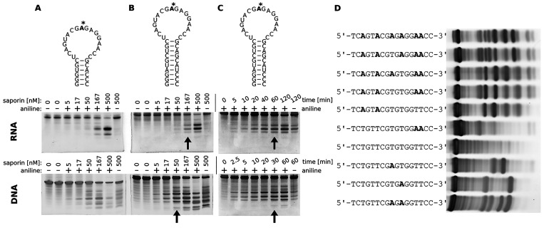
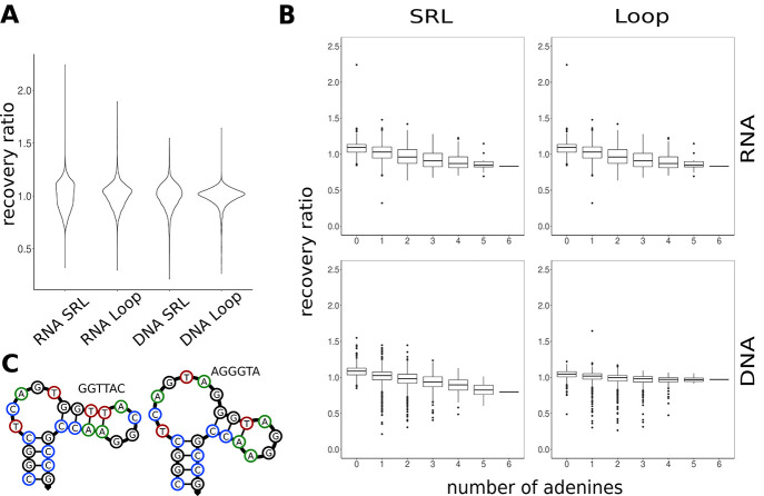
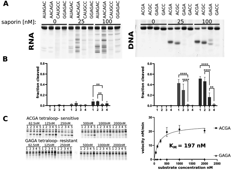
Ribosome-inactivating proteins (RIPs) are RNA:adenosine glycosidases that inactivate eukaryotic ribosomes by depurinating the sarcin-ricin loop (SRL) in 28S rRNA. The GAGA sequence at the top of the SRL or at the top of a hairpin loop is assumed to be their target motif. Saporin is a RIP widely used to develop immunotoxins for research and medical applications, but its sequence specificity has not been investigated. Here, we combine the conventional aniline cleavage assay for depurinated nucleic acids with high-throughput sequencing to study sequence-specific depurination of oligonucleotides caused by saporin. Our data reveal the sequence preference of saporin for different substrates and show that the GAGA motif is not efficiently targeted by this protein, neither in RNA nor in DNA. Instead, a preference of saporin for certain hairpin DNAs was observed. The observed sequence-specific activity of saporin may be relevant to antiviral or apoptosis-inducing effects of RIPs. The developed method could also be useful for studying the sequence specificity of depurination by other RIPs or enzymes.


Conflict of interest statement

The authors declare no competing financial interest.

Figures

Figure 1
Figure 1
Schematic illustration of the process used to treat the DNA and RNA oligos and to construct sequencing libraries. (A) The randomized library may contain sequences that are susceptible or not susceptible to depurination by the RIP being studied. After RIP treatment, some sequences are depurinated (top). These can be cleaved by aniline at the abasic site. Sequences that are resistant to depurination remain intact (bottom). (B) Sequences and secondary structure of both DNA libraries with six randomized bases (N) in the loop. One was based on the ribosomal rRNA sequence of the SRL with the top ACGAGA sequence randomized. The second contained only the six randomized bases in the loop. Both have a stable 11 bp stem and additional flanking 5′ and 3′ constant regions for reverse transcription and PCR. (C) Uncleaved fragments (boxed in red) can be recovered from a denaturing polyacrylamide gel after different treatment times. (D) RNA oligos were reverse transcribed with a primer annealed to the 3′ end (blue). The second strand of cDNA was synthesized with a primer annealed to the other end of the SRL RNA (orange). The cDNA was amplified in a PCR using Illumina UDI primers annealed to sequence newly added in the previous two steps (dark orange and dark blue), yielding the final sequencing library.
Figure 2
Figure 2
Determination of saporin treatment conditions. (A) Cleavage of RNA (OSaH151) and DNA (OSaH152) variants of E73 after treatment with different amounts of saporin for 30 min. The saporin concentrations were 0, 5, 17, 50, 167, and 500 nM. The last two lanes were both incubated with 500 nM saporin, but aniline treatment was omitted in the last lane. The sequence of the RNA oligo is shown on the top with the targeted adenine marked with an asterisk. (B) Similar to panel A, but using oligos with a more stable (longer) stem (OSaH229 and OSaH190). Arrows indicate the condition chosen for kinetic experiments in panel C. (C) The oligos were treated with the amount of saporin indicated by the arrow in panel B for different lengths of time. The arrows indicate the time points deemed appropriate for sequencing. At these points, cleavage bands are visible, but the bulk of the oligo is still unreacted. (D) Nonspecific depurination of 600 nM DNA oligos (OSaH190, -195, -196, -201, -244, -243, -199, and -207–209 from top to bottom, respectively) by 150 nM saporin. All adenine residues in the wild-type SRL sequence (top) are depurinated to some extent. The GAGA pattern is not required for depurination. In panels A–C, canonical and noncanonical base pairs found in the native SRL sequence are not depicted.
Figure 3
Figure 3
Results of the saporin substrate sequencing assays. (A) Two independent treatments of the libraries with saporin were highly reproducible. All variants correlated linearly as shown for the DNA loop library. Judging from the scatter and histogram plots of the abundance ratio across both replicates, the error caused by the method is randomly distributed. The density of the data (blue) closely follows that of a normal distribution (red). (B) Sequence representation of the 100 most efficiently depurinated variants from each library with a sequence logo based on the top 20 variants. Bases are colored green for A, blue for C, orange for G, and red for T (or U). The last column, which is present for only the loop libraries, indicates the possibility of base pairing between randomized positions 1 and 6. Complementary bases at these positions are colored black, while mismatched bases are colored white.
Figure 4
Figure 4
Matrices for the pairwise coupling model. The recovery ratio was used to fit linear regression models that consider pairwise interactions between bases at different positions (see Materials and Methods). The insets show the R2 values (coefficients of determination) between the predicted and observed recovery ratios for each model. Each square shows the linear coefficients of the model that indicate how a particular pair of base combination affects model prediction of the recovery ratio. Blue squares (low linear coefficient) indicate that this base combination promotes depurination by saporin, while red fields (high linear coefficient) indicate that the base combination inhibits depurination. Gray squares show pairs of bases that do not significantly affect depurination by saporin.
Figure 5
Figure 5
Distribution of recovery ratios. (A) Shape of the distribution of recovery ratios for all variants. The distributions of the SRL-based libraries are flatter than those of the loop libraries indicative of a more promiscuous activity. (B) Dependence of recovery on the number of adenine residues. An increasing number of adenine residues reduces the recovery ratios of variants, indicating that adenine is targeted nonspecifically. Interestingly, there are some variants in both DNA libraries that exhibit much lower recovery ratios compared to those of the rest of the population. This indicates that some specific sequence pattern is targeted. (C) Minimum free energy structures of two sensitive variants from the SRL DNA library as predicted by NUPACK. These variants form secondary hairpins with small loops. The small loops closely resemble the sensitive ACG pattern identified in the DNA loop library.
Figure 6
Figure 6
Confirmation of the sequencing results using PAGE. (A) Representative PAGE gels. RNA (500 nM) with randomized AUAGAC (lanes 1, 5, and 9), AACAGA (lanes 2, 6, and 10), CAUGCC (lanes 3, 7, and 11), and GGAGAC (lanes 4, 8, and 12) sequences were treated with the indicated amounts of saporin for 30 min at 37 °C before aniline treatment. DNA oligos OSaH455 (G-ACGA-C in lane lanes 1, 5, and 9), OSaH456 (G-ACGC-C in lanes 2, 6, and 10), OSaH457 (G-GAGA-C in lanes 3, 7, and 11), and OSaH458 (G-GACC-C in lanes 4, 8, and 12) at 500 nM were incubated with the indicated amounts of saporin for 30 min at 37 °C before aniline treatment. Only the sequence in the loop is shown in the figure. (B) Quantification of the fraction cleaved (FL) from PAGE gels using three separate experiments. Shown are the average and standard deviation. Asterisks indicate statistically significant differences at the **p < 0.01 or ****p < 0.0001 level. (C) Kinetic analysis of the most sensitive ACGA tetraloop variant and a tetraloop variant containing the GAGA SRL motif. The indicated amounts of 5′FAM-labeled substrates OSaH525 and OSaH526 were incubated with 20 nM saporin at 37 °C. The gel pictures show cleavage after (1) 0, (2) 5, (3) 10, (4) 15, and (5) 30 min for the sensitive ACGA variant and (1) 0, (2) 30, (3) 50, (4) 90, and (5) 180 min for the GAGA tetraloop variant. Note that the contrast was adjusted in both pictures to better show the cleavage products. The experiment was repeated three times, and the initial reaction velocity estimated after 5 min for the ACGA variant and after 180 min for the GAGA variant. A plot of the average initial velocity against the substrate concentration is shown at the right. Error bars indicate the standard deviation from three experiments. Nonlinear regression was performed to estimate the KM for the sensitive ACGA tetraloop.

Similar articles

Cited by

References

    1. Schrot J.; Weng A.; Melzig M. F. Ribosome-inactivating and related proteins. Toxins (Basel). 2015, 7, 1556–1615. 10.3390/toxins7051556. - DOI - PMC - PubMed
    1. Voorhees R. M.; Schmeing T. M.; Kelley A. C.; Ramakrishnan V. The mechanism for activation of GTP hydrolysis on the ribosome. Science. 2010, 330, 835–838. 10.1126/science.1194460. - DOI - PMC - PubMed
    1. Endo Y.; Mitsui K.; Motizuki M.; Tsurugi K. The mechanism of action of ricin and related toxic lectins on eukaryotic ribosomes. The site and the characteristics of the modification in 28 S ribosomal RNA caused by the toxins. J. Biol. Chem. 1987, 262, 5908–5912. 10.1016/S0021-9258(18)45660-8. - DOI - PubMed
    1. Montanaro L.; Sperti S.; Mattioli A.; Testoni G.; Stirpe F. Inhibition by ricin of protein synthesis in vitro. Inhibition of the binding of elongation factor 2 and of adenosine diphosphate-ribosylated elongation factor 2 to ribosomes. Biochem. J. 1975, 146, 127–131. 10.1042/bj1460127. - DOI - PMC - PubMed
    1. Li X.-P.; Kahn J. N.; Tumer N. E. Peptide Mimics of the Ribosomal P Stalk Inhibit the Activity of Ricin A Chain by Preventing Ribosome Binding. Toxins (Basel). 2018, 10, 371.10.3390/toxins10090371. - DOI - PMC - PubMed

Publication types