. 2022 Aug 23;119(34):e2206240119.
doi: 10.1073/pnas.2206240119. Epub 2022 Aug 15.

De novo designed protein inhibitors of amyloid aggregation and seeding

Affiliations

De novo designed protein inhibitors of amyloid aggregation and seeding

Kevin A Murray et al. Proc Natl Acad Sci U S A. .

Abstract

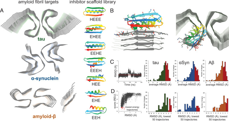
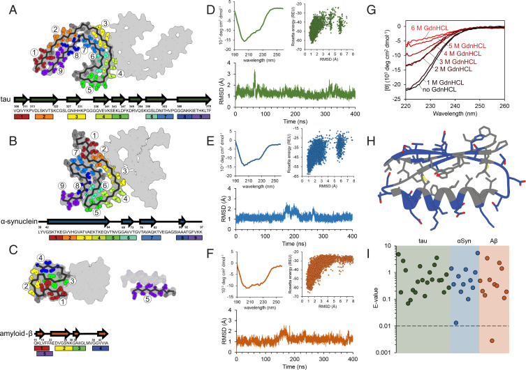
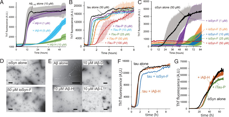
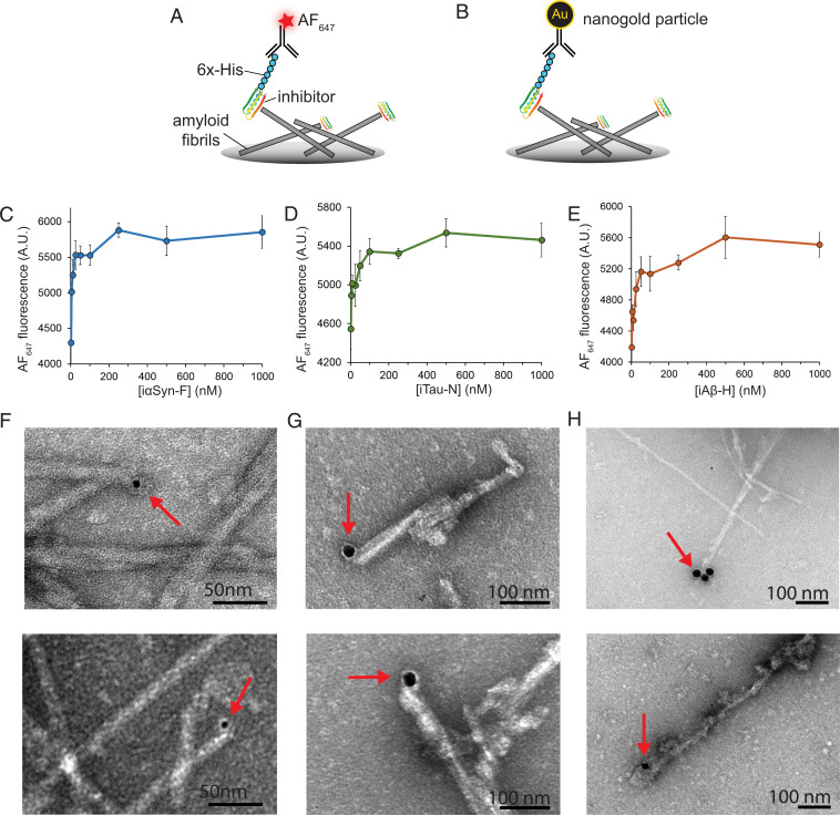
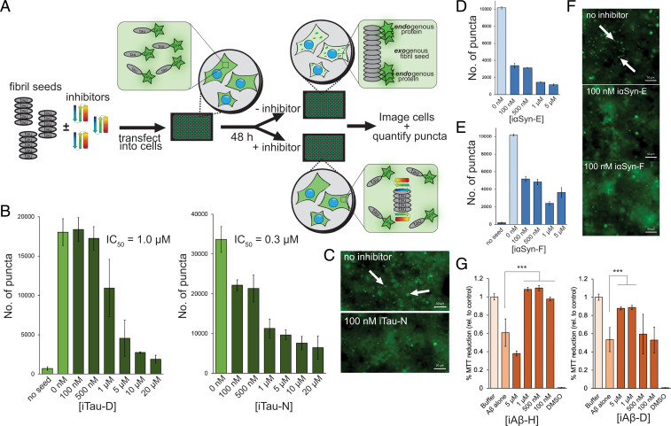
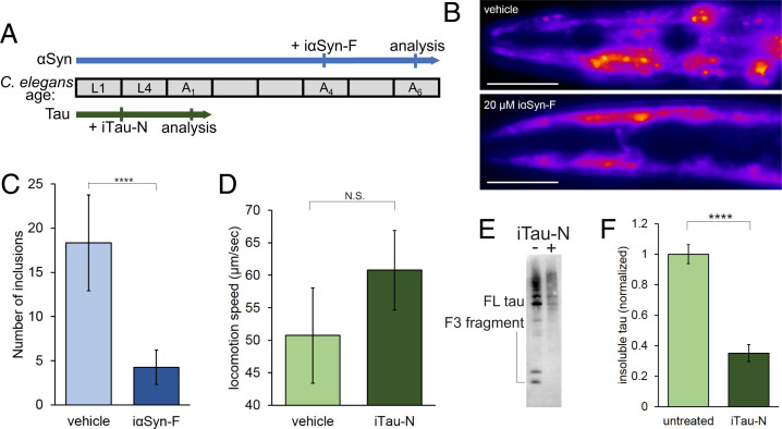
Neurodegenerative diseases are characterized by the pathologic accumulation of aggregated proteins. Known as amyloid, these fibrillar aggregates include proteins such as tau and amyloid-β (Aβ) in Alzheimer's disease (AD) and alpha-synuclein (αSyn) in Parkinson's disease (PD). The development and spread of amyloid fibrils within the brain correlates with disease onset and progression, and inhibiting amyloid formation is a possible route toward therapeutic development. Recent advances have enabled the determination of amyloid fibril structures to atomic-level resolution, improving the possibility of structure-based inhibitor design. In this work, we use these amyloid structures to design inhibitors that bind to the ends of fibrils, "capping" them so as to prevent further growth. Using de novo protein design, we develop a library of miniprotein inhibitors of 35 to 48 residues that target the amyloid structures of tau, Aβ, and αSyn. Biophysical characterization of top in silico designed inhibitors shows they form stable folds, have no sequence similarity to naturally occurring proteins, and specifically prevent the aggregation of their targeted amyloid-prone proteins in vitro. The inhibitors also prevent the seeded aggregation and toxicity of fibrils in cells. In vivo evaluation reveals their ability to reduce aggregation and rescue motor deficits in Caenorhabditis elegans models of PD and AD.

Keywords: alpha-synuclein; amyloid; amyloid-beta; protein design; tau.


Conflict of interest statement

Competing interest statement: D.S.E. is Science Advisory Board chair and equity holder of ADRx, Inc.

Figures

Fig. 1.
Fig. 1.
Computational design of amyloid-inhibiting miniproteins. (A) Amyloid fibrils of three different proteins were used as scaffolds for inhibitor design: tau, αSyn, and Aβ. Atomic structures of the tau paired helical filament derived from Alzheimer’s disease patient brain (Top) (22), the αSyn rod polymorph (Middle) (20), and two forms of Aβ (Bottom) were targeted (14, 41). A library of de novo designed miniproteins was used as inhibitor scaffolds. Seven unique classes of inhibitors were used, each class differing from the arrangement of secondary structural elements (H = α-helix; E = β-sheet). (B) Inhibitor scaffolds were docked to the ends of the fibril structures, capping their growth by preventing further addition of protein monomers. Binding of the miniprotein scaffolds to the fibrils was primarily driven by interacting β-sheets, mimicking the fibril native stacking. (C) The stabilities of top-ranking hits from the docking calculations were assessed by long-range molecular dynamics simulations. Those inhibitors with the lowest average rmsds over time were selected for further testing (red bars). (D) Final screening of inhibitors was performed using Rosetta’s ab initio structure prediction algorithm. The structures of each inhibitor were predicted based on primary sequence alone. The energies of each prediction trajectory were plotted against their rmsd to the original design. Those inhibitors whose lowest energy predictions were the smallest rmsd from the original design were then selected for experimental characterization (red bars).
Fig. 2.
Fig. 2.
Biophysical characterization of designed inhibitors. (AC) Multiple binding sites on each amyloid fibril structure were selected for targeted inhibitor design. The inhibitor scaffolds were systematically docked to different sites along the fibril ends (each number/color corresponding to a unique binding site). The chosen binding sites correspond to particular β-strand segments (shown in arrows) occurring along the protein chain for tau (A) αSyn (B), and Aβ (C). (DF) Initial biophysical characterization of each design consisted of CD spectroscopy (Top Left), ab initio structure prediction (Top Right), and long-range molecular dynamics simulations (Bottom). Inhibitors iTau-N (D), i αSyn-F (E), and iAβ-H (F) are shown to maintain stable folds both computationally and experimentally. (G) To assess the stability of the designed inhibitors, CD measurements were taken after a 20-min incubation with increasing concentrations of the denaturant GdnHCl. iTau-N (shown) remains completely stable in 1 M GdnHCl. GdnHCl denaturation curves do not necessarily show a full cooperative unfolding transition, but may indicate destabilization of the folded miniprotein. (H) The fold of each designed inhibitor is driven by a hydrophobic core region (gray residues) surrounded by an exterior of charged and polar residues (blue) (inhibitor iTau-N shown). (I) Each inhibitor was generated de novo, with no apparent homology to known naturally occurring proteins. BLAST E-values, a metric indicating protein homology, demonstrating the designs are well above the significance threshold of 0.01, for all inhibitors targeting tau (green), αSyn (blue), and Aβ (orange), with the exception of iAβ-L.
Fig. 3.
Fig. 3.
Inhibitors prevent amyloid aggregation by capping fibril ends. (AC) Thioflavin T aggregation kinetics assays for Aβ (A), tau (B), and αSyn (C). Each amyloid protein was aggregated alone and in the presence of increasing concentrations of inhibitor, resulting in reduction in the rate of aggregation or complete abolition of aggregation. (A) The aggregation of Aβ1–42 (10 µM monomer) with the inhibitor iAβ-H. (B) Aggregation of αSyn (50 µM monomer) with inhibitor iαSyn-F nearly eliminates measured aggregation, even at substoichiometric ratios. (C) Tau k18+ (50 µM monomer) with the inhibitor iTau-P (100 µM) produces a fourfold increase in aggregation lag time. (D) Transmission electron micrographs (TEMs) of αSyn (50 µM) aggregated in the absence (Top) or presence (Bottom) of iαSyn-F show the inhibitor prevents the formation of fibrillar aggregates. (Scale bar, 50 nm.) (E) TEM images of Aβ1–42 alone (10 µM) reveal numerous fibrils, whose growth is prevented by the addition of equimolar amounts of iAβ-D, iAβ-H, and iAβ-L. (Scale bar, 100 nm.) (F) Both iαSyn-F and iAβ-H show little effect on tau k18+ aggregation (50 µM k18+ monomer, 50 µM inhibitors). Likewise, iTau-N and iAβ-H have minimal influence on αSyn aggregation (50 µM αSyn monomer, 50 µM inhibitors). All ThT experiments were performed with n = 3 experimental replicates.
Fig. 4.
Fig. 4.
Binding of designed inhibitors to target amyloid fibrils. (A) ELISA binding assays were used to measure inhibitor binding to fibril ends. Inhibitors with uncleaved His-tags were incubated with fibril-coated plates, then labeled with an AF647-conjugated anti-His primary antibody. Fluorescence measurements at different concentrations of inhibitor (iTau-N shown) illustrate binding saturation in the high nanomolar regime. (B) To visualize the designed miniproteins bound to the fibril ends, inhibitors were incubated with fibrils, then labeled with 5 or 20 nm gold nanoparticles (indicated by red arrows), demonstrating a binding mode consistent with their intended design. (CE) ELISA binding curves of iαSyn-F to αSyn fibrils (C), iTau-N to tau k18+ fibrils seeded with AD brain-derived fibrils (D), and iAβ-H to Aβ1–42 fibrils (E). (FH) Transmission electron micrographs of amyloid fibrils bound with miniprotein inhibitors labeled with gold nanoparticles. (F) iαSyn-E bound to αSyn fibrils. (G) iTau-N bound to tau k18+ fibrils seeded with AD brain-derived fibrils. (H) iAβ-H bound to Aβ1–42 fibrils. In all three samples, inhibitors are observed binding primarily to the tips of the fibrils, consistent with their intended design. Additional TEM images are available in SI Appendix, Fig. 5.
Fig. 5.
Fig. 5.
Inhibition of amyloid seeding and toxicity in cells. (A) To assess the effects of the designed inhibitors on amyloid seeding within cells, a HEK293T biosensor cell assay was used for both tau and αSyn. Fibril seeds (tau shown) are incubated overnight with inhibitors, then transfected into the biosensor cells. The cells overexpress amyloid protein (either tau k18 or αSyn) fused to YFP/CFP. At baseline, the cells show diffuse fluorescence, but the endogenous fluorescent amyloid protein can be incorporated into the transfected exogenous fibril seeds, resulting in visible fluorescent puncta. Addition of inhibitor caps the fibrils, preventing the incorporation of the YFP/CFP-labeled amyloid protein and the subsequent formation of puncta. (BE). Inhibitors of tau and alpha-synuclein inhibit intracellular seeding in biosensor cells. Number of puncta on the y axis refers to number of fluorescent intracellular aggregates per experimental well of a 96-well plate. (B). Inhibitors iTau-D and iTau-N cause a significant reduction in the number of fluorescent puncta in cells transfected with AD patient brain extract containing tau fibrils. Minimal aggregation occurs in the absence of transfected fibril seeds. (C) Fluorescent microscopy images of biosensor cells with and without 100 nM iTau-N. Seeded aggregates can be visualized by discrete bright puncta (indicated by white arrows). (DF) Inhibitors iαSyn-E (D) and iαSyn-F (E) greatly reduce aggregated puncta in biosensor cells expressing fluorescently labeled αSyn. (F) Similar to tau aggregates, αSyn aggregates can be visualized as intracellular fluorescent puncta and quantified (white arrows). (G) MTT dye reduction assays were used to assess the capacity of inhibitors to mitigate Aβ aggregate-induced cytotoxicity in N2a neuronal cells. Aβ aggregates alone (1 µM) resulted in ∼40% cell death compared to buffer control. This toxicity can be rescued by addition of either iAβ-H or iAβ-D. (Scale bars, 50 µm.) All error bars represent SD. All statistical analyses were performed using a one-way Analysis of Variance (ANOVA) (***P = 0.002; N.S., nonsignificant).
Fig. 6.
Fig. 6.
Inhibitors prevent amyloid aggregation in C. elegans model strains. (A) Two strains of C. elegans were used to test the in vivo efficacy of the designed miniprotein inhibitors (αSyn: DDP1, tau: BR5706). A timeline of worm lifespan through larval stages (L) and adult days (A) is shown, indicating age of worms during inhibitor treatment and subsequent analysis. (B and C). Alpha-synuclein strain DDP1 overexpresses fluorescently labeled αSyn, which aggregates in the worm head region as adults. (B) Vehicle-treated worms display numerous fluorescent puncta as day 6 adults, which are diminished with the addition of iαSyn-F. (Scale bars, 100 µm.) (C) Quantification of head region αSyn aggregates reveals a large reduction in inhibitor-treated worms. n = 15 worms were used for each experimental condition. (DF). Tau strain BR5706 coexpresses full-length tau with the proaggregation V337M mutation and the F3 fragment of the tau microtubule binding domain with the proaggregation K280 deletion. (D). The movement of treated tau worms was tracked, and average locomotion speed across 30-s intervals was measured. Treatment iTau-N leads to an increase in speed, indicating a recovery of locomotion deficits. n = 15 worms were used for each experimental condition. (E) Tau Western blot of RIPA-soluble worm extracts with and without inhibitor iTau-N treatment. Bands of full-length (FL) and the F3 fragment are indicated. (F) Western blot quantification shows a significant reduction of insoluble tau species in the iTau-N–treated C. elegans compared to vehicle control. Experiment was performed in triplicate, with n > 100 worms for each experimental condition. All error bars represent SD. All statistical analyses were performed using an unpaired t test (****P < 0.0001; N.S., nonsignificant).

Similar articles

Cited by

References

    1. Eisenberg D., Jucker M., The amyloid state of proteins in human diseases. Cell 148, 1188–1203 (2012). - PMC - PubMed
    1. Bloom G. S., Amyloid-β and tau: The trigger and bullet in Alzheimer disease pathogenesis. JAMA Neurol. 71, 505–508 (2014). - PubMed
    1. Spillantini M. G., Crowther R. A., Jakes R., Hasegawa M., Goedert M., alpha-Synuclein in filamentous inclusions of Lewy bodies from Parkinson’s disease and dementia with lewy bodies. Proc. Natl. Acad. Sci. U.S.A. 95, 6469–6473 (1998). - PMC - PubMed
    1. Wang Q., Yu X., Li L., Zheng J., Inhibition of amyloid-β aggregation in Alzheimer’s disease. Curr. Pharm. Des. 20, 1223–1243 (2014). - PubMed
    1. Bulic B., et al. , Development of tau aggregation inhibitors for Alzheimer’s disease. Angew. Chem. Int. Ed. Engl. 48, 1740–1752 (2009). - PubMed

Publication types

MeSH terms