. 2022 Aug 23;119(34):e2207134119.
doi: 10.1073/pnas.2207134119. Epub 2022 Aug 15.

DYF-5/MAK-dependent phosphorylation promotes ciliary tubulin unloading

Affiliations

DYF-5/MAK-dependent phosphorylation promotes ciliary tubulin unloading

Xuguang Jiang et al. Proc Natl Acad Sci U S A. .

Abstract

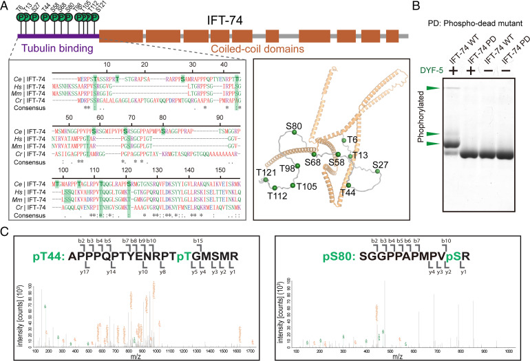
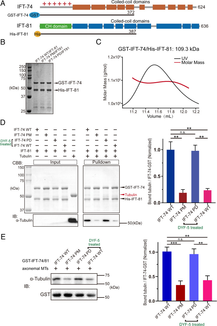
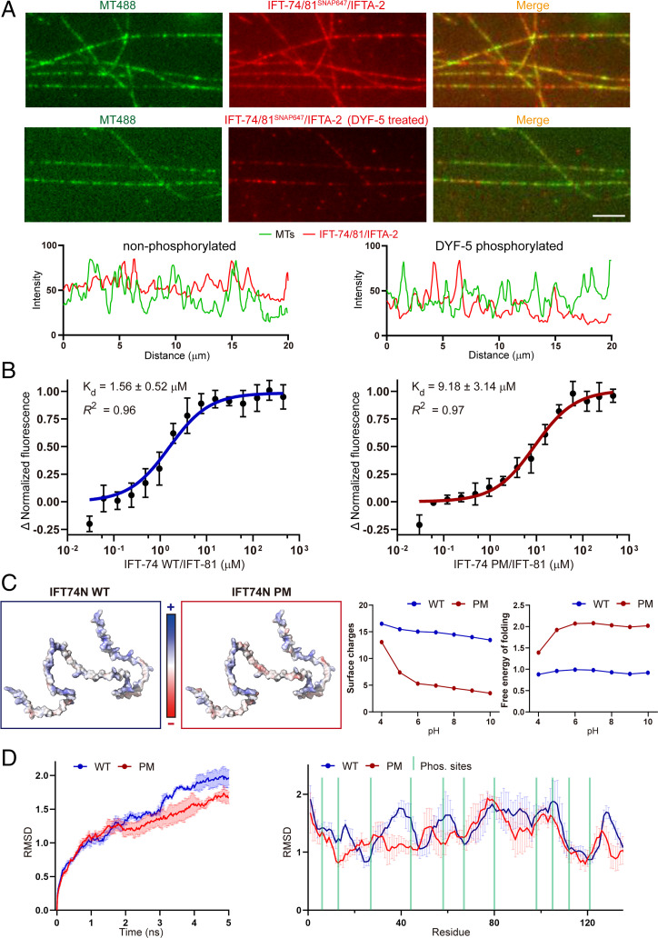
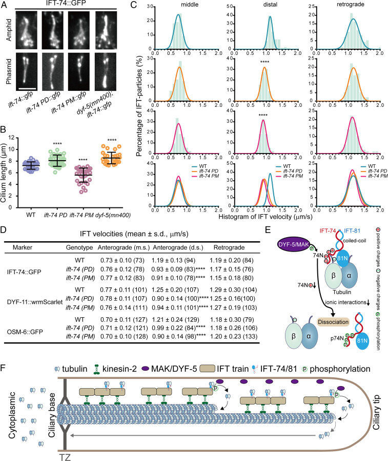
Cilia are microtubule-based organelles that power cell motility and regulate sensation and signaling, and abnormal ciliary structure and function cause various ciliopathies. Cilium formation and maintenance requires intraflagellar transport (IFT), during which the kinesin-2 family motor proteins ferry IFT particles carrying axonemal precursors such as tubulins into cilia. Tubulin dimers are loaded to IFT machinery through an interaction between tubulin and the IFT-74/81 module; however, little is known of how tubulins are unloaded when arriving at the ciliary tip. Here, we show that the ciliary kinase DYF-5/MAK phosphorylates multiple sites within the tubulin-binding module of IFT-74, reducing the tubulin-binding affinity of IFT-74/81 approximately sixfold. Ablation or constitutive activation of IFT-74 phosphorylation abnormally elongates or shortens sensory cilia in Caenorhabditis elegans neurons. We propose that DYF-5/MAK-dependent phosphorylation plays a fundamental role in ciliogenesis by regulating tubulin unloading.


Conflict of interest statement

The authors declare no competing interest.

Figures

Fig. 1.
Fig. 1.
DYF-5 phosphorylates the tubulin-binding region of IFT-74. (A) Schematic diagrams (Top), homologous sequence alignment (Bottom Left), and cartoon structural representation (Bottom Right) showing the identified phosphorylation sites in C. elegans IFT-74. The phosphorylation residues and their possible corresponding homologous sites were labeled with green shadows in the alignment. The structural coordinate of IFT-74 from AlphaFold Protein Structure Database (ID: A0A2C9C2L6) was used to generate the structural figure. (B) Phos-tag kinase assays validating the determined phosphorylation sites. Purified WT and phospho-dead (PD; Ser-Ala/Thr-Ala) mutant IFT-74 (1-372) protein was treated with DYF-5 kinase and evaluated by Phos-tag SDS-PAGE. The phosphorylation-induced bandshifts are indicated by green arrowheads. (C) Representative LC-MS/MS results of the determination of IFT-74 phosphorylation sites.
Fig. 2.
Fig. 2.
IFT-74 phosphorylation by DYF-5 disrupts the binding of tubulin with IFT-74/81. (A) Schematic diagrams showing the IFT-74/81 constructs used for biochemical analysis, which are tagged by GST (GST-IFT-74 1–372) or 6×His (His-IFT-81 1–387). (B) SDS-PAGE result of the purified IFT-74/81 heterodimer proteins. (C) SEC-MALS analysis of the IFT-74/81 recombinant protein. (D) Pull-down assay evaluating the interaction of tubulin with WT, phospho-mutant, and DYF-5-treated IFT-74/81 proteins. (E) Pull-down assay analyzing tubulin-IFT-74/81 binding using C. reinhardtii axonemal MTs. The assays were performed with three replicates and the quantifications are shown at the right. PM: phospho-mimic mutant; PD: phospho-dead mutant. Statistical significance compared with indicated genotype is based on Student's t-test, **P < 0.01, ***P < 0.001, n.s., no significance.
Fig. 3.
Fig. 3.
IFT-74 phosphorylation diminishes its tubulin-binding affinity via rearrangement of surface potentials. (A) TIRF MT-colocalization assay evaluating the interaction between MTs and IFT-74/81/IFTA-2 protein with or without DYF-5 treatment. Representative fluorescence intensity shown (Bottom) was measured by a 20-μm line randomly drawn on MTs. Scale bar, 5 µm. (B) Microscale thermophoresis assay dissecting the binding affinity of tubulin with WT and phospho-mimic IFT-74/81 proteins. (C) Analysis of the phosphorylation-induced changes of IFT-74N charges and folding free energy. (D) Molecular dynamic analysis of the phosphorylation-induced changes of IFT-74N structural stability/flexibility. AlphaFold2-based structural coordinate of IFT-74 (ID: A0A2C9C2L6) was used for the analysis. IFT-74N: IFT-74 N terminus.
Fig. 4.
Fig. 4.
Phosphorylation of IFT-74 controls cilium length via regulating intraciliary tubulin transport. (A) Amphid (Top) and phasmid (Bottom) cilia morphology in ift-74 PM and PD mutants in comparison with WT (N2) and dyf-5 null mutant (mn400) animals. Cilia were imaged with GFP-tagged WT or mutated IFT-74; IFT-74 PD::GFP and IFT-74 PM::GFP were tracked, and the IFT-74::GFP was tracked in the WT animals or dyf-5(mn400) null mutant worms. (Scale bar: 5 μm.) (B) Cilium length in WT, dyf-5(mn400), and ift-74 PM/PD animals (mean ± SD; n = 30; ****P < 0.0001). IFT-74::GFP was used as the cilium marker. (C) Histogram of IFT velocities in WT and ift-74 mutant animals. (Left) Anterograde IFT along the middle segments. (Middle) Anterograde IFT along the distal segments. (Right) Retrograde IFT. IFT-74::GFP was used as the IFT marker for the measurement. Each plot was fit by a Gaussian distribution. Comparisons were performed between the WT and mutants. ****P < 0.0001. (D) Statistics of IFT velocities of WT and mutant animals measured independently using IFT-74::GFP, DYF-11::wrmScarlet, and OSM-6::GFP as ciliary IFT markers. m.s.: middle segment; d.s.: distal segment; ****P < 0.0001. The n values are given in the parentheses. PM: phospho-mimic mutant; PD: phospho-dead mutant. (E) Model representation of DYF-5/MAK–induced IFT-74/81–tubulin dissociation via IFT-74N phosphorylation. (F) Model illustrating the regulation of intraciliary tubulin transport by DYF-5/MAK–mediated IFT-74 phosphorylation. IFT-74/81 binds and transports tubulin toward ciliary middle and tip, where DYF-5/MAK phosphorylates the IFT-74 tubulin-binding region to release bound tubulin for ciliary construction and maintenance. TZ: transition zone.

References

    1. Mitchell D. R., Evolution of cilia. Cold Spring Harb. Perspect. Biol. 9, a028290 (2017). - PMC - PubMed
    1. Reiter J. F., Leroux M. R., Genes and molecular pathways underpinning ciliopathies. Nat. Rev. Mol. Cell Biol. 18, 533–547 (2017). - PMC - PubMed
    1. Kozminski K. G., Johnson K. A., Forscher P., Rosenbaum J. L., A motility in the eukaryotic flagellum unrelated to flagellar beating. Proc. Natl. Acad. Sci. U.S.A. 90, 5519–5523 (1993). - PMC - PubMed
    1. Rosenbaum J. L., Witman G. B., Intraflagellar transport. Nat. Rev. Mol. Cell Biol. 3, 813–825 (2002). - PubMed
    1. Taschner M., Lorentzen E., The intraflagellar transport machinery. Cold Spring Harb. Perspect. Biol. 8, a028092 (2016). - PMC - PubMed

Publication types

MeSH terms

Substances

LinkOut - more resources