. 2022 Aug 23;119(34):e2207641119.
doi: 10.1073/pnas.2207641119. Epub 2022 Aug 15.

Structure of a cholinergic cell membrane

Affiliations

Structure of a cholinergic cell membrane

Nigel Unwin. Proc Natl Acad Sci U S A. .

Abstract

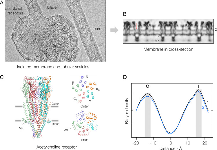
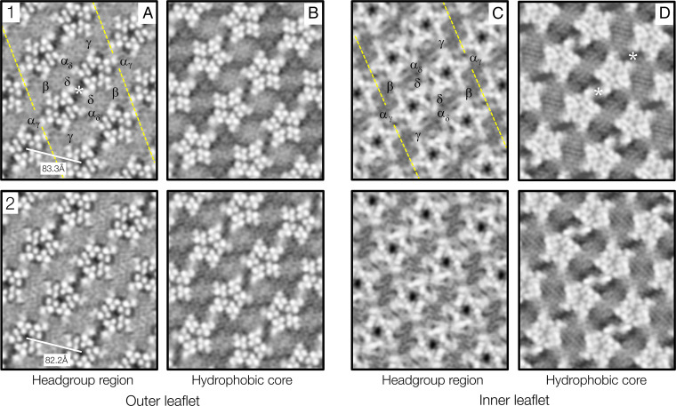
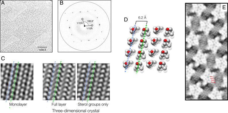
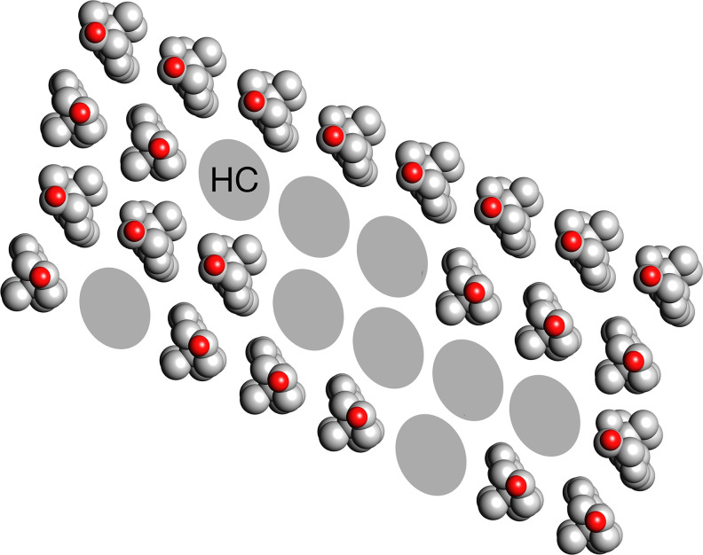
Cell membranes are complex assemblies of proteins and lipids making transient or long-term associations that have yet to be characterized at a molecular level. Here, cryo-electron microscopy is applied to determine how phospholipids and cholesterol arrange between neighboring proteins (nicotinic acetylcholine receptors) of Torpedo cholinergic membrane. The lipids exhibit distinct properties in the two leaflets of the bilayer, influenced by the protein surfaces and by differences in cholesterol concentration. In the outer leaflet, the lipids show no consistent motif away from the protein surfaces, in keeping with their assumed fluidity. In the inner leaflet, where the cholesterol concentration is higher, the lipids organize into extensive close-packed linear arrays. These arrays are built from the sterol groups of cholesterol and the initial saturated portions of the phospholipid hydrocarbon chains. Together, they create an ordered ∼7 Å-thick "skin" within the hydrophobic core of the bilayer. The packing of lipids in the arrays appears to bear a close relationship to the linear cholesterol arrays that form crystalline monolayers at the air-water interface.

Keywords: acetylcholine receptor; cholesterol; cryo-EM; lipid bilayer; phospholipid.


Conflict of interest statement

The author declares no competing interest.

Figures

Fig. 1.
Fig. 1.
Overview and density profile of the lipid bilayer. (A) Isolated cell membrane and budding acetylcholine receptor tubes. In intact tissue, the receptors typically form dimer ribbons packed tightly side to side (14), a regular arrangement that is lost during membrane extraction, but is restored in the budding tubes (15). Since the protein organizes the same way in both contexts, the tube membranes may recapitulate precisely the region of the cell membrane at the synapse where receptors are most densely packed (16). (B) Cross-section determined from segments of tubes having the same lattice dimensions and curvature. The phospholipid headgroup regions in the outer (O) and inner (I) leaflets give rise to a pair of parallel bands in the spaces between individual receptors. MX identifies a helix of the receptor (see C), which substitutes for some of the phospholipid headgroups (17); the arrow points to the receptor’s central water-filled pore. (C) Structure of receptor and cross-sectional 12 Å-thick slabs at the levels of the gray lines, which identify the peaks of density associated with the phospholipid headgroups (17). The receptor is a heteropentamer (stoichiometry: αδ, αγ, β, γ, δ), which includes four TM helices (M1-M4) and a transverse submembrane helix, MX, in each subunit. (D) Mean lipid densities in maps 1 and 2 at successive radii across the lipid bilayer. The shaded columns mark the positions of the two density peaks, which are at different distances from the central low-density trough.
Fig. 2.
Fig. 2.
Lipid-protein organization in the outer and inner leaflets of maps 1 and 2. (A) and (C) are at levels corresponding to the peaks of density in Fig. 1D; (B) and (D) are 7 Å further into the interior of the bilayer. The broken yellow lines identify the well-documented ribbon of δ subunit-linked receptor dimers (14, 15, 18, 24), one of which is labeled in each leaflet to indicate the subunit arrangement. The receptors are ∼1 Å further apart along the direction of the tube axis (horizontal) in map 1 than in map 2. The asterisks mark the locations of previously described cholesterol microdomains (17). Inverted contrast.
Fig. 3.
Fig. 3.
Linear arrays of cholesterol and phospholipids. (A) Image of a cholesterol monolayer film (blotchy areas) on a thin carbon support. (B) Fourier transform showing diffraction spots and contrast transfer function-modulated background intensities; the ring is at a resolution of 3.6 Å. (C) Filtered image of the monolayer film (Left) and projection structures calculated from the closely related triclinic crystal layer (Right): complete layer and with the isooctyl chains removed. (D) Packing of cholesterol in the triclinic crystal layer (26): the molecules align with slightly different tilts along rows, wherein they face alternately in opposite directions. Equivalent rows in C and D are identified by blue and green dashed lines. (E) Detail from Fig. 2D, showing close matching of the 6.2 Å row periodicities (red bars) and the periodicities present in the cell membrane.
Fig. 4.
Fig. 4.
Ordering of the hydrocarbon chains imposed by cholesterol. It is proposed that the cholesterol monomers, no longer stable under saturating conditions in a fluid hydrocarbon environment, self-assemble in linear arrays as in the monolayer films. Side-to-side interactions also occur involving not only other sterol groups but also, in a random way, the paired hydrocarbon chains (HC) of the phospholipids. Because the density maps average over many different sterol/hydrocarbon combinations, continuous lines appear, which are roughly the same distance apart as are the linear arrays of cholesterol in the monolayer films (Fig. 3E).

Similar articles

Cited by

References

    1. Singer S. J., Nicolson G. L., The fluid mosaic model of the structure of cell membranes. Science 175, 720–731 (1972). - PubMed
    1. Gennis R. B., Biomembranes: Molecular Structure and Function (Springer-Verlag New York Inc., 1989).
    1. Bretscher M. S., Asymmetrical lipid bilayer structure for biological membranes. Nat. New Biol. 236, 11–12 (1972). - PubMed
    1. Branton D., Fracture faces of frozen membranes. Proc. Natl. Acad. Sci. U.S.A. 55, 1048–1056 (1966). - PMC - PubMed
    1. Levine Y. K., Wilkins M. H. F., Structure of oriented lipid bilayers. Nat. New Biol. 230, 69–72 (1971). - PubMed

Publication types