. 2022 Oct;298(10):102375.
doi: 10.1016/j.jbc.2022.102375. Epub 2022 Aug 13.

Crystal structures of Schistosoma mansoni histone deacetylase 8 reveal a novel binding site for allosteric inhibitors

Affiliations

Crystal structures of Schistosoma mansoni histone deacetylase 8 reveal a novel binding site for allosteric inhibitors

Fulvio Saccoccia et al. J Biol Chem. 2022 Oct.

Abstract

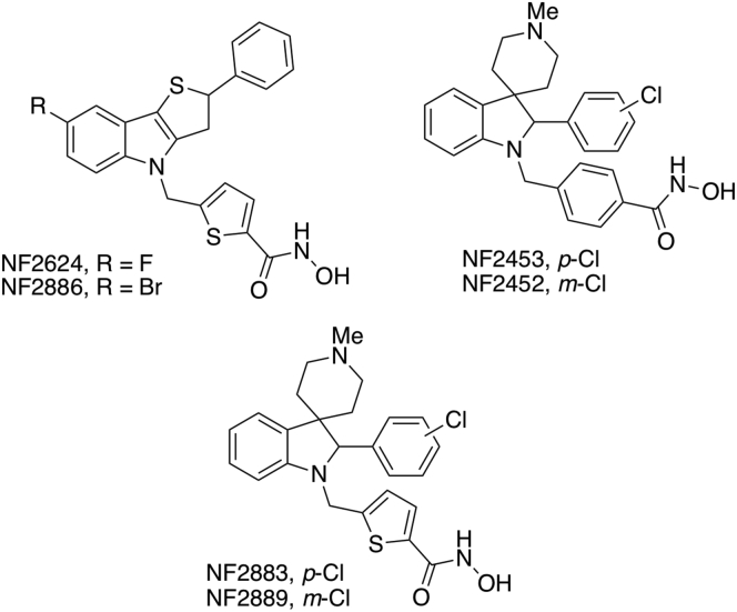
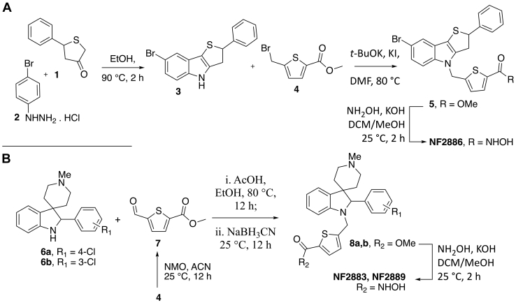
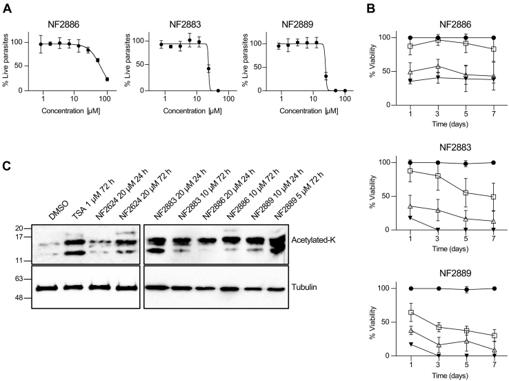
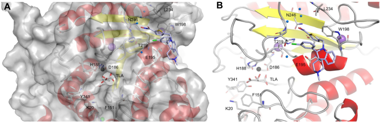
Parasitic diseases cause significant global morbidity and mortality particularly in the poorest regions of the world. Schistosomiasis, one of the most widespread neglected tropical diseases, affects more than 200 million people worldwide. Histone deacetylase (HDAC) inhibitors are prominent epigenetic drugs that are being investigated in the treatment of several diseases, including cancers and parasitic diseases. Schistosoma mansoni HDAC8 (SmHDAC8) is highly expressed in all life cycle stages of the parasite, and selective inhibition is required in order to avoid undesirable off-target effects in the host. Herein, by X-ray crystal structures of SmHDAC8-inhibitor complexes, biochemical and phenotypic studies, we found two schistosomicidal spiroindoline derivatives binding a novel site, next to Trp198, on the enzyme surface. We determined that by acting on this site, either by mutation of the Trp198 or by compound binding, a decrease in the activity of the enzyme is achieved. Remarkably, this allosteric site differs from the human counterpart; rather, it is conserved in all Schistosoma species, as well as Rhabidoptera and Trematoda classes, thus paving the way for the design of HDAC8-selective allosteric inhibitors with improved properties.

Keywords: HDAC8; Schistosoma mansoni; crystal structure; drug discovery; histone deacetylase inhibitor; inhibition mechanism; molecular evolution; parasites’ phylogeny.


Conflict of interest statement

Conflict of interest The authors declare that they have no conflicts of interest with the contents of this article.

Figures

Figure 1
Figure 1
Structures of the thieno[3,2-b]indole and spiroindoline derivatives HDAC inhibitors investigated in the present work.
Figure 2
Figure 2
Synthesis of the thieno[3,2-b]indole and spiroindoline derivatives HDAC inhibitors investigated in the present work.A, synthesis of NF2886. B, synthesis of NF2883 and NF2889.
Figure 3
Figure 3
Viability assays and histoneK-acetylation on parasites.A, dose-response curves of the inhibitors on schistosomula. The y-axis indicates the percentage of viability normalized against DMSO (100%) and gambogic acid 10 μM (0%). Each point represents the average and SD of three independent experiments. The calculated LD50 (μM) was as follows: NF2886, 59.9 ± 4.9; NF2883, 26.2 ± 1.1; NF2889 26.2 ± 1.6. B, dose-response curve of the inhibitors on adult schistosome pairs. DMSO (vehicle) was used as negative control (100% viability – full circle), the indicated compounds were assayed at 10 (open square), 20 (open triangle), and 50 μM (inverted full triangle). Data are expressed as a percent severity score (viability) relative to DMSO. Each point represents the average ± standard error of three independent experiments. C, effects of selected compounds on histones K-acetylation. Representative immunoblot of the histone-enriched protein fractions extracted from S. mansoni adult worm pairs incubated with antiacetylated lysine antibody (acetylated-K) or antitubulin. Worm pairs were treated for 24 or 72 h at the concentration indicated for each compound. DMSO (vehicle) and 1 μM of the HDAC pan inhibitor, TSA, were used as negative and positive controls, respectively. DMSO, dimethyl sulfoxide.
Figure 4
Figure 4
Cartoon representations of SmHDAC8 and NF2889 binding.A, binding of NF2889 on the surface of SmHDAC8 on the new site; the active site is also shown. NF2889 is depicted as light blue sticks; loops between helices are depicted in gray; secondary structure elements are in red and yellow; highlighted in sticks are the side chains of the residues involved in binding interaction with NF2889. B, the active site is in the background where zinc ion (depicted as a gray sphere), TLA (standing for Na/K-tartrate), and some residues involved in catalysis are also highlighted. Water molecules involved in hydrogen bonds are represented as blue spheres.
Figure 5
Figure 5
Fluorescence emission spectra of SmHDAC8 in complexes with NF2886 and NF2889.A, WT, excitation 280 nm; B, W198A mutant, excitation 280 nm; C, WT, excitation 298 nm; D, W198A, excitation 298 nm. NF2886 (gray cross marker) and NF2889 (gray squares). DMSO is included for comparison, as control (full circles).
Figure 6
Figure 6
HDAC8 phylogenetic tree and conservation ofthe novelsite.A, maximum-likelihood phylogenetic tree inference of orthologs of SmHDAC8 based on sequences retrieved from WormBase ParaSite (entry WBGT00950000411223) and Ensembl Metazoa (see Experimental procedures). Details on protein sequences used are given in Supporting Information S8. Sequences belonging to the Schistosoma genus are labeled in red; Rhabditophora class and other Trematodes are in purple; Cestoda are in blue; Nematoda are in green, with Enoplea class in brown. Amphimedon queenslandica (Porifera) sequence was inserted as an outgroup and used to root the tree (bottom, black). Nodes report the confidence obtained by both fast SH-aLRT and ultrafast bootstrap with 5000 replicates each. B, the region of the multiple sequence alignment corresponding to the novel site in SmHDAC8 (entry Smp_091990) is highlighted with red line alongside with residues contributing to the loop β5-β6; the position corresponding to W198 in SmHDAC8 is boxed in red.
Figure 6
Figure 6
HDAC8 phylogenetic tree and conservation ofthe novelsite.A, maximum-likelihood phylogenetic tree inference of orthologs of SmHDAC8 based on sequences retrieved from WormBase ParaSite (entry WBGT00950000411223) and Ensembl Metazoa (see Experimental procedures). Details on protein sequences used are given in Supporting Information S8. Sequences belonging to the Schistosoma genus are labeled in red; Rhabditophora class and other Trematodes are in purple; Cestoda are in blue; Nematoda are in green, with Enoplea class in brown. Amphimedon queenslandica (Porifera) sequence was inserted as an outgroup and used to root the tree (bottom, black). Nodes report the confidence obtained by both fast SH-aLRT and ultrafast bootstrap with 5000 replicates each. B, the region of the multiple sequence alignment corresponding to the novel site in SmHDAC8 (entry Smp_091990) is highlighted with red line alongside with residues contributing to the loop β5-β6; the position corresponding to W198 in SmHDAC8 is boxed in red.
Figure 7
Figure 7
Structural superposition of DMSO-bound (gray cartoon), NF2624 (red ribbon), NF2886 (purple ribbon), NF2883 (yellow ribbon), and NF2889 (cyan ribbon) complexes (bottom-left). Metals are depicted as spheres: zinc, in gray, potassium, in violet. Inhibitors were omitted for clarity. The figure highlights how the folding of loops surrounding the active site are affected by inhibitors binding; in particular, the binding of NF2889 determines the largest variation in the position of loop 2A/B (residues 80–85 and 97–106, respectively) and loop β5-β6 (residues 210–243). Interestingly, any mobile loop contains some of the key residues, which are directly or indirectly involved in catalysis, in particular loop 1 (residues 16–24) contains K20; loop 2 which contributes with binding rail couple, Y99 and D100; loop β8-α12 (residues 337–342), β7-α11 (residues 281–317), and loop β5-β6 (residues 210–243) contain all the key residues involved in catalysis, D186, H188, D286, and Y341. Best-fit structural superposition was achieved by the Bio3d package within R software (35). Side panels report the trajectories of interpolated structures obtained after PCA for the first three PCs. The largest variations are highlighted and labeled according to loop numbering as reported in Marek et al., 2013 (15). DMSO, dimethyl sulfoxide; PCA, principal component analysis.

References

    1. McManus D.P., Dunne D.W., Sacko M., Utzinger J., Vennervald B.J., Zhou X.N. Schistosomiasis. Nat. Rev. Dis. Prim. 2018;4:1–19. - PubMed
    1. Cioli D., Pica-Mattoccia L., Basso A., Guidi A. Schistosomiasis control: praziquantel forever? Mol. Biochem. Parasitol. 2014;195:23–29. - PubMed
    1. Leger E., Webster J.P. Hybridizations within the genus schistosoma: implications for evolution, epidemiology and control. Parasitology. 2017;144:65–80. - PubMed
    1. Hailu G.S., Robaa D., Forgione M., Sippl W., Rotili D., Mai A. Lysine deacetylase inhibitors in parasites: past, present, and future perspectives. J. Med. Chem. 2017;60:4780–4804. - PubMed
    1. Oger F., Dubois F., Caby S., Noël C., Cornette J., Bertin B., et al. The class I histone deacetylases of the platyhelminth parasite Schistosoma mansoni. Biochem. Biophys. Res. Commun. 2008;377:1079–1084. - PubMed

Publication types

MeSH terms

LinkOut - more resources