Review
. 2022 Aug 15;13(8):706.
doi: 10.1038/s41419-022-05153-5.

ER stress and UPR in Alzheimer's disease: mechanisms, pathogenesis, treatments

Affiliations
Review

ER stress and UPR in Alzheimer's disease: mechanisms, pathogenesis, treatments

Amir Ajoolabady et al. Cell Death Dis. .

Abstract

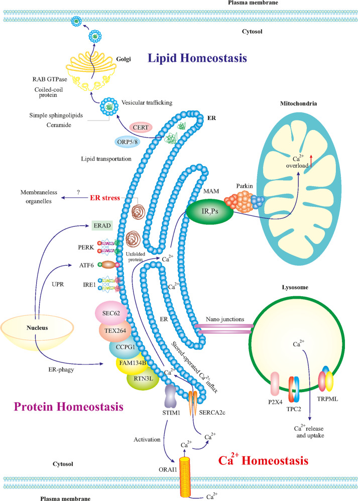
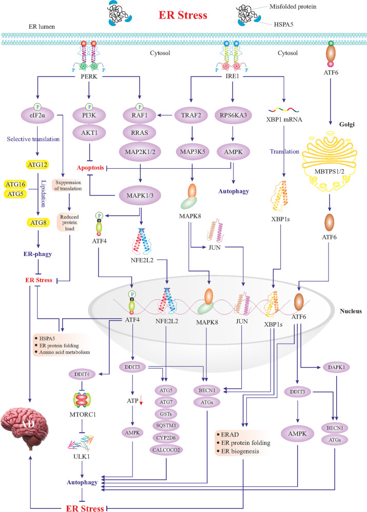
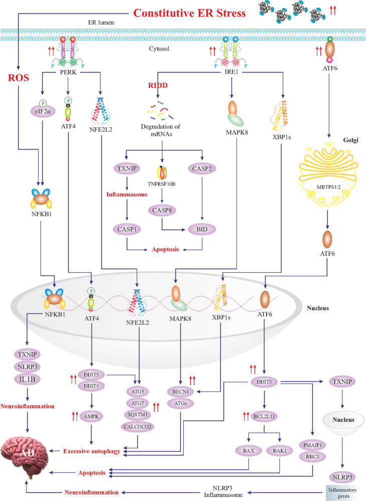
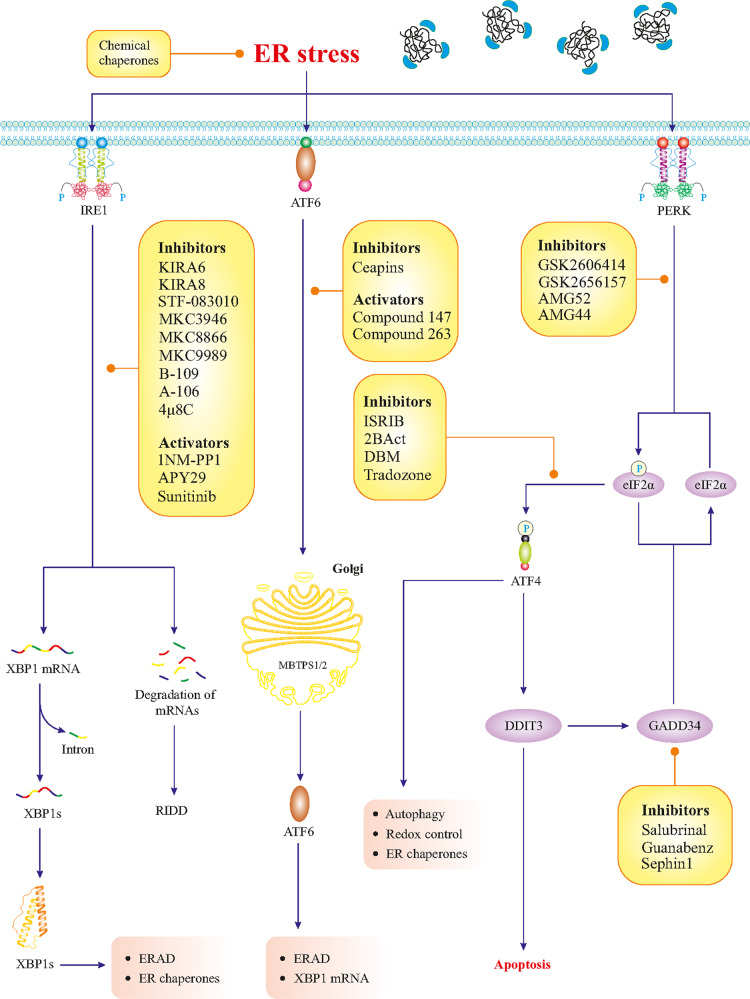
Alzheimer's disease (AD) is a devastating neurodegenerative disorder characterized by gradual loss of memory and cognitive function, which constitutes a heavy burden on the healthcare system globally. Current therapeutics to interfere with the underlying disease process in AD is still under development. Although many efforts have centered on the toxic forms of Aβ to effectively tackle AD, considering the unsatisfactory results so far it is vital to examine other targets and therapeutic approaches as well. The endoplasmic reticulum (ER) stress refers to the build-up of unfolded or misfolded proteins within the ER, thus, perturbing the ER and cellular homeostasis. Emerging evidence indicates that ER stress contributes to the onset and development of AD. A thorough elucidation of ER stress machinery in AD pathology may help to open up new therapeutic avenues in the management of this devastating condition to relieve the cognitive dementia symptoms. Herein, we aim at deciphering the unique role of ER stress in AD pathogenesis, reviewing key findings, and existing controversy in an attempt to summarize plausible therapeutic interventions in the management of AD pathophysiology.


Conflict of interest statement

The authors declare no competing interests.

Figures

Fig. 1
Fig. 1. The ER and lipid, protein, and Ca2+ homeostasis in neurons.
The ER is in direct or indirect communication with other organelles such as mitochondria, other organelles, plasma membrane, phagosomes, lysosomes, and endosomes. Such communications regulate the metabolism and homeostasis of lipids, proteins, and Ca2+. ER-produced lipids including sphingolipids and ceramide are trafficked to the Golgi via vesicles, mediated by CERT and ORP5/8 proteins. In Golgi, RAB GTPases and coiled-coil proteins mediate the final trafficking of the lipids to the plasma membrane. ER stress can influence mitochondria functions and vice versa and the contact points mitochondrial associated membranes (MAM) contain crucial proteins for neuronal physiology. For example, Ca2+ transportation to the mitochondria is influenced by MAM, which may lead to mitochondrial Ca2+ overload, reduced energy capacity, and oxidative stress in the neuron. Besides, nano-junctions between the ER and lysosomes modulate Ca2+ signaling in the ER. ER-mediated activation of the ORAI1 transporter leads to the influx of intracellular Ca2+ to the ER. In addition, it is thought that ER stress is affected by ER communication with membrane-less organelles. The roles of the UPR and ER-phagy (autophagy of the ER) for the turnover of the different components in cell homeostasis and their dysregulation in disease conditions such as AD is currently receiving more attention.
Fig. 2
Fig. 2. Mild ER stress and adaptive UPR signaling.
Mild ER stress triggers adaptive UPR signaling composed of three main branches; PERK, IRE1, and ATF6. As shown in the figure, the PERK–eIF2α axis promotes selective translation of some genes such as ATG12, which along with ATG16 and ATG5 induces lipidation and activation of ATG8, resulting in autophagy of the ER (ER-phagy). PERK-mediated phosphorylation of eIF2α also suppresses the translation to reduce protein overload in the ER. Likewise, the PERK–PI3K–AKT1 axis blocks apoptosis, and the PERK–RAF1–RRAS–MAP kinases cascade activates two cardinal transcription factors, ATF4 and NFE2L2, which transactivate genes encoding proteins involved in autophagy. mTOR complex 1 (mTORC1) is a negative regulator of autophagy via suppression of the ULK1 signaling complex to beclin1(BECN1). Inhibition of mTORC1 by DDIT4 can in turn activate autophagy. NFE2L2-induced genes encode components of autophagy machinery further fueling autophagy. NFE2L2 also upregulates antioxidant genes including CYP2D6 and CALCOCO2. Among ATF4-upregulated genes are those encoding ER chaperones such as HSPA5 and other enzymes to facilitate protein folding in the ER. Activation of the IRE1 branch during the UPR leads to the activation of some key transcription factors (see figure). Thus IRE–TRAF2 axis can activate i MAPK8 and JUN, which relocate to the nucleus and upregulate ATGs and BECN1 genes. IRE1-mediated activation of the AMPK also boosts autophagy and blocks apoptosis. Most importantly, IRE1 via its inherent RNAse activity produces mRNA encoding the transcription factor XBP1s, which in the nucleus upregulates autophagy-associated genes and proteins involved in the ERAD. During the UPR, ATF6 is processed in the Golgi to produce the active transcription factor which in turn can also upregulate genes encoding chaperones and ERAD proteins as well as DAPK1 and DDIT3 genes, with a role in autophagy. Overall, the adaptive UPR suppresses ER stress via induction of corrective autophagy, inhibition of apoptosis, and activation of the ERAD. There is also an upregulation of ER chaperones, inhibition of additional protein translation, and an enhancement of ER capacity all serving to boost the correct folding of proteins.
Fig. 3
Fig. 3. Constitutive ER stress and maladaptive UPR signaling.
Constitutive ER stress triggers maladaptive UPR signaling characterized by excessive activation of the UPR branches. Hyperactivated PERK signaling leads to the activation of NFKB1, igniting neuroinflammation. Prolonged phosphorylation of eIF2alpha causes a block in the synthesis of crucial synaptic and other proteins necessary for neuronal functions. In addition, hyperactivated ATF4 results in an excessive upregulation of DDIT3 and DDIT4 genes with an enhanced expression of autophagy genes, which is detrimental to neurons. Likewise, NFE2L2-induced hyper-transactivation of autophagy genes will lead to excessive autophagy. Overactive IRE1 will lead to massive degradation of mRNAs, being referred to as the RIDD (regulated Ire1-dependent decay), and subsequent induction of apoptosis and neuroinflammation. MAPK8 contributes to excessive autophagy via an enhanced upregulation of autophagy genes and ATF6- and ATF4-transactivated DDIT3 upregulates pro-apoptotic genes, leading to neuronal cell death. DDIT3-induced upregulation of the TXNIP gene, encoding a transcription factor, upregulates neuroinflammatory genes, and promotes the formation of the NLRP3 inflammasome, leading to neuroinflammation. Overall, a maladaptive UPR is characterized by excessive autophagy, apoptosis, and severe neuroinflammation, worsening the pathology observed in AD.
Fig. 4
Fig. 4. Pharmacological targeting of the UPR.
The UPR branches can be targeted and modulated by small pharmaceutical compounds. The list presented here is based on the currently available literature.

Similar articles

Cited by

References

    1. Takano T, Xu C, Funahashi Y, Namba T, Kaibuchi K. Neuronal polarization. Development. 2015;142:2088–93. doi: 10.1242/dev.114454. - DOI - PubMed
    1. Innocenti GM, Vercelli A, Caminiti R. The diameter of cortical axons depends both on the area of origin and target. Cereb Cortex. 2014;24:2178–88. doi: 10.1093/cercor/bht070. - DOI - PubMed
    1. Jan Y-N, Jan LY. Dendrites. Genes Dev. 2001;15:2627–41. doi: 10.1101/gad.916501. - DOI - PubMed
    1. Bélanger M, Allaman I, Magistretti PJ. Brain energy metabolism: focus on astrocyte-neuron metabolic cooperation. Cell Metab. 2011;14:724–38. doi: 10.1016/j.cmet.2011.08.016. - DOI - PubMed
    1. Tydlacka S, Wang C-E, Wang X, Li S, Li X-J. Differential activities of the ubiquitin–proteasome system in neurons versus glia may account for the preferential accumulation of misfolded proteins in neurons. J Neurosci. 2008;28:13285–95. doi: 10.1523/JNEUROSCI.4393-08.2008. - DOI - PMC - PubMed