. 2022 Aug 15;13(8):707.
doi: 10.1038/s41419-022-05101-3.

PKR and TLR3 trigger distinct signals that coordinate the induction of antiviral apoptosis

Affiliations

PKR and TLR3 trigger distinct signals that coordinate the induction of antiviral apoptosis

Wenjie Zuo et al. Cell Death Dis. .

Erratum in

Abstract

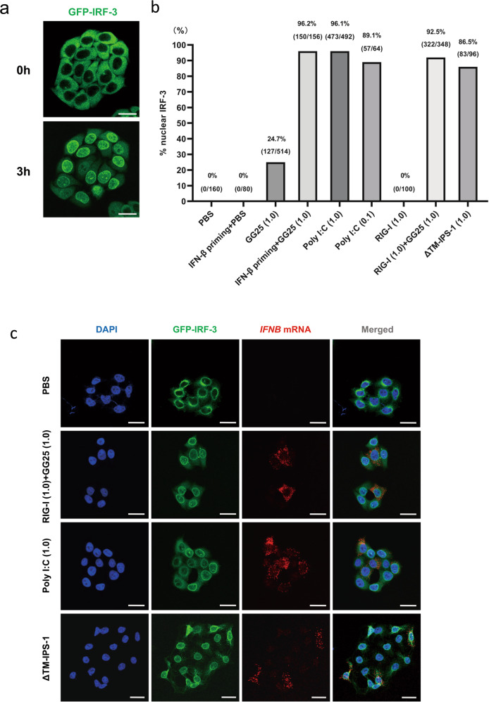
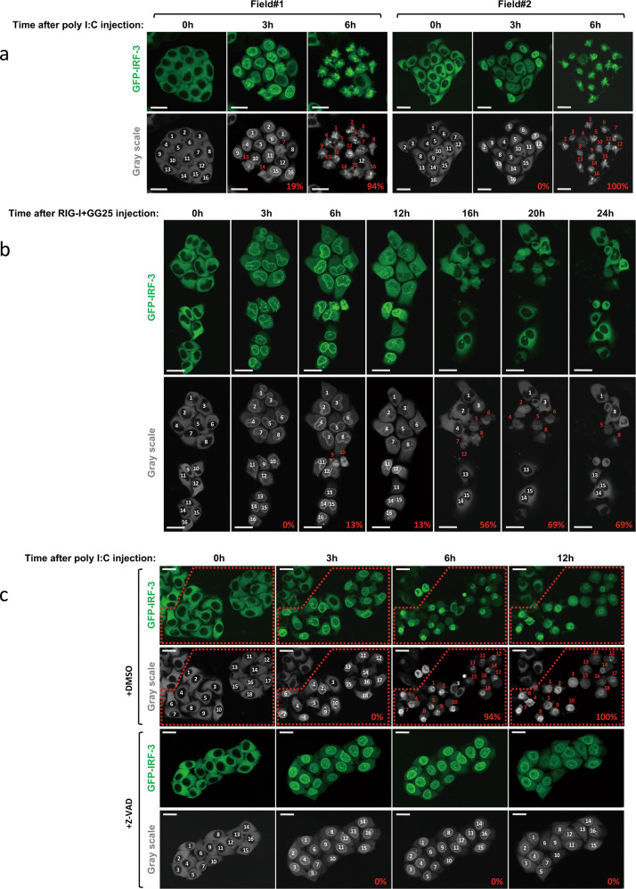
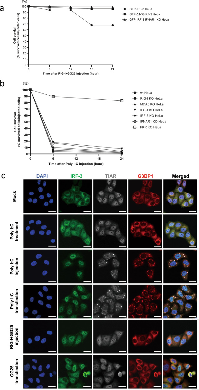
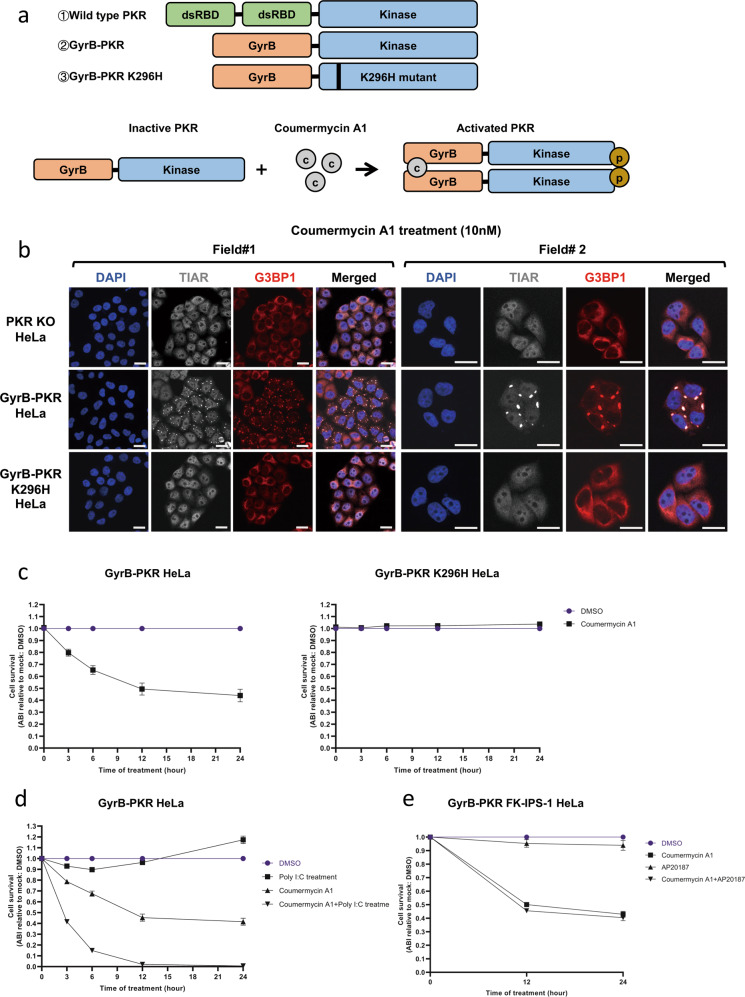
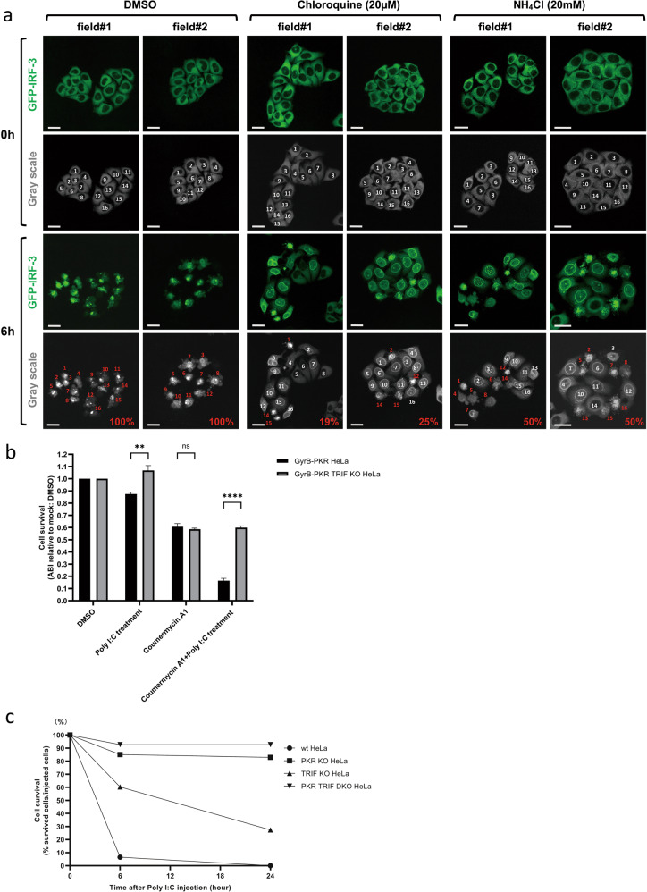
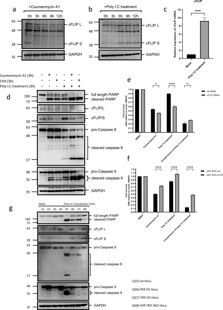
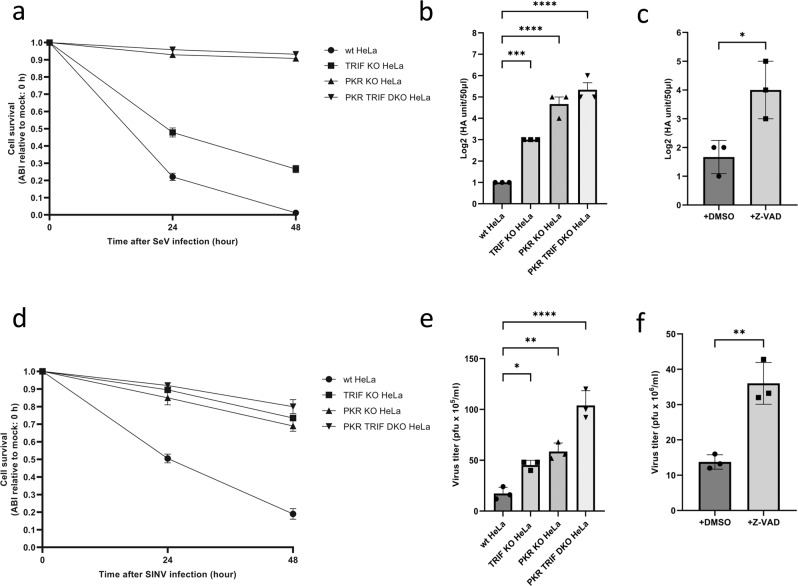
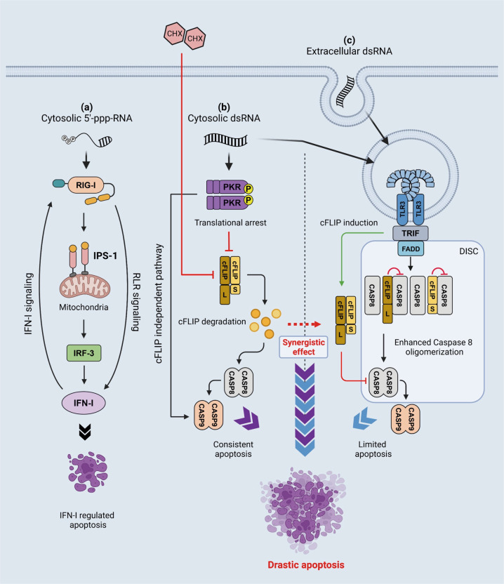
RIG-I-like receptors (RLRs), protein kinase R (PKR), and endosomal Toll-like receptor 3 (TLR3) sense viral non-self RNA and are involved in cell fate determination. However, the mechanisms by which intracellular RNA induces apoptosis, particularly the role of each RNA sensor, remain unclear. We performed cytoplasmic injections of different types of RNA and elucidated the molecular mechanisms underlying viral dsRNA-induced apoptosis. The results obtained revealed that short 5'-triphosphate dsRNA, the sole ligand of RIG-I, induced slow apoptosis in a fraction of cells depending on IRF-3 transcriptional activity and IFN-I production. However, intracellular long dsRNA was sensed by PKR and TLR3, which activate distinct signals, and synergistically induced rapid apoptosis. PKR essentially induced translational arrest, resulting in reduced levels of cellular FLICE-like inhibitory protein and functioned in the TLR3/TRIF-dependent activation of caspase 8. The present results demonstrated that PKR and TLR3 were both essential for inducing the viral RNA-mediated apoptosis of infected cells and the arrest of viral production.


Conflict of interest statement

The authors declare no competing interests.

Figures

Fig. 1
Fig. 1. The cytoplasmic RNA/protein injection induces prompt and potent cellular responses.
a GFP-IRF-3 HeLa cells were injected with poly I:C (1 μg/μl) and observed live for the localization of IRF-3 at the indicated times after the injection. The injection was performed for a colony of cells, each cell was followed for the nuclear translocation of IRF-3, and the % of nuclear IRF-3 was calculated (number of cells with nuclear IRF-3/number of injected cells). b GFP-IRF-3 HeLa cells were injected with the indicated RNA/protein. GG25: 5’-ppp-RNA; poly I:C: long poly I:C; RIG-I: recombinant RIG-I protein; ΔTM-IPS-1: recombinant IPS-1 protein devoid of the transmembrane domain. The amount of injected RNA or protein (μg/μl) is indicated at the bottom of the graph. IFN-β priming indicates the pretreatment of cells with IFN-β (1000 U/ml for 12 h) prior to the injection. At 3 h, injected cells were observed live for the localization of IRF-3, and % nuclear IRF-3 was calculated as in (a) and indicated at the top of each bar. c GFP-IRF-3 HeLa cells were injected with the indicated RNA/protein. Cells were fixed after 3 h and observed for IRF-3 (green) or the expression of IFNB mRNA (FISH, red). Scale bar = 25 µm.
Fig. 2
Fig. 2. Induction of the nuclear translocation of IRF-3 and subsequent cell death by the cytoplasmic injection of RNA/protein.
a GFP-IRF-3 HeLa cells were injected with poly I:C (1 μg/μl) and observed live for the localization of IRF-3 at the indicated times after the injection. Injected cells were numbered (white) and followed; cells with red numbers exhibited morphological cell death; % of dead cells was calculated (as depicted in grayscale images). b GFP-IRF-3 HeLa cells were injected with RIG-I and GG25 (1 μg/μl each) and observed as in (a). c GFP-IRF-3 HeLa cells were injected with poly I:C (1 μg/μl) in the absence (DMSO) or presence of Z-VAD (20 µΜ) and observed as in (a). Z-VAD was added 3 h prior to the injection and kept in the culture medium. Injected cells are indicated in the red dotted box. Scale bar = 25 µm.
Fig. 3
Fig. 3. Diverse mechanisms of cell death induced by GG25 and poly I:C.
a GFP-IRF-3 HeLa, GFP-Δ1-58IRF-3 HeLa, and GFP-IRF-3 IFNAR1 KO HeLa cells were injected with RIG-I and GG25 (1 μg/μl each), observed live for cell death, and quantified for % cell survival at the indicated time points. b HeLa cells and indicated KO HeLa cells were injected with poly I:C (1 μg/μl), observed for cell death, and % cell survival was quantified. c GFP-IRF-3 HeLa cells were mock treated, treated with poly I:C (5 μg/ml in culture medium for 3 h), injected with poly I:C (1 μg/μl for 3 h), transfected with poly I:C (0.5 μg/ml in culture medium with lipofectamine for 3 h), injected with RIG-I and GG25 (1 μg/μl each for 3 h), or transfected with GG25 (4 μg/ml in culture medium with lipofectamine for 3 h). Cells were fixed and stained for TIAR and G3BP1 with respective antibodies for microscopy. Scale bar = 25 µm.
Fig. 4
Fig. 4. Synergistic induction of cell death by PKR and the exogenous poly I:C treatment.
a Schematic representation of PKR and GyrB-PKR fusion proteins and the activation mechanism of GyrB-PKR fusion by coumermycin A1. b Induction of SG by the coumermycin A1 treatment. PKR KO HeLa, GyrB-PKR HeLa, and GyrB-PKR K296H HeLa cells were treated with coumermycin A1 (10 nM for 3 h) and observed for the SG marker TIAR (gray) and G3BP1 (red), as in Fig. 3c. Scale bar = 25 µm. c GyrB-PKR HeLa and GyrB-PKR K296H HeLa cells were mock treated (DMSO) or treated with coumermycin A1 (10 nM) for the indicated times and examined for cell survival. d GyrB-PKR HeLa cells were treated with the indicated chemicals and examined for cell survival at each time point. e GyrB-PKR FK-IPS-1 HeLa cells were treated with the indicated chemicals and examined for cell survival at each time point. Cell survival in (ce) was examined by the Amido black assay (“Materials and methods”) and presented as Amido black intensity (ABI) relative to the mock. Data are represented as the means ± SEM of three independent experiments.
Fig. 5
Fig. 5. Involvement of TLR3 signaling in cell death induced by cytoplasmic poly I:C.
a GFP-IRF-3 HeLa cells were treated with DMSO, chloroquine (20 μM for 6 h), or NH4Cl (20 mM for 6 h) and then injected with poly I:C (1 μg/μl). GFP images of live cells were taken 0 and 6 h after the poly I:C injection. In each field, cells were numbered, assessed as dead (red) or alive (white), and % cell death was calculated. Scale bar = 25 µm. b GyrB-PKR HeLa and GyrB-PKR TRIF KO HeLa cells were treated with the indicated chemicals for 6 h and examined for cell survival. The means ± SEM of three independent experiments are shown; data were analyzed by a two-way ANOVA followed by Tukey’s multiple comparisons test; **P < 0.01, ****P < 0.0001; ns not significant. c Wild-type, PKR KO, TRIF KO, and PKR TRIF DKO HeLa cells were injected with poly I:C (1 μg/μl), observed live for cell death, and % cell survival at the indicated time points was quantified.
Fig. 6
Fig. 6. PKR activation resulted in the downregulation of cFLIP and promoted the endosomal poly I:C-induced activation of caspases 8 and 9.
a GyrB-PKR HeLa cells were treated with coumermycin A1 for the indicated times and examined for the levels of cFLIP L, cFLIP S, and GAPDH by immunoblotting. b GyrB-PKR HeLa cells were treated with poly I:C (5 μg/ml) and examined for the levels of cFLIP L, cFLIP S, and GAPDH by immunoblotting. c GyrB-PKR HeLa cells were left untreated or treated with poly I:C (5 µg/ml, 6 h). Cells were harvested and the relative expression of the cFLIP gene was examined by RT-qPCR. The means + SEM of three independent experiments are shown; data were analyzed by an unpaired t test; ***P < 0.001. d GyrB-PKR HeLa cells were treated with the indicated chemicals (10 nM coumermycin A1; 10 µg/ml CHX; 5 μg/ml poly I:C) for 3 h and examined for the indicated proteins by immunoblotting. e GyrB-PKR HeLa cells were transfected with control (ctl) or specific siRNA for cFLIP for 48 h, treated with the indicated chemicals as in (d), and examined for cell survival. f GyrB-PKR HeLa cells were transfected with the control (pEF-BOS vec) or expression vector for cFLIP (pEF-BOS cFLIP) for 48 h, treated with the indicated chemicals as in (d), and examined for cell survival. g Wild-type, PKR KO, TRIF KO, and PKR TRIF DKO HeLa cells were transfected with poly I:C (0.5 µg/ml) for 12 h and examined for the indicated proteins by immunoblotting. Cell survival in (e, f) is presented as ABI relative to the mock, and the means ± SEM of three independent experiments are shown; data were analyzed by a two-way ANOVA followed by Tukey’s multiple comparisons test; *P < 0.05, ****P < 0.0001; ns not significant. Refer to full-length western blot images in Supplemental Materials.
Fig. 7
Fig. 7. Roles of PKR and TLR3/TRIF in virus-induced cell death and viral yield.
a Wild-type, PKR KO, TRIF KO, and PKR TRIF DKO HeLa cells were mock treated or infected with Sendai virus (SeV) for 48 h, and quantified for cell survival. b HA yield in the culture supernatant of cells infected in (a) were examined. c Wild-type HeLa cells were infected with SeV with or without Z-VAD (50 μM) in culture medium for 72 h, and the culture supernatant was examined for HA yield. d The same set of cells in (a) were mock treated or infected with Sindbis virus (SINV) for 48 h and quantified for cell survival. e Viral titers in the culture supernatant of cells in (d) were examined by a plaque assay. f Wild-type HeLa cells were infected with SINV with or without Z-VAD (50 μM) in culture medium for 72 h, and viral titers in the culture supernatant were examined by the plaque assay. The means ± SEM of three independent experiments are shown; data in (b, d) were analyzed by a one-way ANOVA followed by Dunnett’s multiple comparisons test; data in (c, f) were analyzed by an unpaired t test; *P < 0.05, **P < 0.01, ***P < 0.001, ****P < 0.0001.
Fig. 8
Fig. 8. Schematic view of cell death signaling induced by 5’-ppp-RNA, cytosolic dsRNA, and extracellular dsRNA.
a Cytosolic short 5’-ppp-dsRNA is sensed by RIG-I and activates signaling, leading to the induction of IFN-I and limited apoptosis with slow kinetics. This apoptosis is dependent on genes regulated by IRF-3 and IFN-I. b Cytosolic dsRNA introduced by the injection or transfection activates TLR3 through endosomal entrapment as above. In contrast to endosomal dsRNA, cytosolic dsRNA activates PKR, which results in the induction of SG and translational shutdown, leading to the downregulation of cFLIP and apoptosis through the activation of caspase 8. In addition, PKR was reported to promote the mitochondrial pathway in order to activate caspase 9; therefore, robust apoptosis was induced via caspases 8/9. c Extracellular dsRNA is incorporated by endocytosis and sensed by TLR3. TLR3 signals TRIF and activates signaling, leading to the induction of IFN-I and the expression of cFLIP in order to negatively regulate caspase 8. The TLR3 signaling complex also recruits FADD to activate DISC containing caspase 8; however, due to the effects of cFLIP, apoptosis is suppressed. When cells are treated with extracellular dsRNA and CHX, caspase 8 promotes apoptosis because of the downregulation of cFLIP.

Similar articles

Cited by

References

    1. Janeway CA, Medzhitov R. Innate immune recognition. Annu Rev Immunol. 2002;20:197–216. - PubMed
    1. Akira S, Uematsu S, Takeuchi O. Pathogen recognition and innate immunity. Cell. 2006;124:783–801. - PubMed
    1. Satoh T, Kato H, Kumagai Y, Yoneyama M, Sato S, Matsushita K, et al. LGP2 is a positive regulator of RIG-I- and MDA5-mediated antiviral responses. Proc Natl Acad Sci USA. 2010;107:1512–7. - PMC - PubMed
    1. Kang D, Gopalkrishnan RV, Wu Q, Jankowsky E, Pyle AM, Fisher PB. mda-5: an interferon-inducible putative RNA helicase with double-stranded RNA-dependent ATPase activity and melanoma growth-suppressive properties. Proc Natl Acad Sci USA. 2002;99:637–42. - PMC - PubMed
    1. Yoneyama M, Kikuchi M, Natsukawa T, Shinobu N, Imaizumi T, Miyagishi M, et al. The RNA helicase RIG-I has an essential function in double-stranded RNA-induced innate antiviral responses. Nat Immunol. 2004;5:730–7. - PubMed

Publication types

MeSH terms