. 2022 Aug 15;5(1):822.
doi: 10.1038/s42003-022-03778-y.

Long non-coding RNA DARS-AS1 promotes tumor progression by directly suppressing PACT-mediated cellular stress

Affiliations

Long non-coding RNA DARS-AS1 promotes tumor progression by directly suppressing PACT-mediated cellular stress

Liuqing Yang et al. Commun Biol. .

Abstract

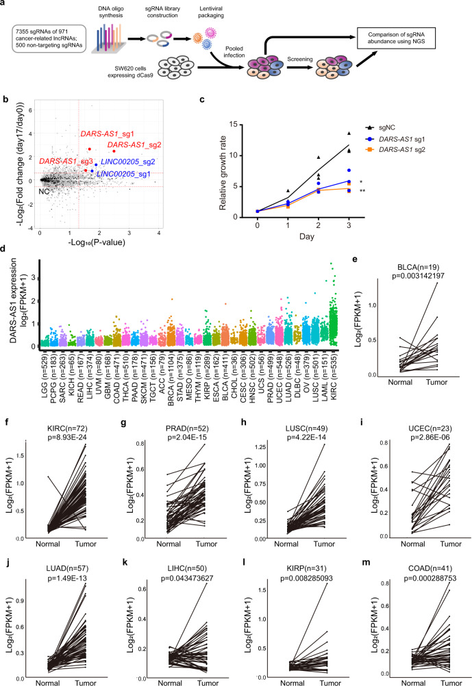
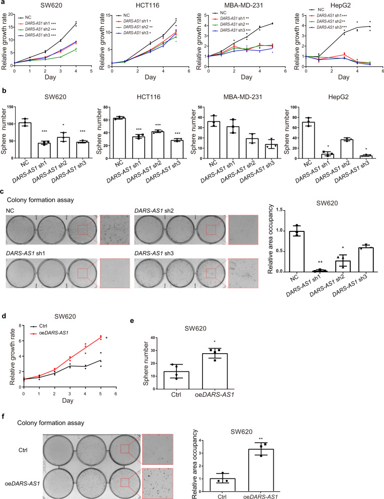
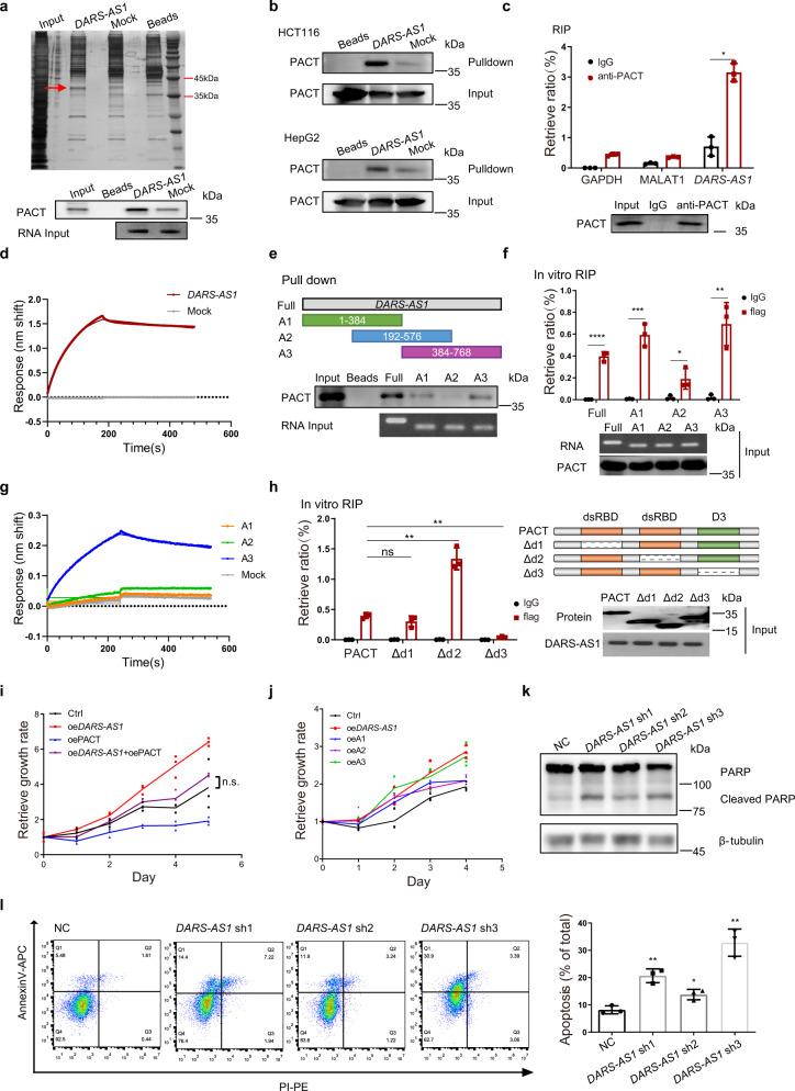
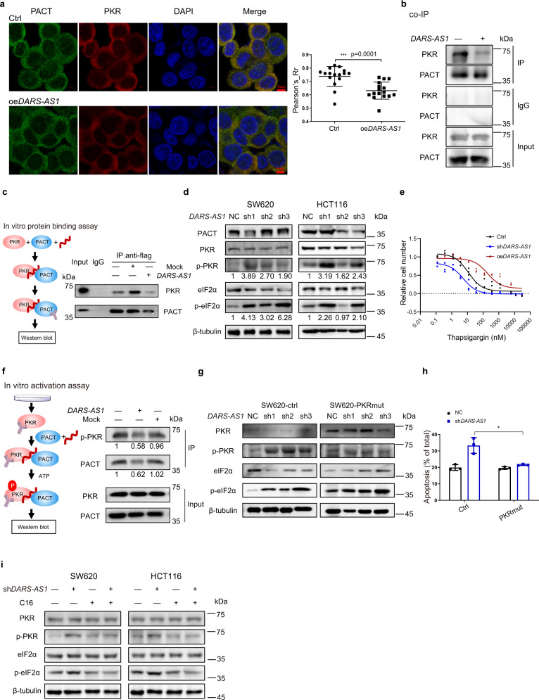
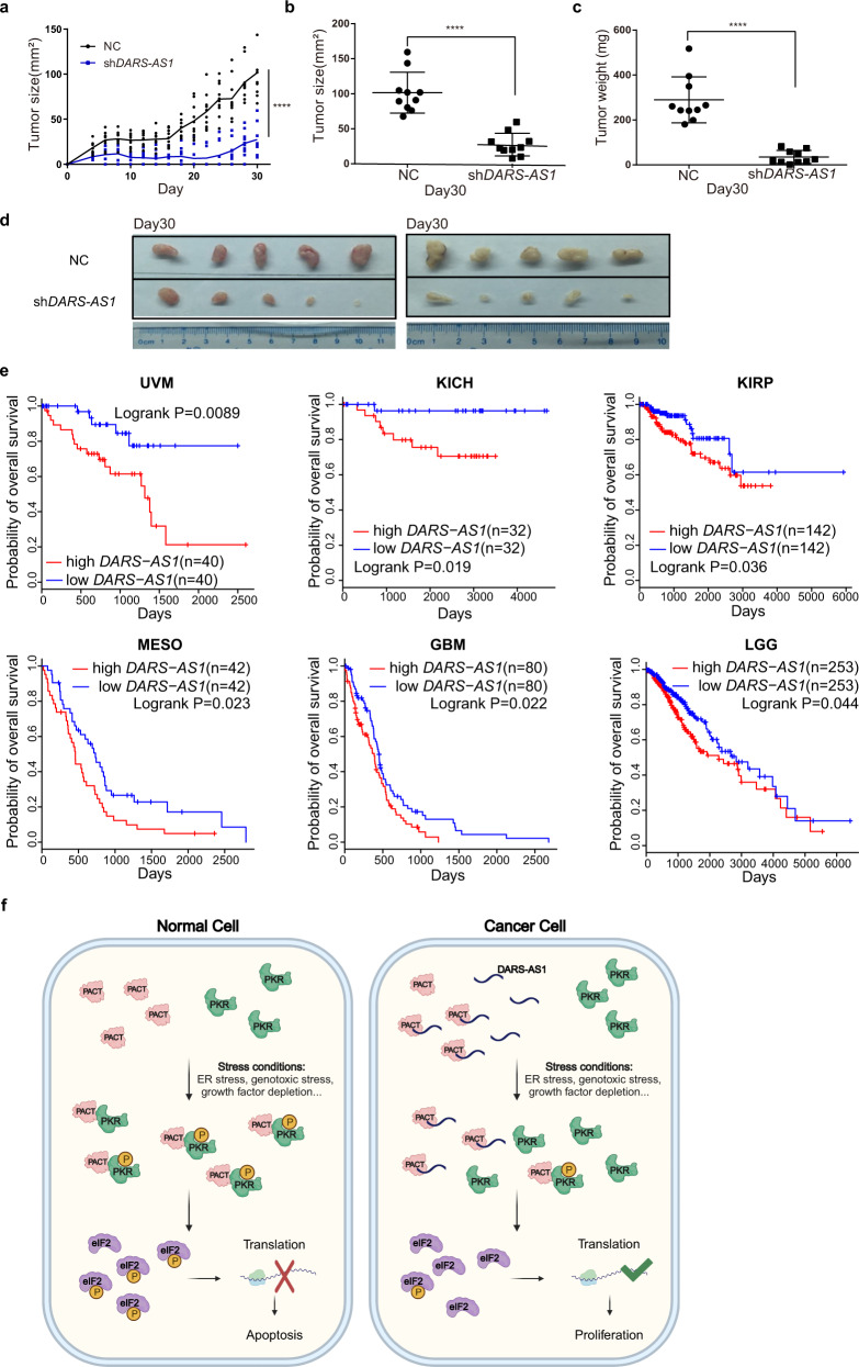
Cancer cells evolve various mechanisms to overcome cellular stresses and maintain progression. Protein kinase R (PKR) and its protein activator (PACT) are the initial responders in monitoring diverse stress signals and lead to inhibition of cell proliferation and cell apoptosis in consequence. However, the regulation of PACT-PKR pathway in cancer cells remains largely unknown. Herein, we identify that the long non-coding RNA (lncRNA) aspartyl-tRNA synthetase antisense RNA 1 (DARS-AS1) is directly involved in the inhibition of the PACT-PKR pathway and promotes the proliferation of cancer cells. Using large-scale CRISPRi functional screening of 971 cancer-associated lncRNAs, we find that DARS-AS1 is associated with significantly enhanced proliferation of cancer cells. Accordingly, knocking down DARS-AS1 inhibits cell proliferation of multiple cancer cell lines and promotes cancer cell apoptosis in vitro and significantly reduces tumor growth in vivo. Mechanistically, DARS-AS1 directly binds to the activator domain of PACT and prevents PACT-PKR interaction, thereby decreasing PKR activation, eIF2α phosphorylation and inhibiting apoptotic cell death. Clinically, DARS-AS1 is broadly expressed across multiple cancers and the increased expression of this lncRNA indicates poor prognosis. This study elucidates the lncRNA DARS-AS1 directed cancer-specific modulation of the PACT-PKR pathway and provides another target for cancer prognosis and therapeutic treatment.


Conflict of interest statement

The authors declare no competing interests.

Figures

Fig. 1
Fig. 1. DARS-AS1 is characterized as an oncogenic lncRNA.
a Schematic representation of screening using the CRISPRi system. b Enrichment of sgRNAs after screening. The horizontal dotted lines represent log2(Fold change) = ±0.58. The vertical dotted line represents p value = 0.05. Black dots are non-targeting sgRNAs (marked as NC). Red dots are sgRNAs targeting DARS-AS1. Blue dots are sgRNAs targeting LINC00205, a previously reported oncogenic lncRNA. Fold change = (normalized reads day 17)/(normalized reads day 0). c Knockdown DARS-AS1 by sgRNAs inhibits cell growth. Error bars represent ±SD in triplicate experiments. *p ≤ 0.05, **p ≤ 0.01, by two-tailed Student’s t test. d Expression of DARS-AS1 in tumors (TCGA datasets). em Expression of DARS-AS1 in paired normal and tumor samples from BLCA, KIRC, PRAD, LUSC, UCEC, LUAD, LIHC, KIRP and COAD patients, respectively (TCGA datasets). p values were obtained by paired two-tailed Student’s t test.
Fig. 2
Fig. 2. Knockdown of DARS-AS1 suppresses cell proliferation in multiple cancers.
Impacts on cell proliferation (a) and sphere formation (b) of SW620, HCT116, MBA-MD-231, and HepG2 cells by control shRNA and DARS-AS1-shRNAs. c Impacts on colony formation of SW620 cells by control shRNA and DARS-AS1-shRNAs. Cell proliferation (d), sphere formation (e) and colony formation (f) of DARS-AS1-overexpressing SW620 cells. Data shown are means ± SD in triplicates experiments. *p ≤ 0.05, **p ≤ 0.01 and ***p ≤ 0.001, by two-tailed Student’s t test.
Fig. 3
Fig. 3. DARS-AS1 directly interacts with PACT.
a RNA pull-down assay identifies DARS-AS1 interacting with PACT in SW620 cells. Upper, silver staining of associated proteins. Lower, immunoblot with anti-PACT antibodies. b RNA pull-down assays were performed in HCT116 (upper) and HepG2 (lower) cells. Immunoblot detected the enrichment of PACT. c RNA immunoprecipitation (RIP) assays were performed using the indicated antibodies in SW620 cells. d The binding curves of PACT with full length DARS-AS1 or control RNA were obtained by biolayer interferometry (BLI). RNAs were immobilized on streptavidin biosensors. 1 μM PACT were used for measuring the association. e RNA pull-down assays were performed using biotinylated full length DARS-AS1 or truncations (upper). Immunoblot shows the retrieved PACT (lower). f Purified flag-tagged PACT was incubated with biotinylated full length DARS-AS1 or truncations (as showed in e) for in vitro RIP assays. Retrieved RNAs were validated by RT-qPCR. g The relative affinities of different RNA fragments for PACT were obtained by biolayer interferometry. 100 nM RNA and 1 μM PACT were used in each assay. h In vitro RIP assays were performed using purified intact or truncated flag-tagged PACT. Retrieved RNA was validated by RT-qPCR. i Cell growth rate of SW620 cells overexpressing DARS-AS1, PACT, or both. j Overexpression of full length or truncated DARS-AS1 in SW620 cells shows different impacts on cell growth. k Cell apoptosis was detected by immunoblot with anti-PARP antibodies. l Knockdown DARS-AS1 induces SW620 cell apoptosis, as revealed by flow cytometry. Data shown are means ± SD in triplicates experiments. *p ≤ 0.05, **p ≤ 0.01, ***p ≤ 0.001, ****p < 0.0001, by two-tailed Student’s t test.
Fig. 4
Fig. 4. DARS-AS1 blocks PACT-mediated PKR activation.
a The colocalization of PACT and PKR in control cells or DARS-AS1-overxpression cells were observed by fluorescence confocal microscope. Cell nucleus was stained by DAPI. The statistic results were obtained by 16 photos. b Co-immunoprecipitation (co-IP) was performed using anti-PACT antibody in cell lysates of SW620 control cells or DARS-AS1-overexpressing cells. c Flag-tagged PACT, purified PKR, and in vitro transcribed DARS-AS1 or mock RNA were incubated for in vitro protein binding assay. Immunoprecipitation used anti-flag antibody. d Immunoblot using indicated antibodies was performed in SW620 and HCT116 cells transfected with control shRNAs or DARS-AS1-shRNAs, followed by serum starvation. e The expression level of DARS-AS1 alters cell sensitivity of thapsigargin. SW620 cells were transfected with DARS-AS1 shRNAs, DARS-AS1-overexpression plasmids or control plasmids. Cells were treated by thapsigargin for 48 h and detected cell viability by MTS reagents. f In vitro transcribed DARS-AS1 or mock RNA and purified PACT were used for in vitro activation assay and immunoblot detection. g Immunoblot using indicated antibodies were performed in SW620-ctrl cells (left) or cells overexpressing PKR mutant (right). These cells were then transfected with control shRNAs or DARS-AS1-shRNAs, followed by serum starvation. h Inactive mutant PKR compensated SW620 cell apoptosis induced by DARS-AS1, as revealed by flow cytometry. i Immunoblot using indicated antibodies were performed in SW620 (left) or HCT116 cells (right). Cells transfected with control shRNAs or DARS-AS1-shRNAs were treated by serum starvation, suppling with 100 nM PKR inhibiter C16 or DMSO. Scale bars =5 µm. Data shown are means ± SD in triplicates experiments. *p ≤ 0.05, by two-tailed Student’s t test.
Fig. 5
Fig. 5. DARS-AS1 promotes tumor proliferation in vivo and correlates with poor prognosis.
ad Effect of DARS-AS1-knockdown on colorectal tumorigenesis in nude mice. Growth curve (a), tumor size (b), weight (c) and tumor image (d) are shown. Error bars represent ±SEM. n = 10. ****p < 0.0001, by two-tailed Student’s t test. e Kaplan–Meier analyses of the correlations between DARS-AS1 expression level and overall survival of patients with UVM, KICH, KIRP, MESO, GBM, and LGG. High, patients in the top 50% of DARS-AS1 expression levels; low, patients in the bottom 50% of DARS-AS1 expression levels. p values were determined using a log rank test. f Proposed model of DARS-AS1 in regulating PACT-PKR pathway and tumor growth.

Similar articles

Cited by

References

    1. Wang G, Yang Z-Q, Zhang K. Endoplasmic reticulum stress response in cancer: molecular mechanism and therapeutic potential. Am. J. Transl. Res. 2010;2:65–74. - PMC - PubMed
    1. Dicks, N., Gutierrez, K., Michalak, M., Bordignon, V. & Agellon, L. B. Endoplasmic reticulum stress, genome damage, and cancer. Front. Oncol.5, 11 (2015). - PMC - PubMed
    1. O’Malley J, Kumar R, Inigo J, Yadava N, Chandra D. Mitochondrial stress response and cancer. Trends Cancer. 2020;6:688–701. doi: 10.1016/j.trecan.2020.04.009. - DOI - PMC - PubMed
    1. Lin SC, et al. The mitochondrial deoxyguanosine kinase is required for cancer cell stemness in lung adenocarcinoma. EMBO Mol. Med. 2019;11:20. - PMC - PubMed
    1. Haupt Y, Maya R, Kazaz A, Oren M. Mdm2 promotes the rapid degradation of p53. Nature. 1997;387:296–299. doi: 10.1038/387296a0. - DOI - PubMed

Publication types

MeSH terms