

Imaging of groin pain in athletes: patterns of injury at MRI and gender differences therein
- PMID: 35971036
- PMCID: PMC10250268
- DOI: 10.1007/s11845-022-03126-3
Imaging of groin pain in athletes: patterns of injury at MRI and gender differences therein
Abstract
Aim: The purpose of our study was to review a large cohort of athletes of all levels presenting with groin pain who underwent investigation with MRI and to determine what the commonest patterns of injury were. We aimed to explore whether particular findings were commonly found in association and whether measurable gender differences exist in the incidence of specific injuries.
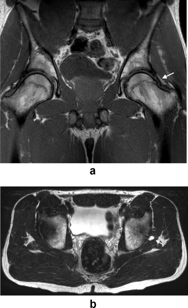
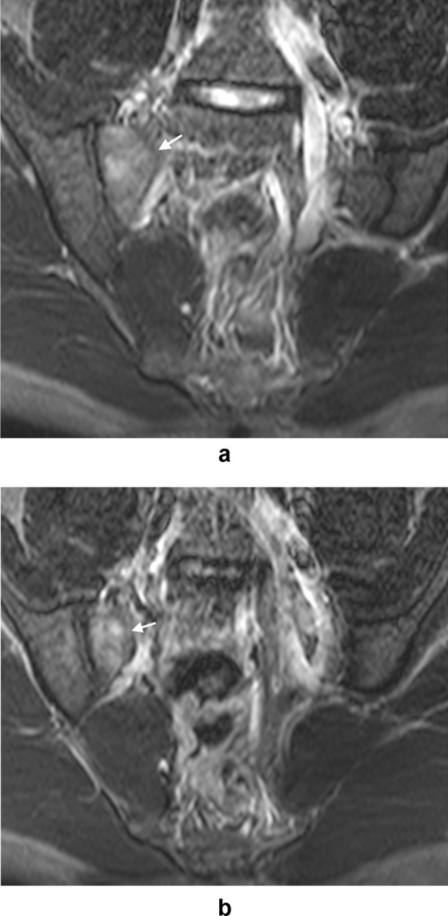
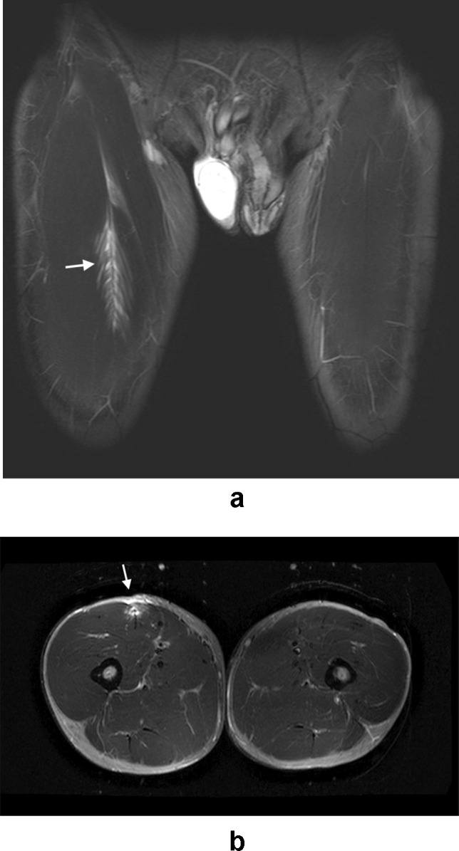
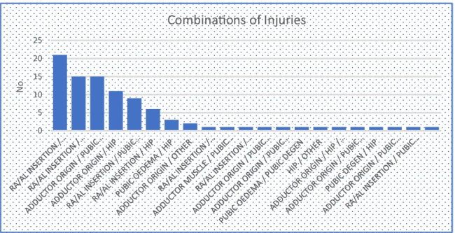
Materials and methods: Imaging records were reviewed to identify MRI studies of the pelvis performed for the investigation of groin pain in patients who were active in sports/athletic pursuits. Findings were classified and recorded as follows: injury to the common rectus abdominis/adductor longus origin, injury to the short adductor muscles, pubic bone oedema, pubic symphysis degenerative changes, hip joint injury and 'other'. The prevalence of specific injuries in female athletes compared to males was analysed using relative risk ratios.
Results: A total of 470 athletes underwent MRI for the investigation of groin pain during the study period. Forty-six were female, and 424 were male. Female athletes were significantly less likely to have rectus abdominis-adductor longus (RR = 0.31, p = .017), short adductor (RR = 0.14, p = .005) or hip (RR = 0.41, p = .003) injuries. Pubic bone degenerative changes were much more common in female athletes (RR = 7.37, p = .002).
Conclusion: Significant gender differences exist in the frequency with which specific injuries are observed. Female athletes are also significantly underrepresented; this is likely a multifactorial phenomenon; however, the possibility of unconscious referrer bias must be considered.
Keywords: Athletic groin pain; Athletic imaging; Femoroacetabular impingement; Groin pain imaging; Sports imaging; Sports medicine.
© 2022. The Author(s).
Conflict of interest statement
The authors declare no competing interests.
Figures
Similar articles
-
MRI findings in athletic groin pain: correlation of imaging with history and examination in symptomatic and asymptomatic athletes.Skeletal Radiol. 2025 Apr;54(4):841-850. doi: 10.1007/s00256-024-04603-9. Epub 2024 Feb 2. Skeletal Radiol. 2025. PMID: 38302788 Free PMC article.
-
Imaging of Groin Pain: Magnetic Resonance and Ultrasound Imaging Features.Sports Health. 2017 Sep/Oct;9(5):428-435. doi: 10.1177/1941738117694841. Epub 2017 Mar 8. Sports Health. 2017. PMID: 28850315 Free PMC article. Review.
-
The cleft sign may be an independent factor of magnetic resonance imaging findings associated with a delayed return-to-play time in athletes with groin pain.Knee Surg Sports Traumatol Arthrosc. 2021 May;29(5):1474-1482. doi: 10.1007/s00167-020-06410-w. Epub 2021 Jan 15. Knee Surg Sports Traumatol Arthrosc. 2021. PMID: 33452578
-
Sports-Related Groin Pain Secondary to Symphysis Pubis Disorders: Correlation Between MRI Findings and Outcome After Fluoroscopy-Guided Injection of Steroid and Local Anesthetic.AJR Am J Roentgenol. 2017 Aug;209(2):380-388. doi: 10.2214/AJR.16.17578. Epub 2017 Jun 13. AJR Am J Roentgenol. 2017. PMID: 28609118
-
Imaging of inguinal-related groin pain in athletes.Br J Radiol. 2018 Dec;91(1092):20170856. doi: 10.1259/bjr.20170856. Epub 2018 Jul 25. Br J Radiol. 2018. PMID: 29947268 Free PMC article. Review.
Cited by
-
Anatomical Features in Inguinal-Pubic-Adductor Area That May Contribute to Gender Difference in Susceptibility to Groin Pain Syndrome.J Pers Med. 2024 Aug 14;14(8):860. doi: 10.3390/jpm14080860. J Pers Med. 2024. PMID: 39202051 Free PMC article. Review.
-
MR imaging spectrum of adolescent pubic symphyseal injuries/athletic pubalgia.Pediatr Radiol. 2024 Jul;54(8):1270-1280. doi: 10.1007/s00247-024-05946-0. Epub 2024 May 13. Pediatr Radiol. 2024. PMID: 38736018
References
-
- Morelli V, Smith V. Groin injuries in athletes. Am Fam Physician. 2001;64(8):1405–1414. - PubMed
MeSH terms
LinkOut - more resources
Full Text Sources
Medical
