. 2022 Aug 6:2022:4552568.
doi: 10.1155/2022/4552568. eCollection 2022.

A Preliminary Study of Alterations in Iron Disposal and Neural Activity in Ischemic Stroke

Affiliations

A Preliminary Study of Alterations in Iron Disposal and Neural Activity in Ischemic Stroke

Abolfazl Mahmoudi Aqeel-Abadi et al. Biomed Res Int. .

Abstract

Purpose: The study aimed to evaluate the postrehabilitation changes in deep gray matter (DGM) nuclei, corticospinal tract (CST), and motor cortex area, involved in motor tasks in patients with ischemic stroke.

Methods: Three patients participated in this study, who had experienced an ischemic stroke on the left side of the brain. They underwent a standard rehabilitation program for four consecutive weeks, including transcranial direct current stimulation (tDCS), neuromuscular electrical stimulation (NMES), and occupational therapy. The patients' motor ability was evaluated by Fugl-Meyer assessment-upper extremity (FMA-UE) and Wolf motor function test (WMFT). Multimodal magnetic resonance imaging (MRI) was acquired from the patients by a 3 Tesla machine before and after the rehabilitation. The magnetic susceptibility changes were examined in DGM nuclei including the bilateral caudate (CA), putamen (PT), globus pallidus (GP), and thalamus (TH) using quantitative susceptibility mapping (QSM). Functional MRI (fMRI) in the motor cortex areas was acquired to evaluate the postrehab functional motor activity. The three-dimensional corticospinal tract (CST) was reconstructed using diffusion-weighted imaging (DWI) and diffusion tensor tractography (DTT), and the fractional anisotropy (FA) was measured along the tract. Ultimately, the relationship between the structural and functional changes was evaluated in CST and motor cortex.

Results: Postrehabilitation FMA-UE and WMFT scores increased for all patients compared to the prerehabilitation. QSM analysis revealed increasing in susceptibility values in GP and CA in all patients at the ipsilesional hemisphere. By fMRI analysis, the ipsilesional hemisphere demonstrated an increase in functional activity in motor areas for all 3 patients. In the ipsilesional hemisphere, the fractional anisotropy (FA) was increased in CST in two patients, while the mean diffusivity (MD) was decreased in CA in a patient, in PT and TH in another patient, and in PT in two patients.

Conclusion: This preliminary study demonstrates that the magnetic susceptibility may decrease at some ipsilesional DGM nuclei after tDCS, NMES, and occupational therapy for patients with ischemic stroke, suggesting a drop in the level of iron deposition, which may be associated with an increase in the level of activity in motor cortex after rehabilitation.


Conflict of interest statement

The authors declare that they have no conflicts of interest.

Figures

Figure 1
Figure 1
The basal ganglia and thalamus and their relationship to the motor cortex.
Figure 2
Figure 2
Illustration of STAR-QSM technique. The raw phase was unwrapped using Laplacian-based phase unwrapping. The background field was analyzed with a complex harmonic variable size kernel of V-SHARP attenuation for phase data with a regularization parameter of 0.05 and a maximum radius of 5 mm. Note: STAR-QSM: streaking artifact reduction quantitative susceptibility mapping; V-SHARP: variable-kernel sophisticated harmonic artifact reduction for phase.
Figure 3
Figure 3
Image segmentation pipeline for producing the hybrid contrast images. First, the registration transfer matrix is computed based on magnitude GRE images, and the QSM images are reconstructed based on the phase images. HC images are then produced using T1 and QSM images. At the last step, HC images are segmented using FSL_FIRST, and the masks of DGM regions are finally achieved. Note: DGM: deep gray matter; GRE: gradient-recalled echo; HC: hybrid contrast; MNI: Montreal Neurological Institute; QSM: quantitative susceptibility mapping.
Figure 4
Figure 4
Illustration of the segmented basal ganglia on HC images (axial (a) and coronal (c) views), on QSM images (axial (b) and coronal (d) views), and their 3D rendering (e to h). Note: HC: hybrid contrast; QSM: quantitative susceptibility mapping. Color assignment to brain structures: yellow: caudate nucleus; green: putamen; pink: globus pallidus; cyan: thalamus; red: hippocampus; blue: amygdala; brown: nucleus accumbens.
Figure 5
Figure 5
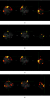
fMRI activation map for patient 1 undergoing the motor hand task, for left hand (unaffected hand) before rehabilitation (a); right hand (affected one) before rehabilitation (b); left hand after rehabilitation (c); and right hand after rehabilitation (d).
Figure 6
Figure 6
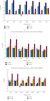
The level of fMRI activity extracted from Brodmann areas 1, 2, 3, 4, and 6 in patients 1, 2, and 3. After rehabilitation, an increase in activity is seen in all the motor cortex areas in patient 1 (a), in all ipsilesional (left) motor cortex areas in patient 2 (b), and in all ipsilesional (left) motor cortex areas, except for BA3aL in patient 3 (c).
Figure 7
Figure 7
T1-w images, FA maps, and MD maps of patient 1 before (pre) and after (post) rehabilitation. Note: FA: fractional anisotropy; MD: mean diffusivity.
Figure 8
Figure 8
Extracted CST in axial (a) and coronal views (b) before (pre) and after (post) rehabilitation for patient 1. Note: CST: corticospinal tract.
Figure 9
Figure 9
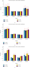
Mean diffusivity (MD) values in deep gray matter nuclei in patients 1, 2, and 3. After rehabilitation, no visible change is observed for patient 1 (a), while a slight decrease is observed in ipsilesional (left) thalamus in patient 2 (b) and in the thalamus and putamen for patient 3 (c).
Figure 10
Figure 10
Fractional anisotropy (FA) values in left and right corticospinal tract (CST) in patients (pt1, pt2, and pt3). After rehabilitation, a slight increase in ipsilesional (left) CST is observed in patients 1 and 3.
Figure 11
Figure 11
The susceptibility values extracted from DGM nuclei areas in patients 1, 2, and 3. After rehabilitation, a slight decrease is observed in ipsilesional (left) hemisphere for the patients 1 and 2 (a) and (b), and in ipsilesional CA and GP for the patient 3 (c). Note: DGM: deep gray matter; PT: putamen; CA: caudate; TH: thalamus; GP: globus pallidus.

References

    1. Tomandl B. F., Klotz E., Handschu R., et al. Comprehensive imaging of ischemic stroke with multisection CT. Radiographics . 2003;23(3):565–592. doi: 10.1148/rg.233025036. - DOI - PubMed
    1. Darwish E. A., Abdelhameed-El-Nouby M., Geneidy E. Mapping the ischemic penumbra and predicting stroke progression in acute ischemic stroke: the overlooked role of susceptibility weighted imaging. Insights Into Imaging . 2020;11(1):1–12. doi: 10.1186/s13244-019-0810-y. - DOI - PMC - PubMed
    1. Lai S.-M., Studenski S., Duncan P. W., Perera S. Persisting consequences of stroke measured by the stroke impact scale. Stroke . 2002;33(7):1840–1844. doi: 10.1161/01.STR.0000019289.15440.F2. - DOI - PubMed
    1. van Kordelaar J., van Wegen E., Kwakkel G. Impact of time on quality of motor control of the paretic upper limb after stroke. Archives of Physical Medicine and Rehabilitation . 2014;95(2):338–344. doi: 10.1016/j.apmr.2013.10.006. - DOI - PubMed
    1. Anderson T. P., Greenberg F. R. Predictive factors in stroke rehabilitation . 1974. - PubMed