. 2022 Oct 26;13(5):e0203222.
doi: 10.1128/mbio.02032-22. Epub 2022 Aug 16.

Evidence for a Widespread Third System for Bacterial Polysaccharide Export across the Outer Membrane Comprising a Composite OPX/β-Barrel Translocon

Affiliations

Evidence for a Widespread Third System for Bacterial Polysaccharide Export across the Outer Membrane Comprising a Composite OPX/β-Barrel Translocon

Johannes Schwabe et al. mBio. .

Abstract

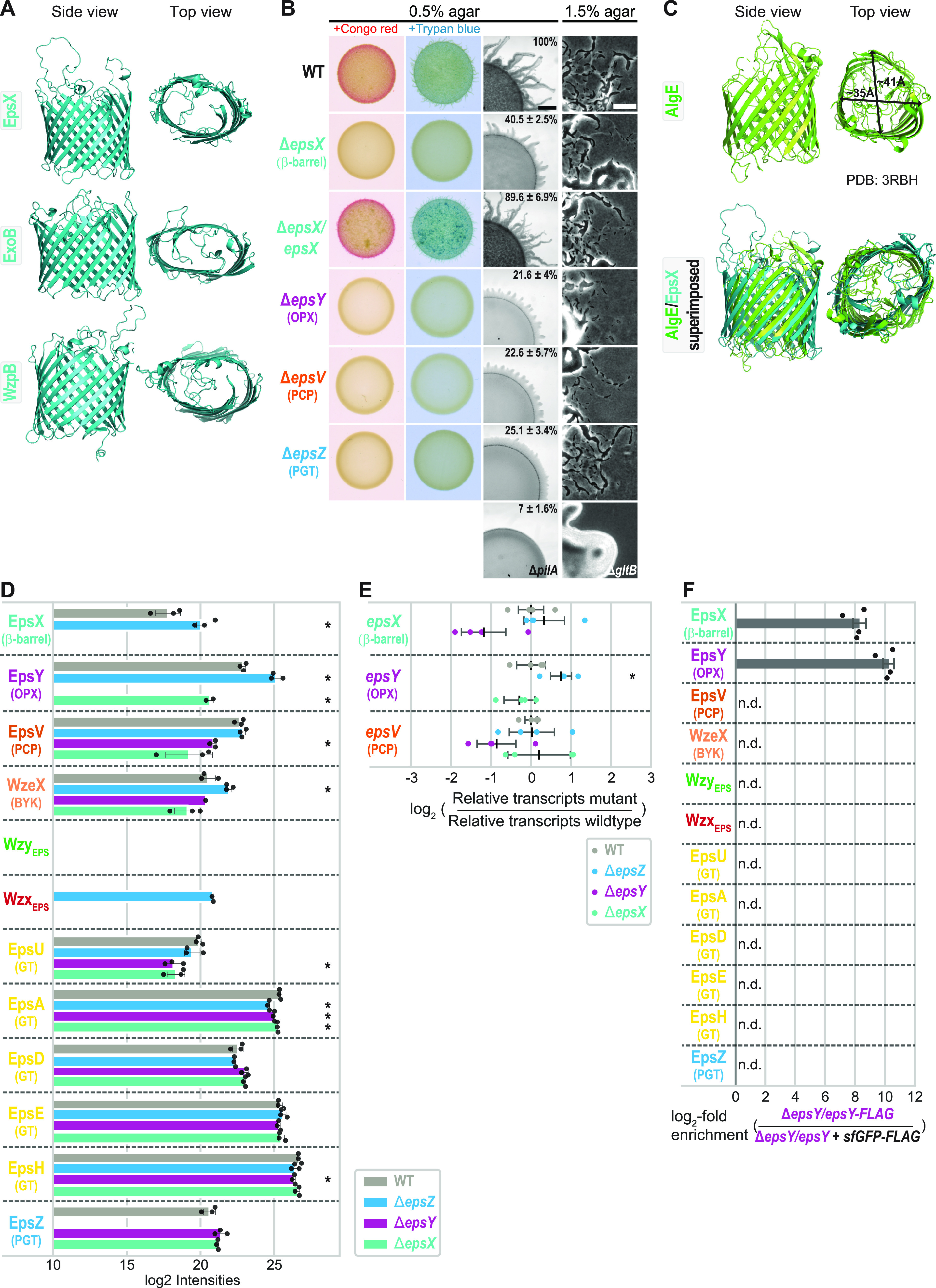
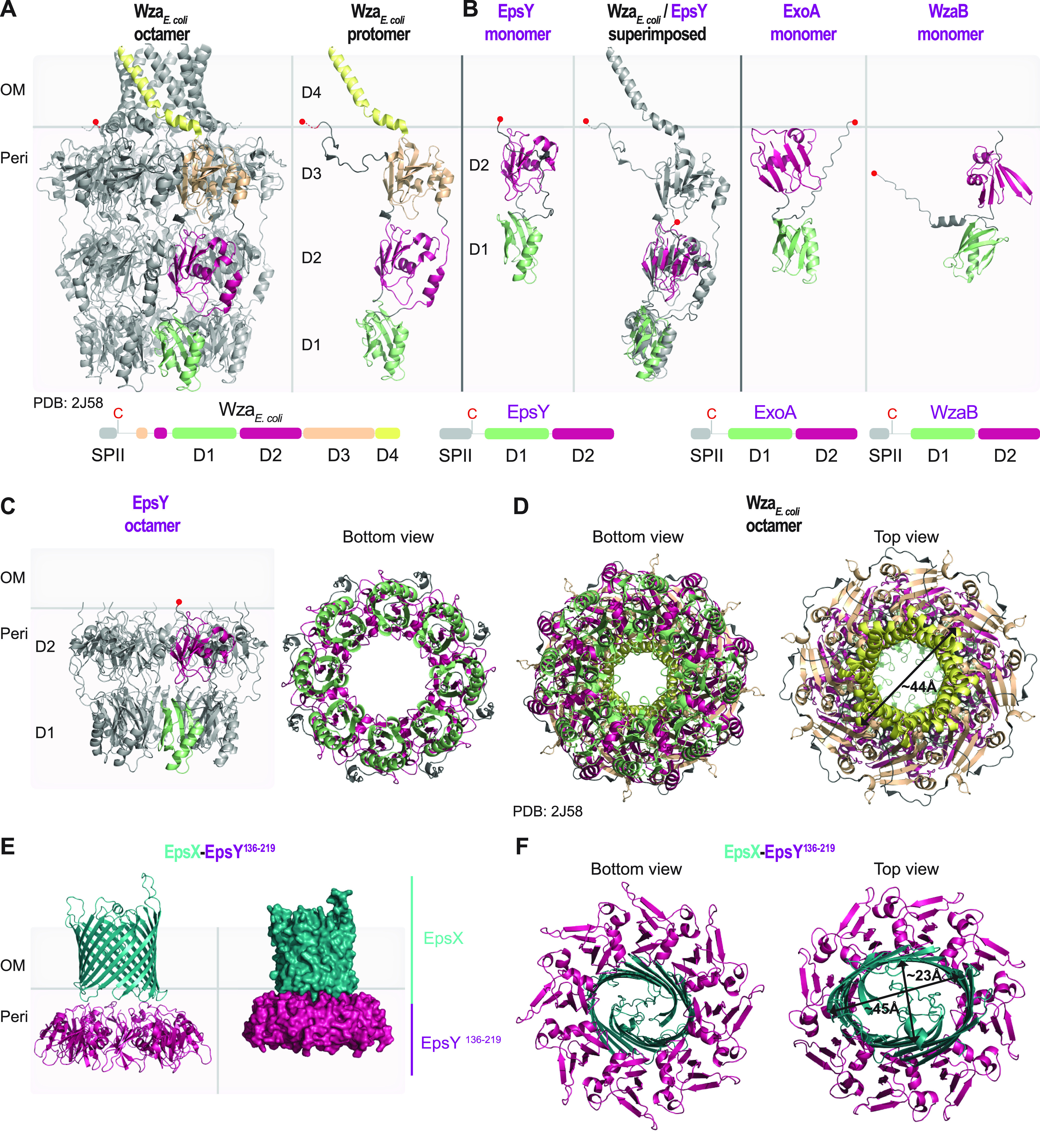
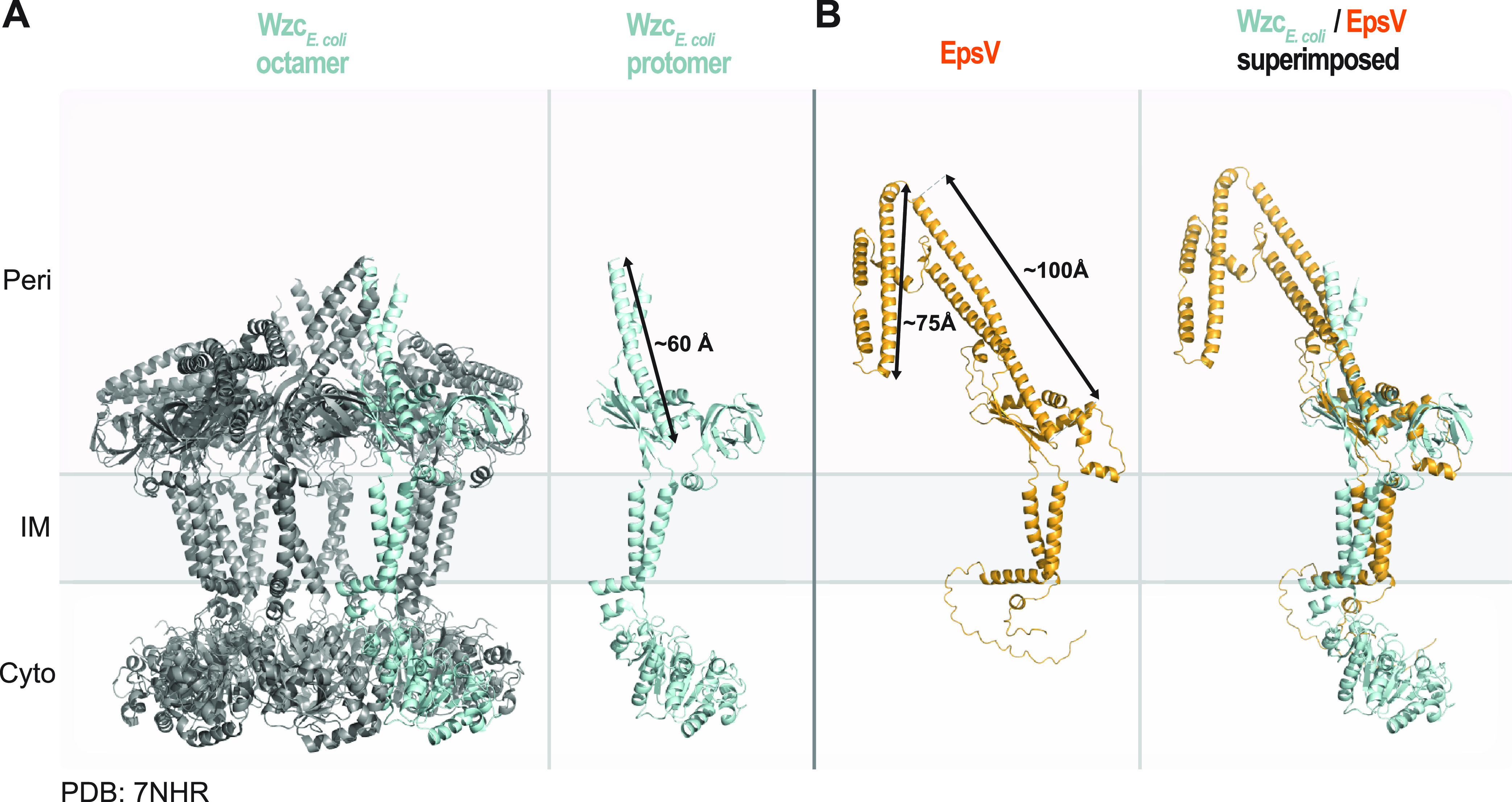
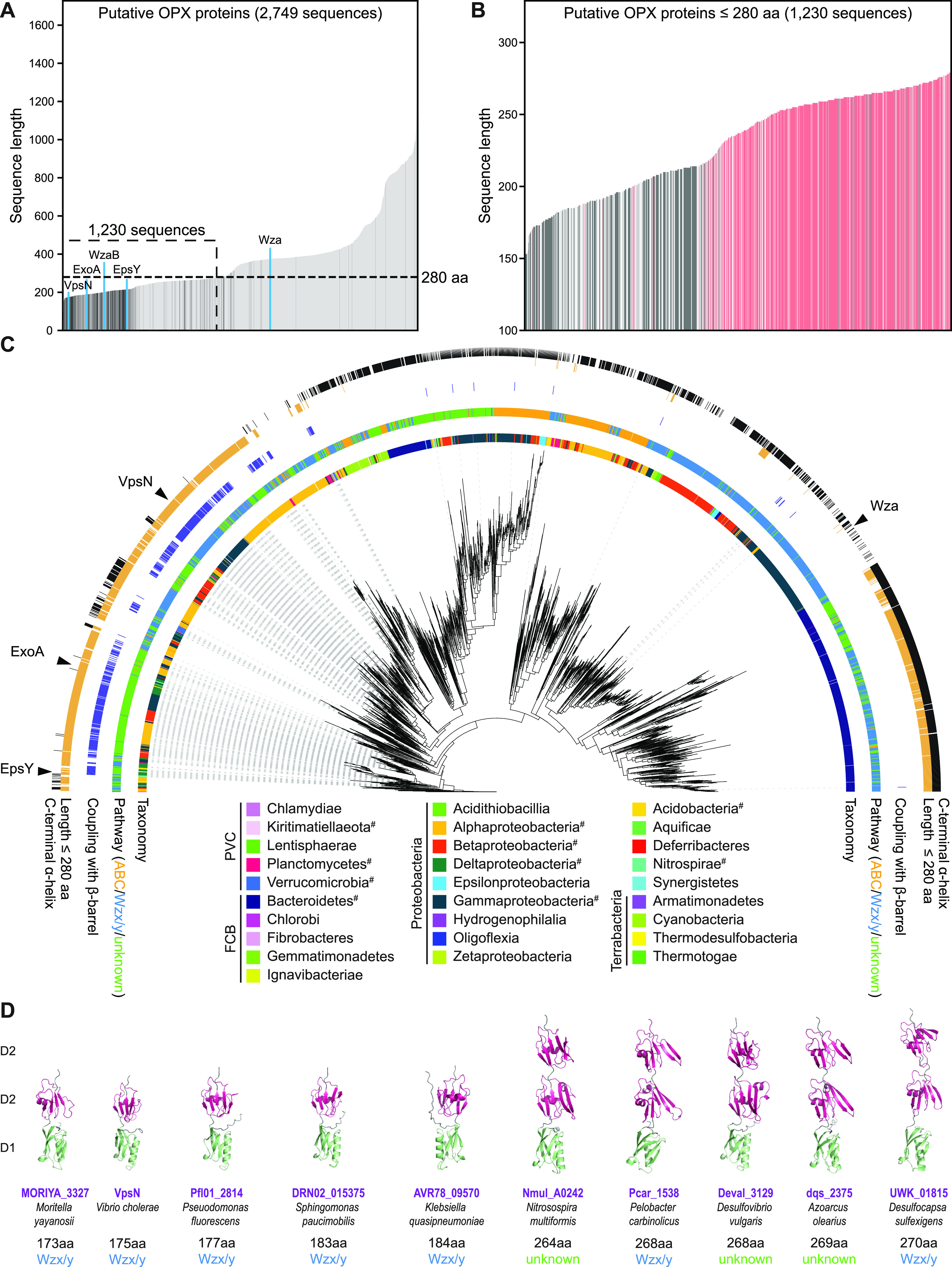
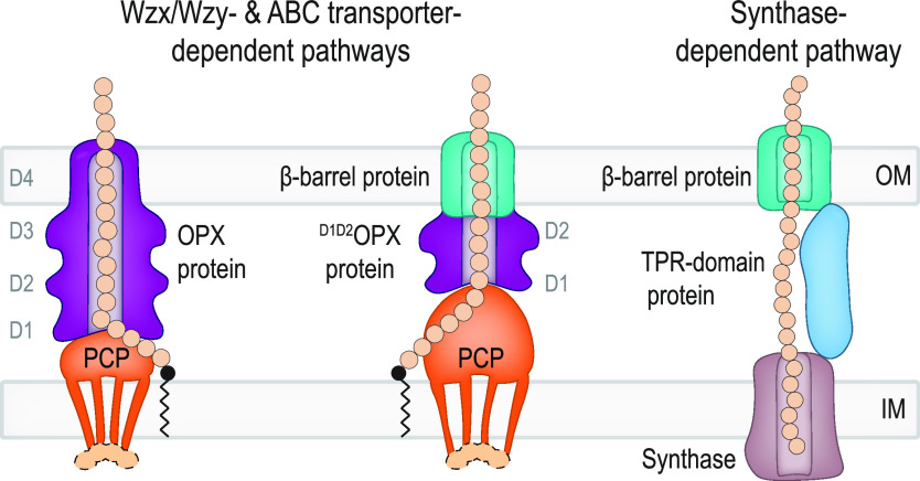
In Gram-negative bacteria, secreted polysaccharides have multiple critical functions. In Wzx/Wzy- and ABC transporter-dependent pathways, an outer membrane (OM) polysaccharide export (OPX) type translocon exports the polysaccharide across the OM. The paradigm OPX protein Wza of Escherichia coli is an octamer in which the eight C-terminal domains form an α-helical OM pore and the eight copies of the three N-terminal domains (D1 to D3) form a periplasmic cavity. In synthase-dependent pathways, the OM translocon is a 16- to 18-stranded β-barrel protein. In Myxococcus xanthus, the secreted polysaccharide EPS (exopolysaccharide) is synthesized in a Wzx/Wzy-dependent pathway. Here, using experiments, phylogenomics, and computational structural biology, we identify and characterize EpsX as an OM 18-stranded β-barrel protein important for EPS synthesis and identify AlgE, a β-barrel translocon of a synthase-dependent pathway, as its closest structural homolog. We also find that EpsY, the OPX protein of the EPS pathway, consists only of the periplasmic D1 and D2 domains and completely lacks the domain for spanning the OM (herein termed a D1D2OPX protein). In vivo, EpsX and EpsY mutually stabilize each other and interact in in vivo pulldown experiments supporting their direct interaction. Based on these observations, we propose that EpsY and EpsX make up and represent a third type of translocon for polysaccharide export across the OM. Specifically, in this composite translocon, EpsX functions as the OM-spanning β-barrel translocon together with the periplasmic D1D2OPX protein EpsY. Based on computational genomics, similar composite systems are widespread in Gram-negative bacteria. IMPORTANCE Bacteria secrete a wide variety of polysaccharides that have critical functions in, e.g., fitness, surface colonization, and biofilm formation and in beneficial and pathogenic human-, animal-, and plant-microbe interactions. In Gram-negative bacteria, export of these chemically diverse polysaccharides across the outer membrane depends on two known translocons, i.e., an outer membrane OPX protein in Wzx/Wzy- and ABC transporter-dependent pathways and an outer membrane 16- to 18-stranded β-barrel protein in synthase-dependent pathways. Here, using a combination of experiments in Myxococcus xanthus, phylogenomics, and computational structural biology, we provide evidence supporting that a third type of translocon can export polysaccharides across the outer membrane. Specifically, in this translocon, an outer membrane-spanning β-barrel protein functions together with an entirely periplasmic OPX protein that completely lacks the domain for spanning the OM. Computational genomics support that similar composite systems are widespread in Gram-negative bacteria.

Keywords: Myxococcus xanthus; OM translocon; OPX proteins; Wzx/Wzy pathway; beta-barrel proteins; capsular polysaccharide; exopolysaccharide; export of polysaccharides; synthase-dependent pathway.


Conflict of interest statement

The authors declare no conflict of interest.

Figures

FIG 1
FIG 1
The 18-stranded β-barrel protein EpsX is an integral part of the EPS pathway. (A) AlphaFold models of EpsX, ExoB, and WzpB. Proteins are oriented based on the N and C termini of OM β-barrel proteins being periplasmic (66). Model ranks 1 are shown. (B) Phenotypic characterization of ΔepsX mutant. Two left columns, cells were placed on 0.5% agar supplemented with 0.5% CTT and Congo red or trypan blue and incubated for 24 h. Two right columns, T4P-dependent motility and gliding motility were tested on 0.5% and 1.5% agar, respectively, supplemented with 0.5% CTT, and images were recorded after 24 h. The ΔpilA mutant, which lacks the major pilin of the T4P (67), and the ΔgltB mutant, which lacks a component of the gliding motility machinery (23), served as negative controls for T4P-dependent and gliding motility, respectively. In the complementation strain, epsX was expressed from the pilA promoter on a plasmid integrated in a single copy at the Mx8 attB site. Scale bars, 1 mm (left) and 50 μm (right). Numbers indicate expansion from the edge of the colony calculated from three biological replicates and relative to that of the WT, where 100% corresponds to 1.4 mm. (C) Comparison of AlgE and EpsX. Upper panel, lateral and top views of the solved structure of AlgE (PDB 3RBH) (11). Arrows indicate the external diameter of the β-barrel. Lower panel, superimposition of the solved structure of AlgE and the EpsX AlphaFold model. EpsX is colored in teal. EpsX aligns to AlgE with a root mean square deviation (RMSD) of 6.035 Å over 1,501 Cα. (D) EpsX and EpsY mutually stabilize each other, and EpsY stabilizes EpsV. Protein amounts in whole-cell proteomes of M. xanthus strains were determined using LFQ mass spectrometry-based proteomics (see Materials and Methods). Normalized log2 intensities of Eps proteins in the indicated strains are shown. Missing bars indicate that the proteins were not detected. Data points represent three biological replicates. Error bars, standard deviation (SD) based in these replicates. *, P < 0.05 (Welch’s test). WzeX is important for EPS synthesis and was proposed to act as the BY kinase partner of EpsV (14, 18). (E) RT-qPCR analysis of epsV, epsY, and epsX transcripts levels. Total RNA was isolated from cells grown as panel D. Data are shown as log2 transcripts in a mutant relative to that of the WT. Individual data points represent four biological replicates with each two technical replicates and are colored according to the strain analyzed. Center marker and error bars represent mean and SD. *, P < 0.05 (Welch’s test). (F) EpsX and EpsY interact. Pulldown experiments on whole-cell lysates of strains expressing EpsY-FLAG or sfGFP-FLAG (negative control). In the EpsY-FLAG strain, epsY-FLAG was expressed ectopically from the pilA promoter on a plasmid integrated in a single copy at the Mx8 attB site. In the sfGFP-FLAG strain, epsY was expressed from the pilA promoter on a plasmid integrated in a single copy at the Mx8 attB site, and sfGFP-FLAG was expressed from the pilA promoter on a plasmid integrated in a single copy at the 18-19 intergenic locus. Samples from four biological replicates were analyzed by LC-MS (see Material and Methods). Log2-fold enrichment of proteins in EpsY-FLAG over sfGFP-FLAG samples was calculated. Columns represent mean log2-fold enrichment (n = 4). Error bars, SD based on these replicates; n.d., proteins detected in neither EpsY-FLAG nor sfGFP-FLAG samples.
FIG 2
FIG 2
Structural characterization of the EpsY D1D2OPX protein alone and in complex with EpsX, its partner 18-stranded β-barrel protein. (A) Structure of WzaE. coli. Left panel, the solved structure of octameric Wza (PDB 2J58) (7). Right panel, an individual Wza protomer. The four domains of Wza are labeled D1 to D4. Light green, D1; dark pink, D2; light orange, D3; yellow, D4. The acylated N-terminal cysteine is indicated by a red circle and placed at the inner leaflet of the OM. Lower panel, domain organization of Wza. (B) AlphaFold model of EpsY. Left panel, lateral view of EpsY monomer as predicted by AlphaFold. Right panel, superimposition of a Wza protomer from the solved structure (gray) and the EpsY model. The EpsY monomer aligns to the Wza protomer with an RMSD of 3.306 Å over 904 Cα. Right panels, AlphaFold models of ExoA and WzaB monomers. In all three AlphaFold models, the two domains are labeled D1 and D2 and colored according to the homologous domains in Wza. The acylated N-terminal cysteine is indicated by a red circle; note that the acylated N-terminal cysteine of WzaB is not modeled “on top” of D2, but the confidence in the relative position of this residue is low (Fig. S3C). Model rank 1 is shown for all structures. Lower panels, domain organization of EpsY, ExoA, and WzaB. SPII, type 2 signal peptide. (C) AlphaFold-Multimer model of octameric EpsY. Left panel, one protomer is colored as described in the legend for panel B. Right panel, bottom view of octameric EpsY with all protomers colored as described for panel B. Model rank 1 is shown. (D) Structure of Wza. All eight protomers are colored as described in the legend for panel A. An arrow indicates the external diameter of the α-helical pore. In the bottom view, the tyrosine residues that form the so-called tyrosine ring are present in the loops extending into the central channel. (E and F) AlphaFold-Multimer model of a heterocomplex of octameric EpsY136-219 and an EpsX monomer. In the heterocomplex, the EpsY136-219 octamer is colored as D2 as described for panel B, and EpsX is colored teal. (E) The right panel is a surface-rendered representation. Model rank 1 is shown. (F) Arrows indicate the diameter of the β-barrel. Model rank 1 is shown.
FIG 3
FIG 3
Structural characterization of the PCP EpsV. (A) Solved structure of octameric WzcE. coli (PDB 7NHR) (30). Left panel, the protein is colored in gray, and one protomer is colored in light blue. Right panel, individual Wzc protomer in class 2 conformation (30). Note that individual protomers have different conformations in the octamer. An arrow indicates the length of the extended α-helical stretch. (B) AlphaFold model of EpsV. Arrows indicate the lengths of the α-helical stretches. Model rank 1 is shown. Right panel, superimposition of a protomer from the solved structure of Wzc and the EpsV model. EpsV aligns to Wzc with an RMSD of 4.610 Å over 727 Cα.
FIG 4
FIG 4
Computational genomics of OPX proteins. (A) Length distribution of 2,749 OPX proteins. OPX proteins encoded within a distance of five genes of a gene encoding a β-barrel protein are shown in dark gray, and the remaining OPX proteins are in light gray. OPX proteins discussed in the text are highlighted in blue. The 1,230 short OPX proteins are indicated based on the upper size limit of ≤280 aa. (B) Length distribution of OPX proteins of ≤280 aa. Four hundred fifty-three OPX proteins with a coupled β-barrel protein and no C-terminal α-helix are in dark gray; 535 OPX proteins with no coupled β-barrel protein but a C-terminal α-helix are in red. Proteins not matching these criteria are in light gray. (C) Maximum-likelihood phylogenetic tree built from the PES motif of the 2,749 OPX proteins. The inner ring is colored based on NCBI taxonomy (https://www.ncbi.nlm.nih.gov/taxonomy/). PVC, FCB, and Terrabacteria are superphyla, and the phylum Proteobacteria is divided into classes. #, this phylum contains an OPX protein(s) of ≤280 aa coupled to a β-barrel protein(s). The second ring indicates the pathway assigned to individual OPX proteins. The third ring indicates, in dark blue, coupling with a β-barrel protein. The fourth ring indicates, in orange, OPX proteins of ≤280 aa. The fifth ring indicates, in black, whether an OPX protein has a C-terminal α-helix. (D) AlphaFold models of indicated OPX proteins. Domains are labeled D1 and D2 and colored according to the homologous domains in Wza (Fig. 2A). Model rank 1 is shown for all structures.
FIG 5
FIG 5
Three different mechanisms for polysaccharide export across the OM. Left schematic, in classical Wzx/Wzy- and ABC transporter-dependent pathways, polysaccharide transfer across the periplasm and OM is mediated by a complex composed of a PCP and an OPX protein with an α-helical barrel in the OM. Middle schematic, in pathways in which the OPX protein lacks domain D4 (D1D2OPX or D1D2D2OPX proteins), a β-barrel protein constitutes the OM part of the composite OPX/β-barrel protein translocon. The D1 to D4 and D1 to D2 rings of the OPX proteins are indicated. In Wzx/Wzy-dependent pathways, the PCP can be associated with a cytoplasmic BY kinase (light orange). Synthesis, polymerization, and translocation of the polysaccharide across the IM are not shown. Right schematic, in synthase-dependent pathways, transfer across the periplasm and the OM depends on a TPR domain-containing protein and a β-barrel protein translocon in the OM.

Similar articles

Cited by

References

    1. Morris G, Harding S. 2009. Polysaccharides, microbial, p 482–494. In Encyclopedia of microbiology. Elsevier. doi:10.1016/B978-012373944-5.00135-8. - DOI
    1. Sutherland IW. 1998. Novel and established applications of microbial polysaccharides. Trends Biotechnol 16:41–46. doi:10.1016/S0167-7799(97)01139-6. - DOI - PubMed
    1. Hall-Stoodley L, Costerton JW, Stoodley P. 2004. Bacterial biofilms: from the natural environment to infectious diseases. Nat Rev Microbiol 2:95–108. doi:10.1038/nrmicro821. - DOI - PubMed
    1. Low KE, Howell PL. 2018. Gram-negative synthase-dependent exopolysaccharide biosynthetic machines. Curr Opin Struct Biol 53:32–44. doi:10.1016/j.sbi.2018.05.001. - DOI - PubMed
    1. Whitfield C, Wear SS, Sande C. 2020. Assembly of bacterial capsular polysaccharides and exopolysaccharides. Annu Rev Microbiol 74:521–543. doi:10.1146/annurev-micro-011420-075607. - DOI - PubMed

Publication types

MeSH terms

Substances

LinkOut - more resources