. 2022 Oct 26;13(5):e0203922.
doi: 10.1128/mbio.02039-22. Epub 2022 Aug 16.

Herpes Simplex Virus 1 Entry Glycoproteins Form Complexes before and during Membrane Fusion

Affiliations

Herpes Simplex Virus 1 Entry Glycoproteins Form Complexes before and during Membrane Fusion

Zemplen Pataki et al. mBio. .

Abstract

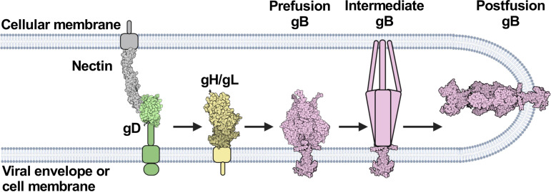
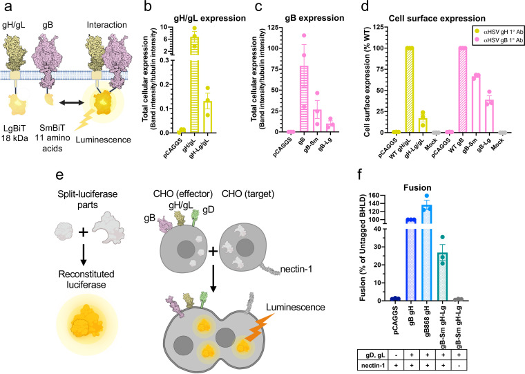
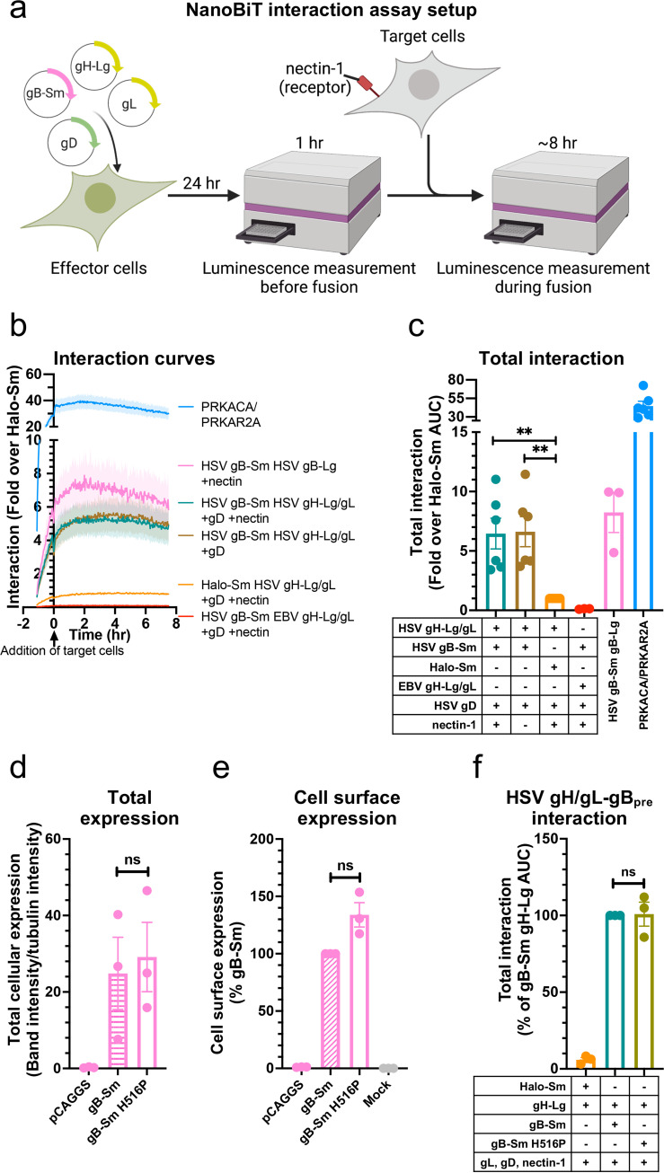
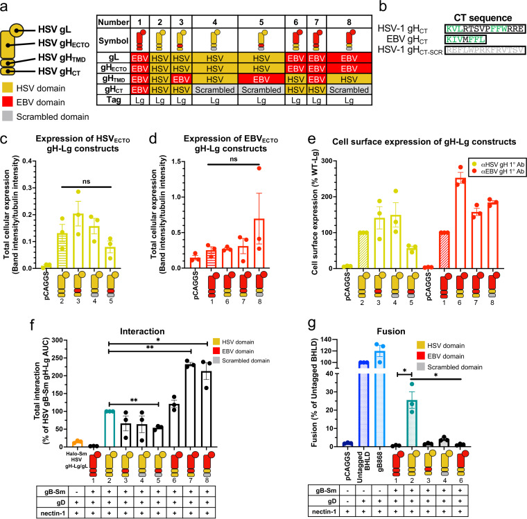
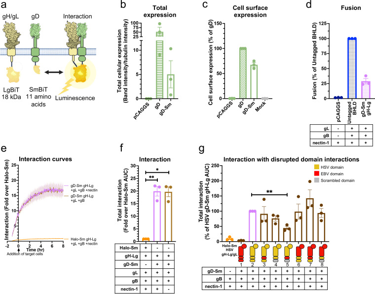
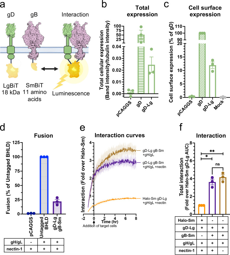
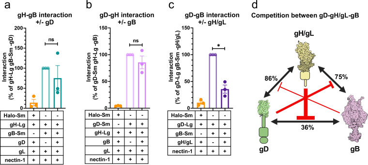
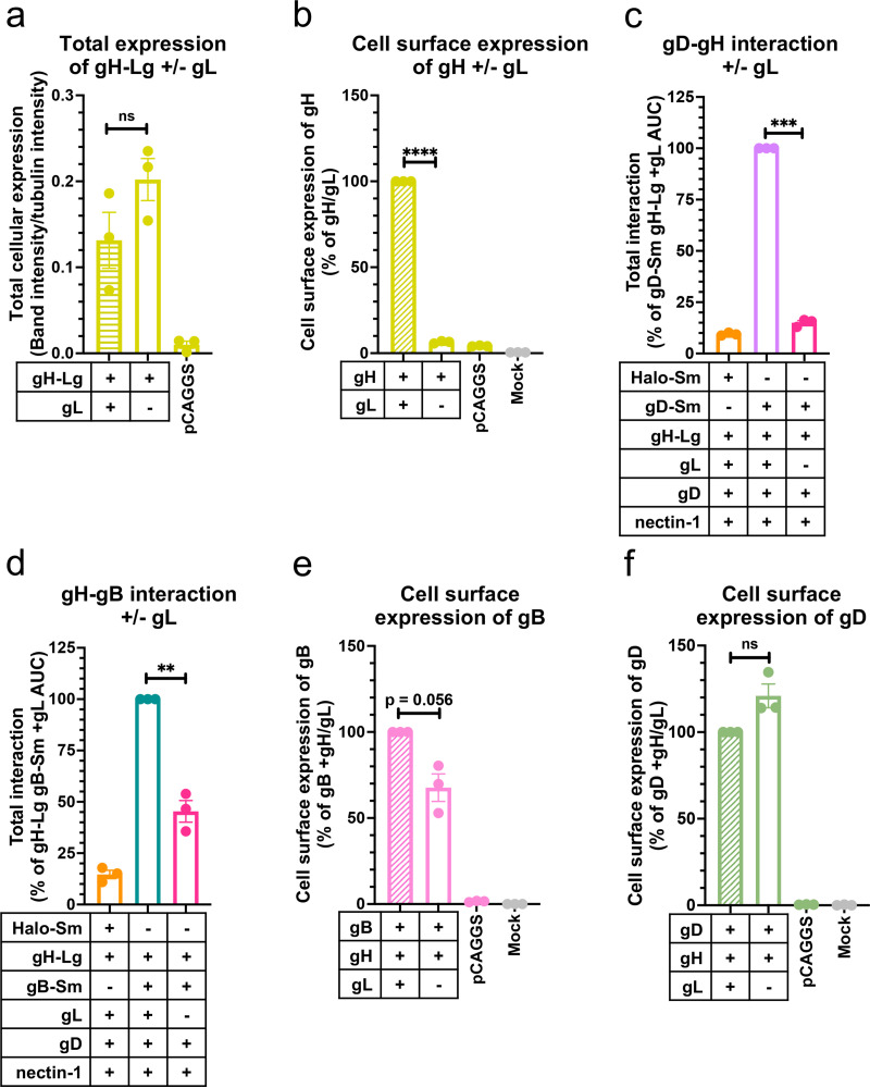
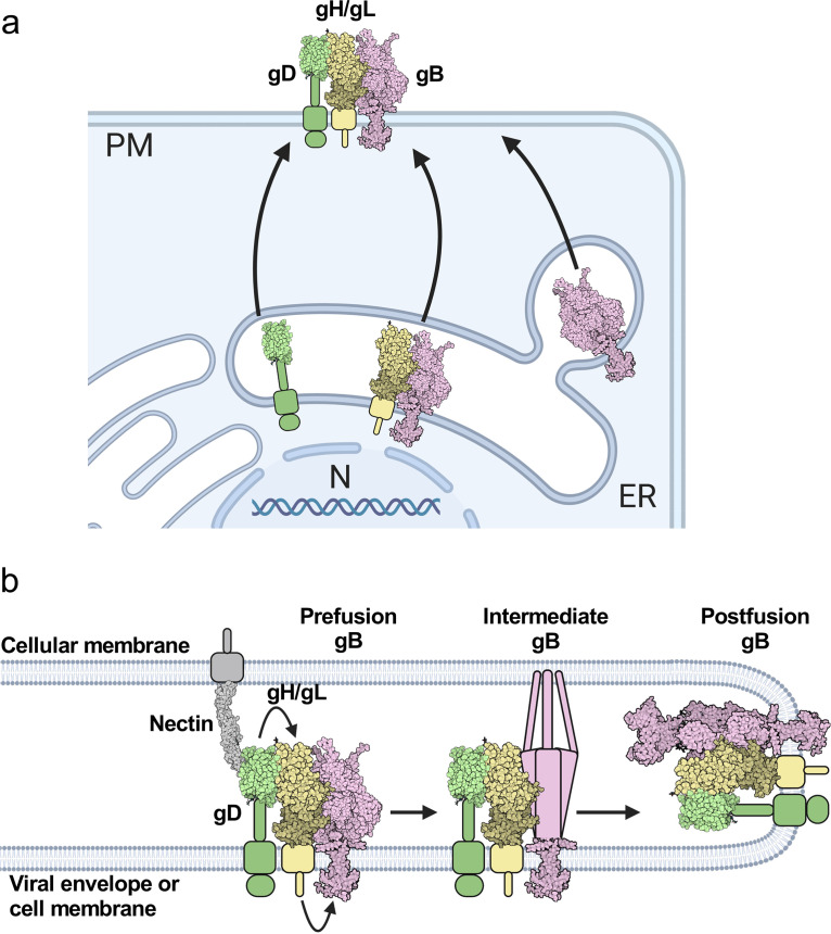
Herpesviruses-ubiquitous pathogens that cause persistent infections-have some of the most complex cell entry mechanisms. Entry of the prototypical herpes simplex virus 1 (HSV-1) requires coordinated efforts of 4 glycoproteins, gB, gD, gH, and gL. The current model posits that the glycoproteins do not interact before receptor engagement and that binding of gD to its receptor causes a "cascade" of sequential pairwise interactions, first activating the gH/gL complex and subsequently activating gB, the viral fusogen. But how these glycoproteins interact remains unresolved. Here, using a quantitative split-luciferase approach, we show that pairwise HSV-1 glycoprotein complexes form before fusion, interact at a steady level throughout fusion, and do not depend on the presence of the cellular receptor. Based on our findings, we propose a revised "conformational cascade" model of HSV-1 entry. We hypothesize that all 4 glycoproteins assemble into a complex before fusion, with gH/gL positioned between gD and gB. Once gD binds to a cognate receptor, the proximity of the glycoproteins within this complex allows for efficient transmission of the activating signal from the receptor-activated gD to gH/gL to gB through sequential conformational changes, ultimately triggering the fusogenic refolding of gB. Our results also highlight previously unappreciated contributions of the transmembrane and cytoplasmic domains to glycoprotein interactions and fusion. Similar principles could be at play in other multicomponent viral entry systems, and the split-luciferase approach used here is a powerful tool for investigating protein-protein interactions in these and a variety of other systems. IMPORTANCE Herpes simplex virus 1 (HSV-1) infects the majority of humans for life and can cause diseases ranging from painful sores to deadly brain inflammation. No vaccines or curative treatments currently exist. HSV-1 infection of target cells requires coordinated efforts of four viral glycoproteins. But how these glycoproteins interact remains unclear. Using a quantitative protein interaction assay, we found that HSV-1 glycoproteins form receptor-independent complexes and interact at a steady level. We propose that the 4 proteins form a complex, which could facilitate transmission of the entry-triggering signal from the receptor-binding component to the membrane fusogen component through sequential conformational changes. Similar principles could be applicable across other multicomponent protein systems. A revised model of HSV-1 entry could facilitate the development of therapeutics targeting this process.

Keywords: HSV-1; NanoBiT protein interaction assay; chimeric protein; domain; glycoprotein; herpesvirus; interaction; membrane fusion; split-luciferase cell-cell fusion assay.


Conflict of interest statement

The authors declare no conflict of interest.

Figures

FIG 1
FIG 1
HSV-1 fusion pathway model. gD (PDB 2C36) (72) binds a receptor (PDB 3U83) (77) on the target cell. gD then activates gH/gL; gH/gL (PDB 3M1C) (78) activates gB (PDB 6Z9M and 5V2S) (12, 15) to refold and cause membrane fusion. gD is thought to be a dimer (72) but is shown here as a monomer for clarity. Figure created with BioRender.com.
FIG 2
FIG 2
Tagging HSV-1 gH/gL and gB with split luciferases to probe their interaction. (a) NanoBiT protein-protein interaction assay approach (49). The interaction between gH/gL and gB was tested by tagging gH and gB with complementary parts of a split luciferase and transfecting into cells. Reconstitution of luciferase reports on fusion. (b to d) Total cellular expression and cell-surface expression of Lg- and Sm- tagged gH and gB by Western blotting and flow cytometry, respectively. R137 and R68 antibodies for gH/gL and gB for Western blotting, respectively. LP11 and R68 antibodies for gH/gL and gB for flow cytometry, respectively. pCAGGS was an empty vector negative control. The mock control was an untransfected negative control, incubated with a nontargeting primary antibody. Columns show the mean. Error bars are the standard error of the mean (SEM). e) Split-luciferase cell-cell fusion assay (51) experimental setup to test whether tagged proteins retain their function. Cells transfected with viral proteins fuse to cells transfected with nectin-1 receptor. Reconstitution of Rluc8 luciferase reports on fusion. (b) Total fusion of gB-Sm and gH-Lg/gL, 8 h after mixing effector and target cells. gB868 was a hyperfusogenic positive-control gB construct. pCAGGS and the condition without nectin-1 receptor are negative controls. Columns show the mean. Error bars are the SEM. Data are three biological replicates from independent experiments. Diagrams and cartoons were created using BioRender.com.
FIG 3
FIG 3
HSV-1 gH/gL and gB interact at a steady level and independently of nectin-1. (a) Interaction assay experimental setup. Cells are transfected with viral proteins required for fusion, including split-luciferase-tagged proteins of interest. Interaction is measured by luminescence before and during fusion. Fusion is induced by the addition of target cells expressing the viral receptor nectin-1. (b) gH/gL and gB interaction over time, with target cells expressing or lacking nectin-1. The Halo-Sm and EBV gH-Lg conditions were negative controls. gB-Sm/gB-Lg and PRKACA/PRKAR2A were positive controls. The shaded regions are the SEM. (c) The interactions are quantified by calculating the area under the curve (AUC). (d and e) Total cellular expression and cell-surface expression of Sm-tagged gB H516P—which locks gB in its prefusion conformation—by Western blotting and flow cytometry, respectively. (f) The interaction between gH/gL and gB H516P. Columns show the mean. Error bars are the SEM. ns, not statistically significant; **, P < 0.01. Data in all panels are three biological replicates from independent experiments. Diagrams were created with BioRender.com.
FIG 4
FIG 4
All HSV-1 gH domains are involved in interactions with HSV-1 gB. (a) Summary of the composition of gH-Lg/gL constructs designed to disrupt domain interactions. (b) Sequence comparison of HSV-1, EBV, and scrambled HSV-1 gH cytotails. Green indicates similar clusters of residues in HSV-1 and EBV. (c and d) Total cellular expression of gH-Lg/gL constructs with disrupted domain interactions compared to HSV gH-Lg/gL (2) or EBV gH-Lg/gL (1). Cartoons represent gH-Lg/gL constructs, indicating which domains are HSV-1 (yellow), EBV (red), or scrambled (gray). R137 and R2267 antibodies were used against constructs with HSV and EBV ectodomains, respectively. (e) Cell surface expression of gH-Lg/gL constructs with disrupted domain interactions. LP11 and AMMO1 antibodies were used against constructs with HSV and EBV ectodomains, respectively. (f) Interaction between gB and gH/gL constructs with disrupted ectodomain, TMD, or CTD interactions. (g) Total fusion of gB-Sm and gH-Lg/gL constructs with disrupted domain interactions 8 h after mixing effector and target cells. Columns show the mean. Error bars are the SEM. *, P < 0.05; **, P < 0.01. Data in all panels are three biological replicates from independent experiments. Cartoons were created using BioRender.com.
FIG 5
FIG 5
HSV-1 gH/gL and gD interact at a steady level and independently of nectin-1, through all domains. (a) NanoBiT interaction assay setup to test gD-gH/gL interactions. (b and c) Total cellular expression and cell surface expression of gD-Sm by Western blotting and flow cytometry, respectively. R7 and DL6 antibodies were used, respectively. (d) Total fusion of gD-Sm and gH-Lg/gL. (e and f) Interaction of gD and gH/gL. Curves or bars represent the mean, and the shaded area or error bars are the SEM. (g) Interaction of gD with gH/gL with disrupted domain interactions. Columns are the mean, and error bars are the SEM. *, P < 0.05; **, P < 0.01. Data in all panels are three biological replicates from independent experiments. Illustrations were created with BioRender.com.
FIG 6
FIG 6
gD and gB interact at a steady level and independently of nectin-1. (a) NanoBiT interaction assay setup to test gD-gB interactions. Created with BioRender.com. (b and c) Total cellular expression and cell surface expression of gD-Lg by Western blotting and flow cytometry, respectively. (d) Total fusion of gD-Lg and gB-Sm. (e and f) Interaction of gD and gB. Curves indicate the mean and the shaded area is the SEM. Columns are the mean and error bars are the SEM. *, P < 0.05; **, P < 0.01. Data in all panels are three biological replicates from independent experiments.
FIG 7
FIG 7
gD, gH/gL, and gB compete with one another for binding. (a) Interaction of gH/gL and gB in the absence versus the presence of gD. (b) Interaction of gD and gH/gL in the absence versus the presence of gB. (c) Interaction of gD and gB in the absence versus the presence of gH/gL. Columns indicate the mean and error bars are the SEM. *, P < 0.05. Data represent three biological replicates from independent experiments in all panels. (d) Binding competition model between gD, gH/gL, and gB. The interaction of each pair was decreased in the presence of the third interacting partner, suggesting interaction inhibition by binding competition. The sizes of the red inhibitory arrows are proportional to the degree of inhibition. Values indicate the percentage of the interaction of two interacting partners that remains after inhibition by the presence of the third interacting partner. Created using BioRender.com.
FIG 8
FIG 8
HSV-1 gL is required for gH-gB and gD-gH interactions and for cell surface expression of gH and gB. (a) Total cellular expression of gH-Lg in the presence versus the absence of gL. (b) gH cell surface expression in the presence versus the absence of gL (60, 70) using R137. (c) gD-gH interaction in the presence versus the absence of gL. (d) gH-gB interaction in the presence versus the absence of gL. (e) gB cell surface expression in the presence versus the absence of gL. (f) gD cell surface expression in the presence versus the absence of gL. Columns are the mean. Error bars are the SEM. **, P < 0.01; ***, P < 0.001; ****, P < 0.0001. Data represent three biological replicates from independent experiments in all panels.
FIG 9
FIG 9
gD-gH/gL-gB trafficking, interaction, and fusion models. (a) Intracellular interactions and trafficking of gD, gH/gL, and gB. gD does not interact with gH in the ER and traffics independently of gH to the plasma membrane. gB interacts with gH in the ER and may traffic with gH/gL to the plasma membrane. Some gB traffics to the plasma membrane without gH/gL. gD, gH/gL, and gB all interact with one another once they leave the ER and compete with one another for binding. gH/gL inhibits binding of the other two binding partners the most, suggesting it binds well to both gD and gB and may position itself between gD and gB in the putative gD-gH/gL-gB complex. gH/gL interacts with gD and gB through all three domains. Cell and glycoprotein sizes are not to scale. (b) New HSV-1 fusion pathway model. gD, gH/gL, and gB are all interacting with each other before fusion. Nectin-1 binds to gD, causing a conformational change (not shown), which activates gH/gL via their ectodomains. gH/gL undergoes a conformational change (not shown) and the gHCT activates the gBCTD. The gB ectodomain refolds and catalyzes membrane fusion. gD, gH/gL, and gB continue to interact. For this model we assume a complex with a 1:1:1 ratio of gD:gH/gL:gB, but the true stoichiometry is unknown.

Similar articles

Cited by

References

    1. Harrison SC. 2008. Viral membrane fusion. Nat Struct Mol Biol 15:690–698. doi:10.1038/nsmb.1456. - DOI - PMC - PubMed
    1. Connolly SA, Jardetzky TS, Longnecker R. 2021. The structural basis of herpesvirus entry. Nat Rev Microbiol 19:110–121. doi:10.1038/s41579-020-00448-w. - DOI - PMC - PubMed
    1. Cohen JI. 2020. Herpesvirus latency. J Clin Invest 130:3361–3369. doi:10.1172/JCI136225. - DOI - PMC - PubMed
    1. Renner DW, Szpara ML. 2018. Impacts of genome-wide analyses on our understanding of human herpesvirus diversity and evolution. J Virol 92:e00908-17. doi:10.1128/JVI.00908-17. - DOI - PMC - PubMed
    1. Looker KJ, Magaret AS, May MT, Turner KM, Vickerman P, Gottlieb SL, Newman LM. 2015. Global and regional estimates of prevalent and incident herpes simplex virus type 1 infections in 2012. PLoS One 10:e0140765. doi:10.1371/journal.pone.0140765. - DOI - PMC - PubMed

Publication types